Loading subject page…


167,935 views

Video Summary: What Is Fluid Mosaic Model
Ever wonder how your iPhone's screen protector stays flexible yet maintains its structure? The fluid mosaic model works similarly-it explains how cell membranes maintain flexibility while controlling what enters and exits cells. Proposed by Singer and Nicholson in 1972, this groundbreaking model revolutionized our understanding of cellular boundaries. From muscle contractions during a marathon to insulin signaling in diabetes patients at Johns Hopkins Hospital, this model explains countless biological processes. Watch the full video on JoVE Coach to master this concept with expert-led visuals and step-by-step explanations.
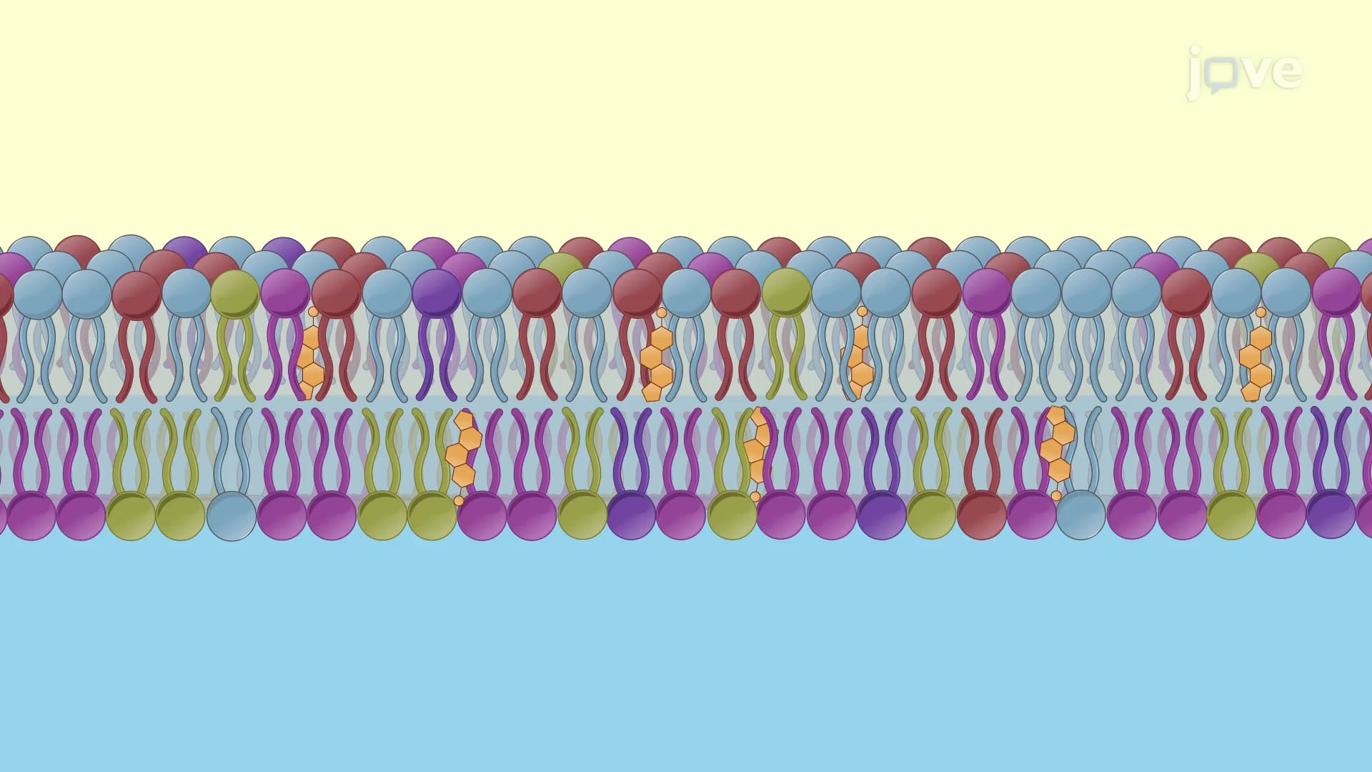
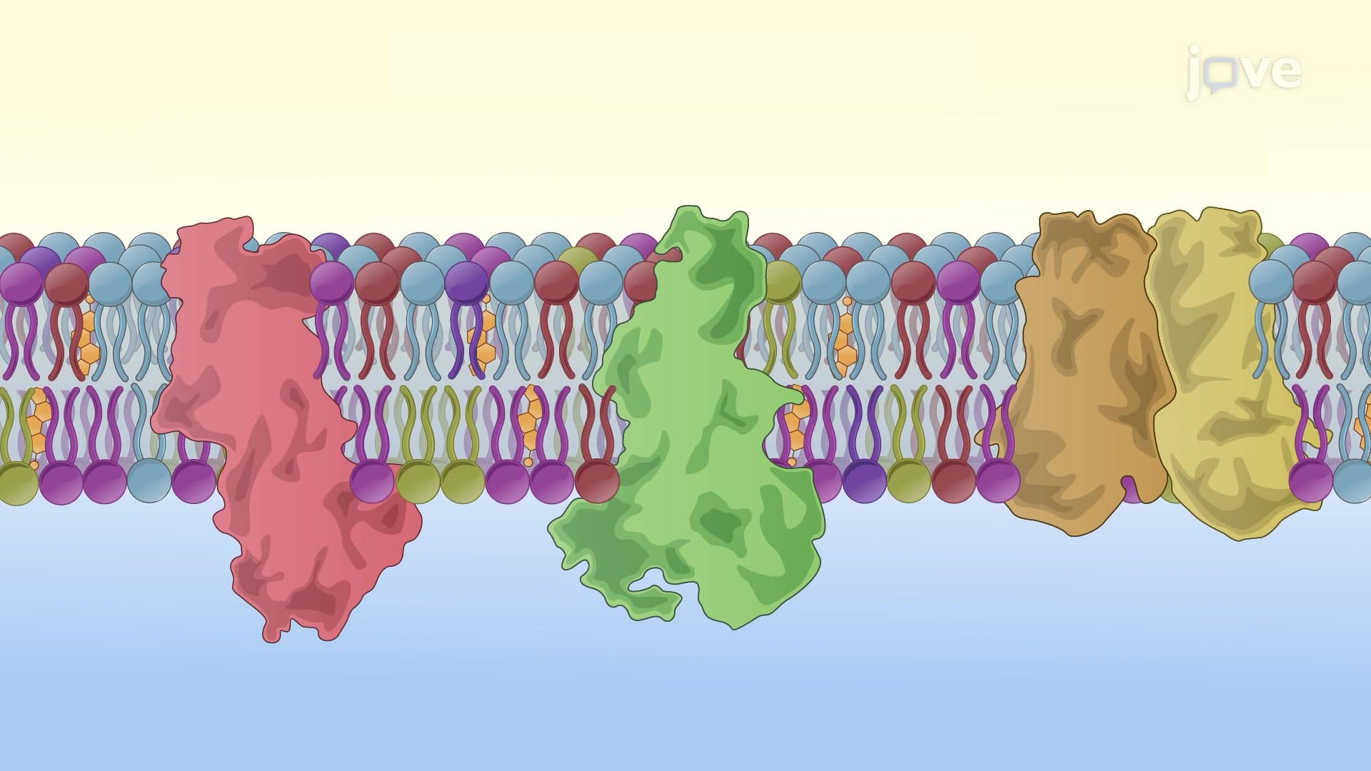
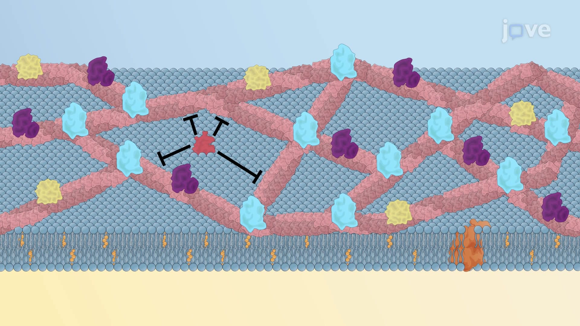
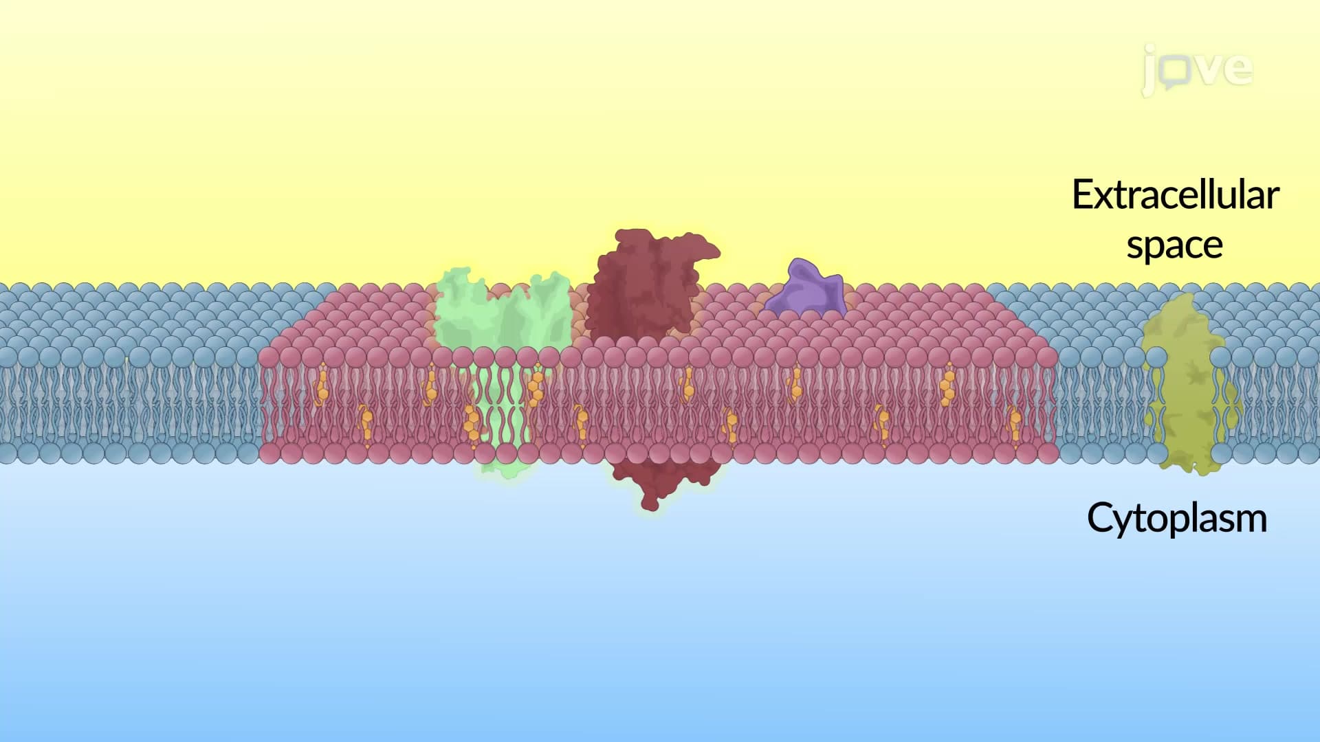
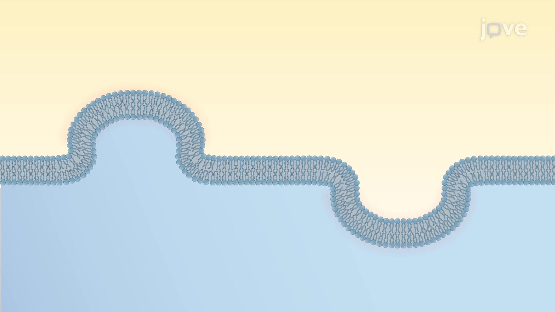
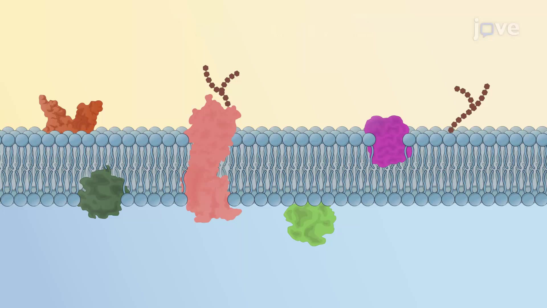
The fluid mosaic model represents one of biology's most elegant structural concepts, describing cell membranes as dynamic, flexible barriers rather than rigid walls. This model fundamentally changed how scientists understand cellular boundaries, comparing them to a "fluid" sea of lipids with a "mosaic" of proteins floating within and on the surface.
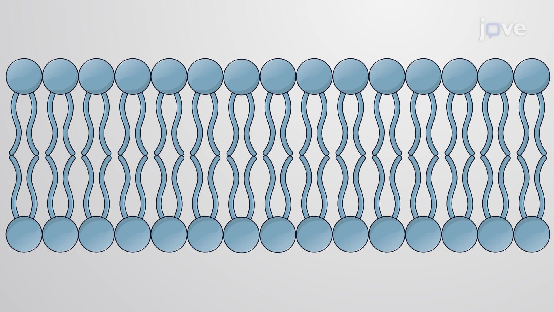
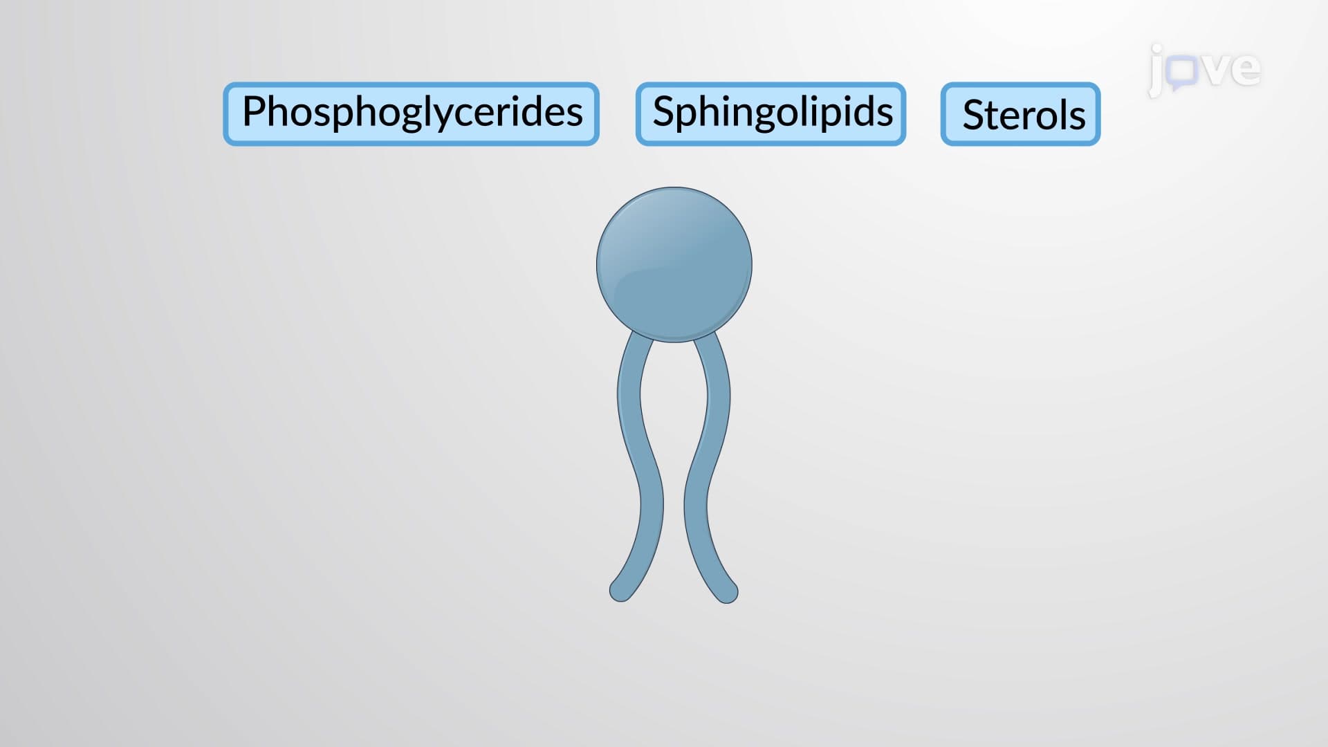
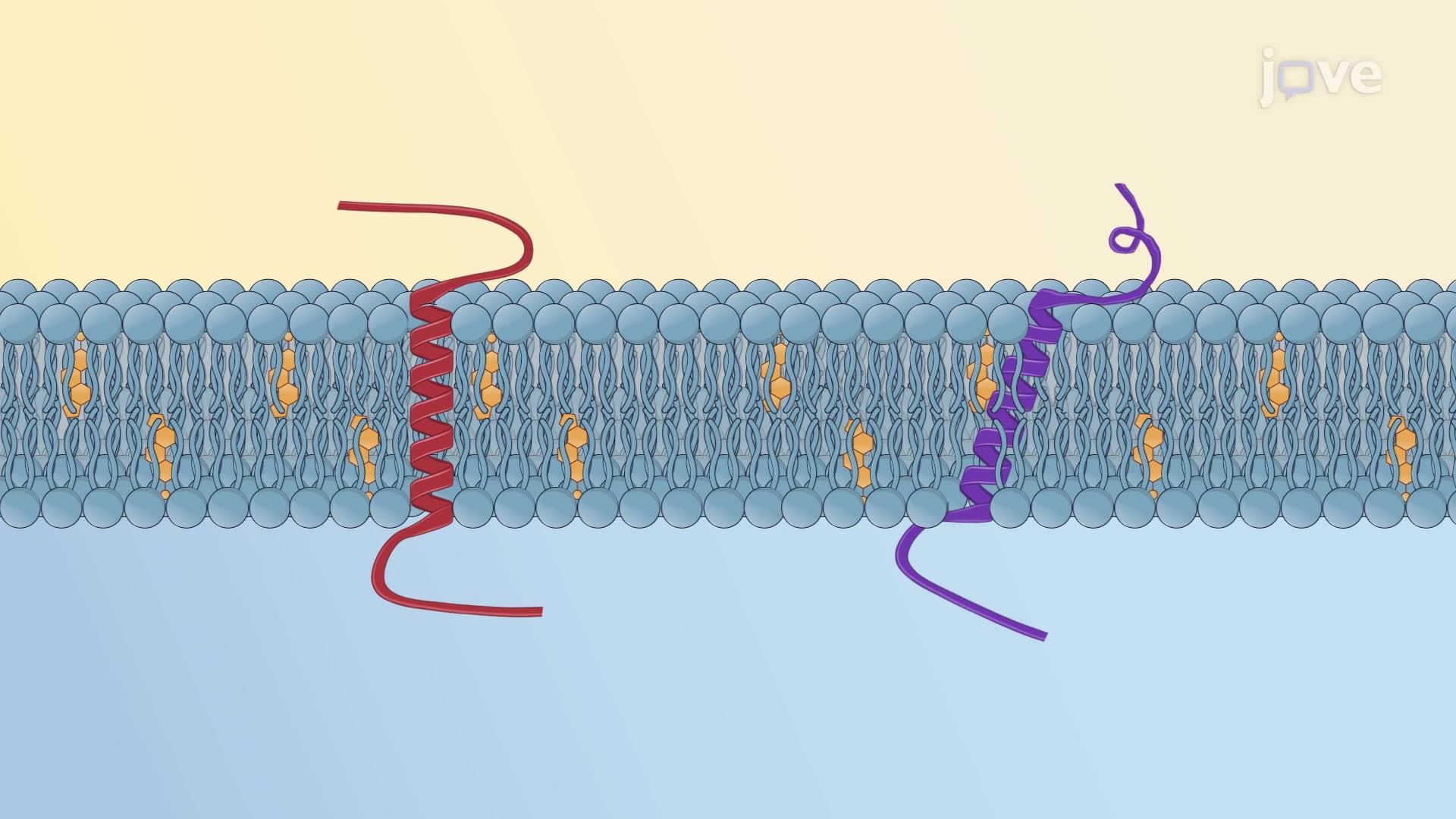
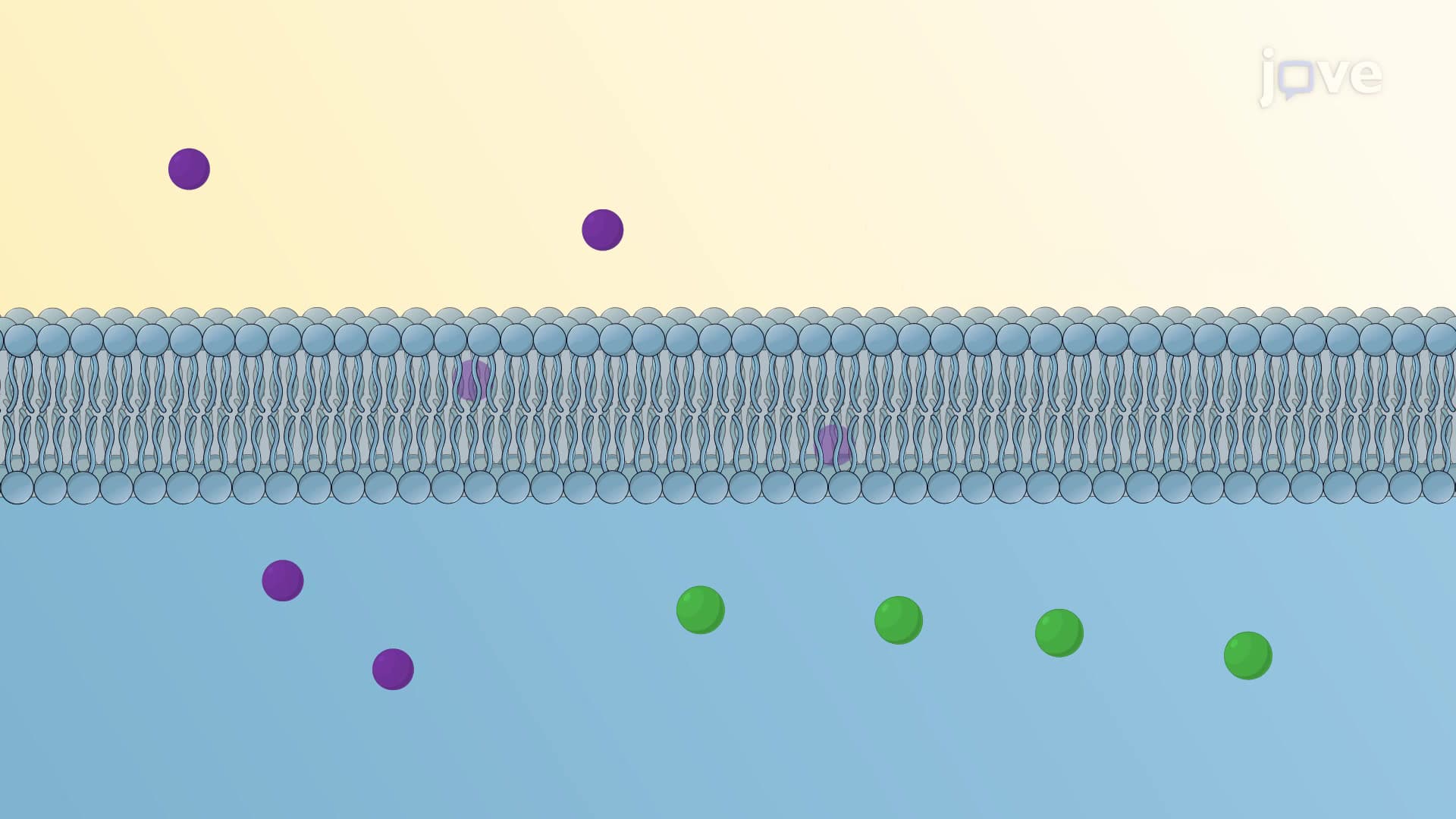
At its core, the phospholipid bilayer forms the membrane's foundation. Each phospholipid molecule contains a hydrophilic (water-loving) phosphate head and two hydrophobic (water-fearing) fatty acid tails. These molecules spontaneously arrange into a double layer, with heads facing the watery environments inside and outside the cell, while tails cluster together in the membrane's interior. This arrangement occurs naturally due to thermodynamic principles-no cellular energy required.
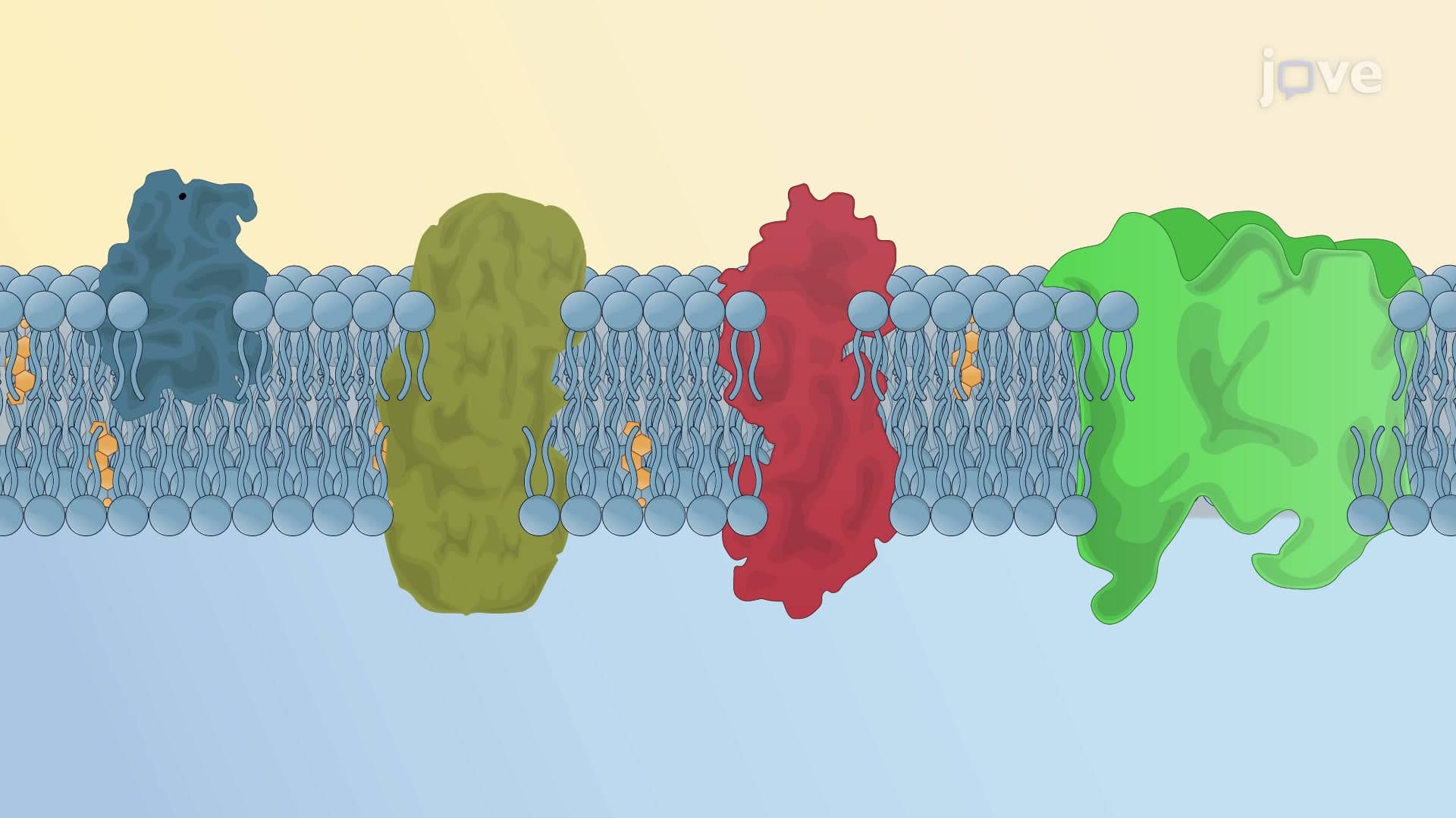
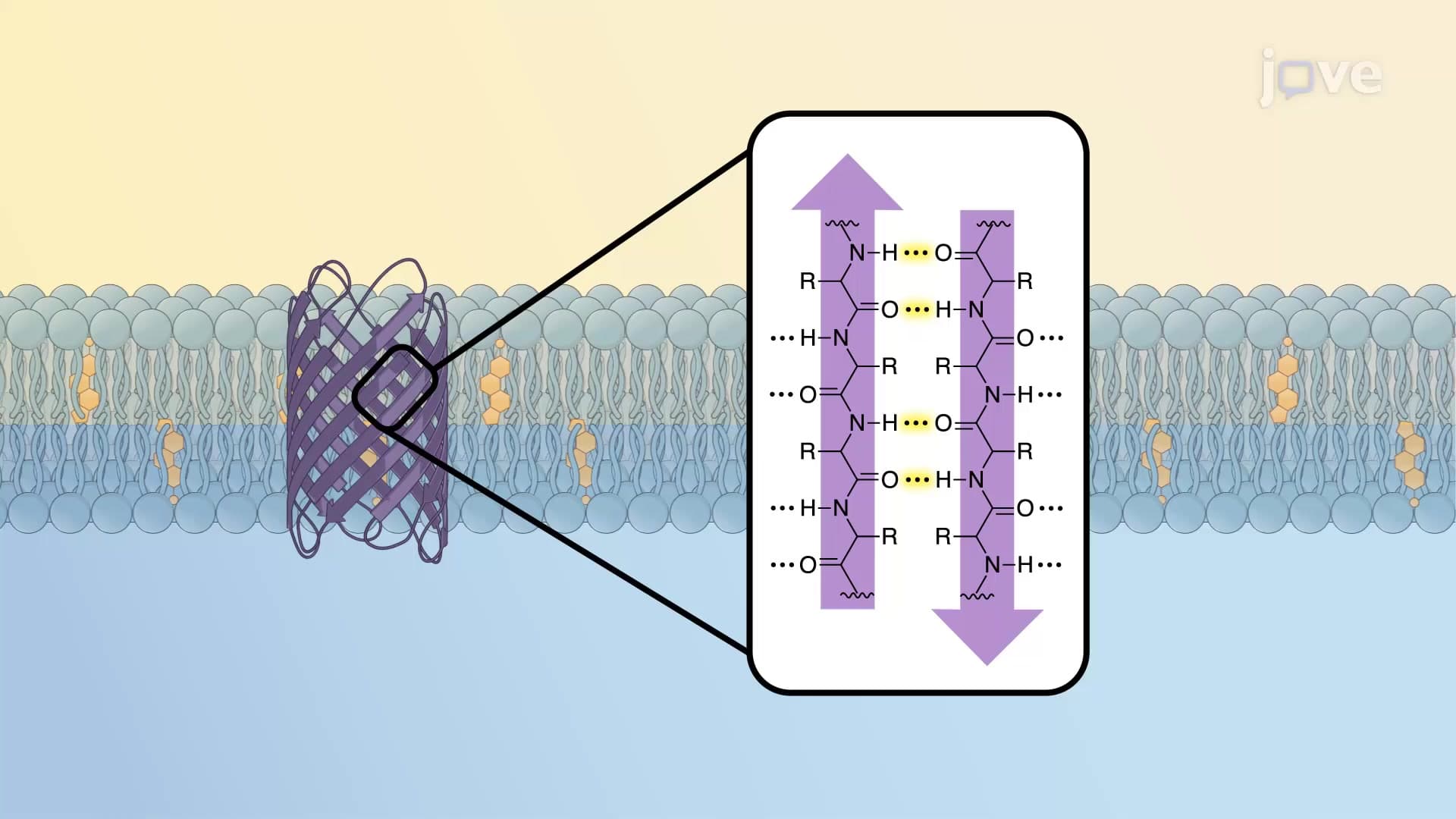
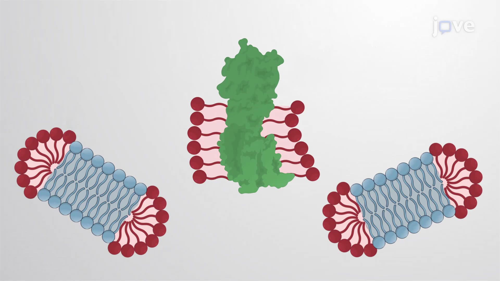
Membrane proteins serve as the membrane's workforce, performing specialized functions that pure lipid layers cannot accomplish. Integral proteins span the entire membrane thickness, often forming channels or transporters that allow specific molecules to cross the cellular barrier. The glucose transporters that move sugar into muscle cells during exercise exemplify integral proteins in action.
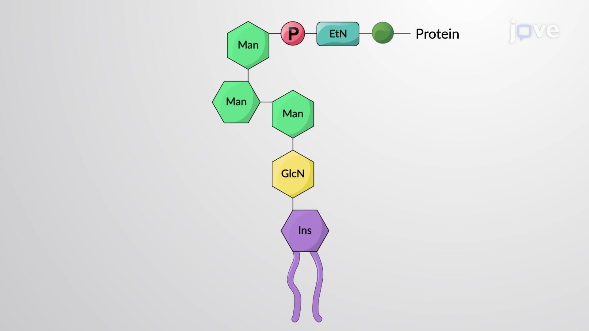
Peripheral proteins attach loosely to the membrane's surface, often serving regulatory roles. These proteins can associate and dissociate based on cellular needs, like traffic controllers managing molecular flow. Many peripheral proteins participate in cell signaling cascades that respond to hormones like insulin or stress hormones released during fight-or-flight responses.
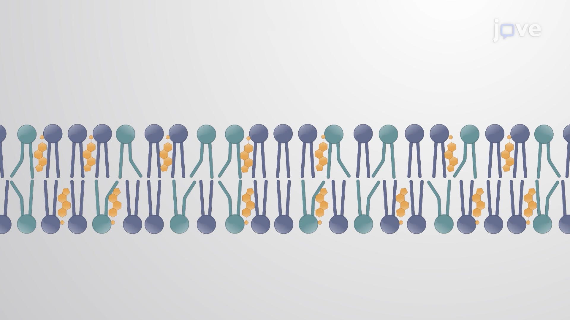
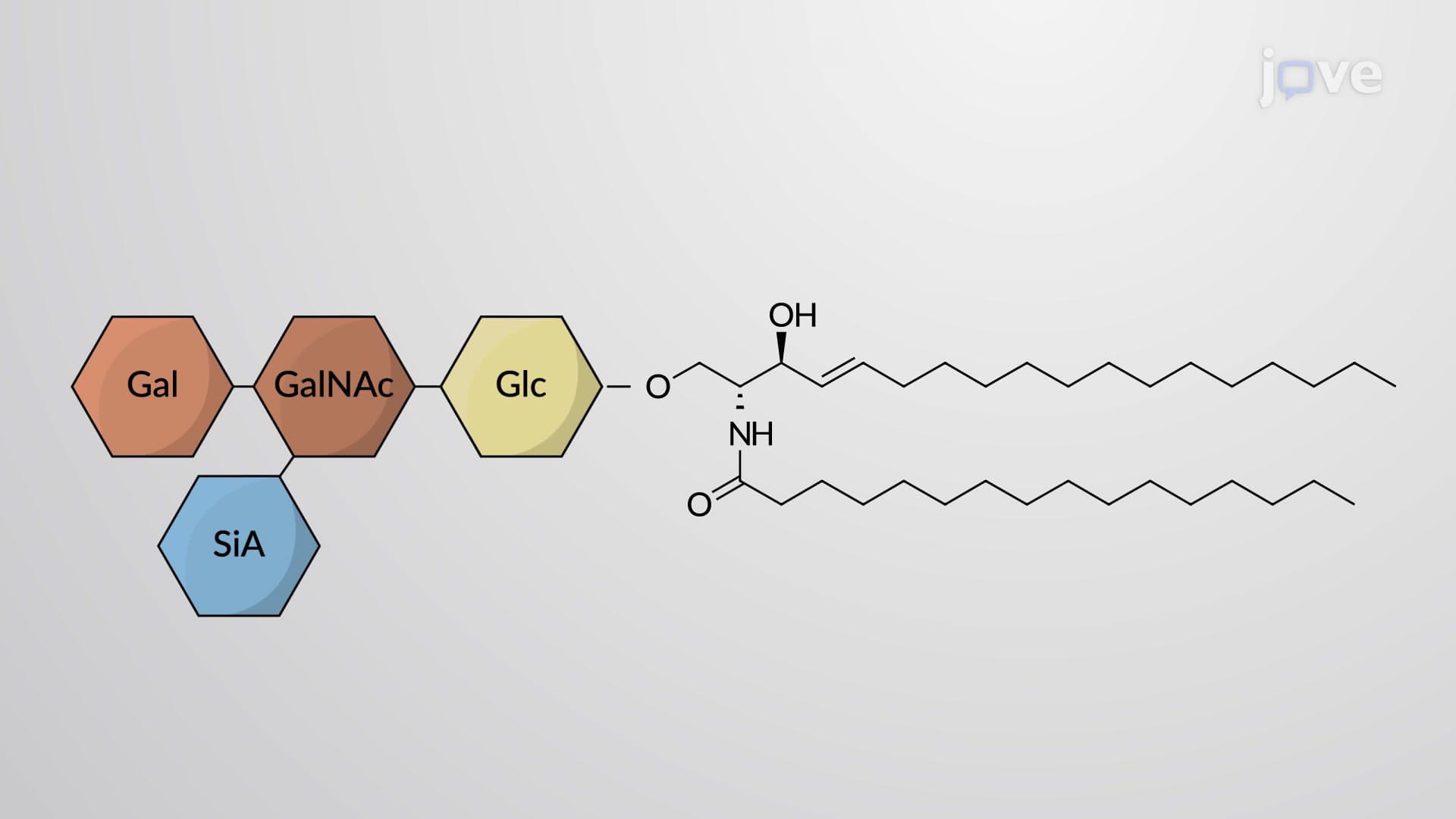
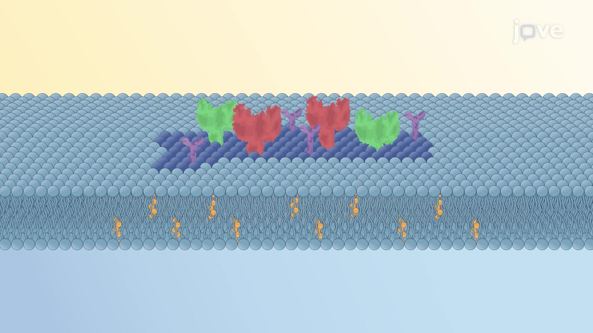
Recent research has expanded the fluid mosaic model beyond Singer and Nicholson's original concept. Scientists now recognize that membranes contain specialized regions called lipid rafts-cholesterol and sphingolipid-rich domains that function like organized workstations within the fluid membrane.
These rafts concentrate specific proteins and facilitate complex cellular processes. For instance, immune system receptors cluster in lipid rafts on T-cell surfaces, enabling rapid response to pathogens. Similarly, nerve cells use lipid rafts to organize ion channels essential for transmitting electrical signals throughout the nervous system.
Understanding the fluid mosaic model proves crucial for success in advanced placement biology, MCAT preparation, and college-level cell biology courses. Exam questions frequently test knowledge of membrane protein types, phospholipid orientation, and factors affecting membrane fluidity. Medical applications include understanding how cholesterol medications work, why certain genetic disorders affect membrane proteins, and how pathogens breach cellular defenses.
Related Micro-courses