Loading video…


 16 Concepts
 168K views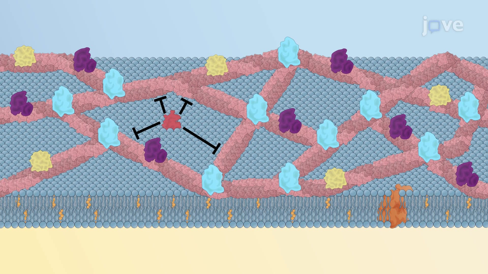

Video Summary: What Is Protein Diffusion
Ever wonder how your cell phone screen responds instantly to your touch? Protein diffusion enables similar rapid communication within cell membranes, allowing proteins to move laterally like dancers on a fluid dance floor. This fundamental process occurs in every cell membrane, from the neurons processing your thoughts to the heart muscle cells beating in your chest. Understanding what is protein diffusion reveals how cellular machinery maintains proper positioning and function. Watch the full video on JoVE Coach to master this concept with expert-led visuals and step-by-step explanations.
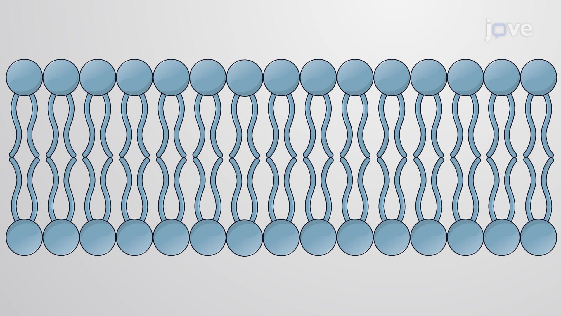
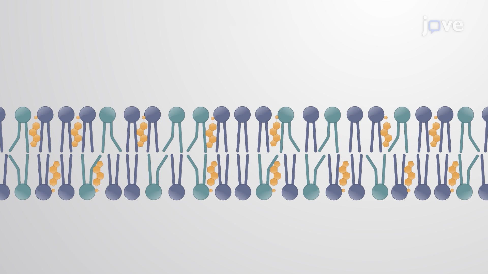
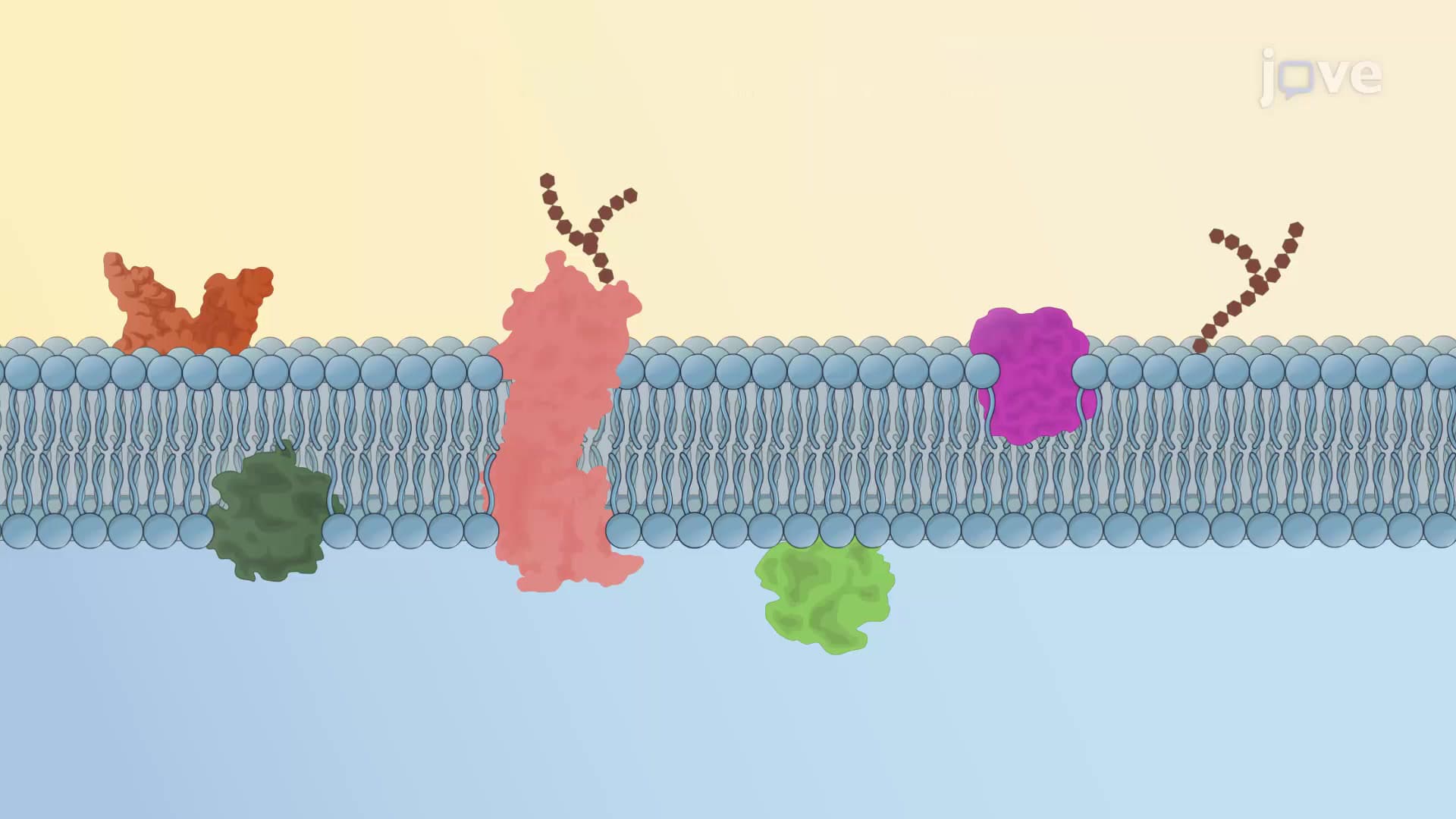
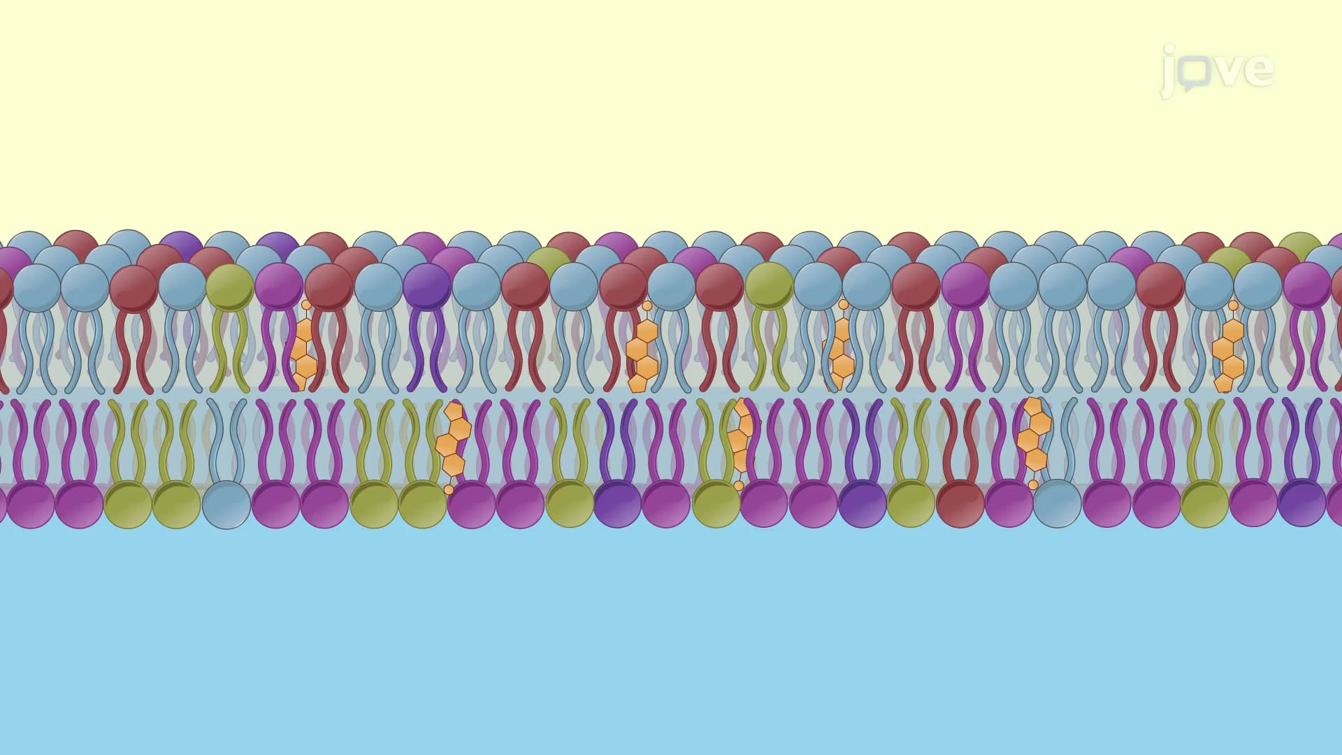
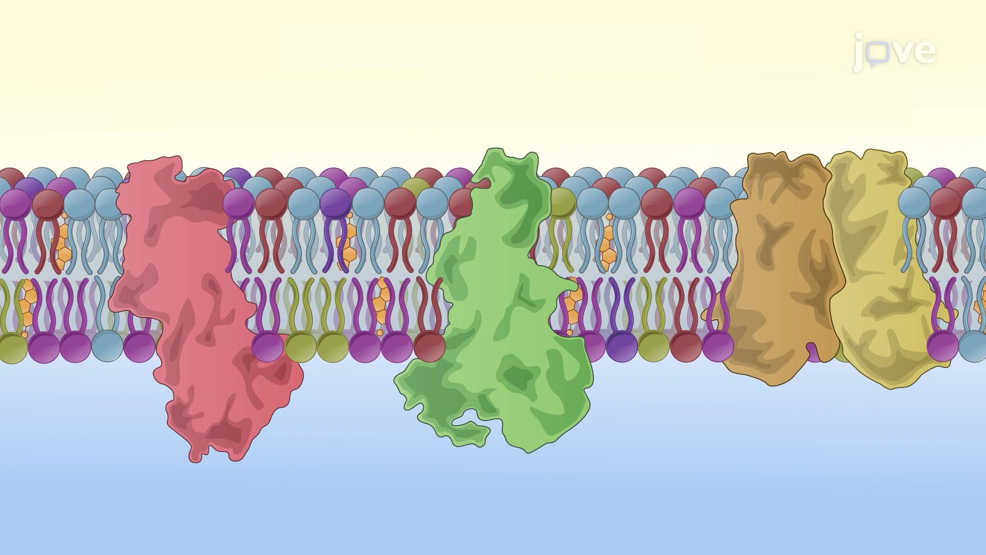
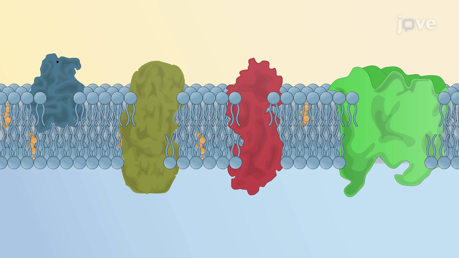
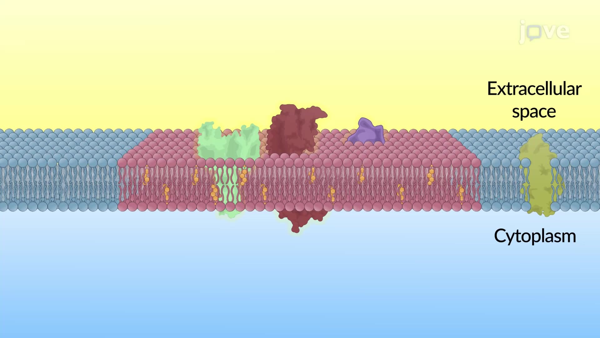
What is protein diffusion fundamentally describes the lateral movement of integral membrane proteins within the fluid mosaic structure of cell membranes. At normal body temperature (37°C), phospholipid bilayers maintain a fluid consistency that permits proteins to move sideways, much like icebergs floating and drifting across an ocean surface. This protein diffusion biology explained concept is crucial for cellular processes including receptor clustering, enzyme complex formation, and signal transduction pathways.
The protein diffusion definition encompasses both free and restricted movement patterns. Free diffusion occurs when proteins move unimpeded through membrane regions, while restricted diffusion happens when cellular structures create barriers or designated pathways. This distinction appears frequently on AP Biology exams and college cell biology assessments, where students must differentiate between these movement patterns.
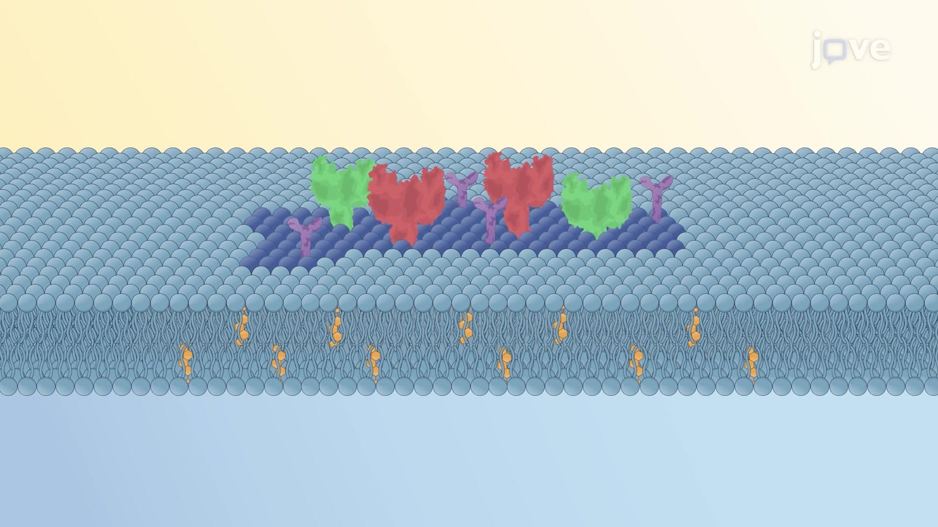
The actin cytoskeleton plays a pivotal role in regulating protein diffusion through the formation of dynamic fence structures. These cytoskeletal networks create distinct membrane compartments called corrals, effectively organizing proteins into functional neighborhoods. For example, in cardiac muscle cells, specific ion channels cluster within corrals to ensure coordinated contraction, while in neurons, neurotransmitter receptors concentrate at synaptic sites.
Actin turnover creates temporary gaps in these cytoskeletal fences, allowing controlled protein movement between compartments. This process resembles opening gates in a pasture system, permitting selective protein migration when cellular conditions require reorganization. Medical students studying for the MCAT often encounter questions about how cytoskeletal disruption affects protein localization and cellular function.
Scientists measure protein diffusion rates using fluorescence recovery after photobleaching (FRAP), a sophisticated technique that provides quantitative data about membrane protein mobility. Researchers first label target proteins with fluorescent markers, then use focused laser beams to photobleach specific membrane regions, creating dark spots devoid of fluorescence.
Recovery time indicates diffusion efficiency-faster recovery suggests higher protein mobility, while slower recovery indicates restricted movement. This methodology helps researchers understand disease mechanisms, particularly in conditions like Alzheimer's disease where membrane protein organization becomes disrupted. College biochemistry courses frequently include FRAP analysis in laboratory exercises and exam questions.
Understanding protein diffusion proves essential in pharmaceutical development and medical diagnostics. Drug companies utilize this knowledge when designing membrane-targeted therapeutics, ensuring proper distribution and efficacy. Additionally, abnormal protein diffusion patterns serve as biomarkers for various diseases, including cancer metastasis and neurological disorders affecting membrane integrity.
Related Micro-courses