Loading subject page…

- Analytical Chemistry
- Mass Spectrometry Fragmentation Methods
- Electrospray Ionization (ESI) Mass Spectrometry
Micro-course: Mass Spectrometry Fragmentation Methods
30,224 views
Learn key concepts and earn your certificate.



Electrospray Ionization (ESI) Mass Spectrometry


00:00
01:12
Video Summary: Electrospray Ionization Esi Mass Spectrometry Explained
Did you know that analyzing a single protein molecule requires turning it into a charged gas particle without destroying it? ESI mass spectrometry explained reveals how scientists at the CDC use this gentle ionization technique to identify disease-causing proteins in patient samples. Unlike harsh traditional methods that shatter large biomolecules, Electrospray Ionization ESI Mass Spectrometry Explained demonstrates how polar solvents and high voltage create intact molecular ions perfect for detecting everything from insulin to DNA fragments. Watch the full video on JoVE Coach to master this concept with expert-led visuals and step-by-step explanations.
- Understand how Electrospray Ionization ESI Mass Spectrometry Explained differs from conventional electron-impact ionization for analyzing large biomolecules
- Identify the key components required for ESI: polar solvents, high voltage capillary, and vacuum chamber conditions
- Analyze why soft ionization methods preserve molecular integrity in proteins and nucleic acids during mass spectrometry analysis
- Compare the advantages of ESI over traditional ionization techniques when examining high molecular weight compounds
- Evaluate how solvent choice and ionic additives affect the resulting mass spectrum peaks and molecular ion detection
- Apply knowledge of charge addition to interpret ESI mass spectra, including M+23 peaks from sodium ion association
- Recognize the relationship between droplet evaporation and gaseous ion formation in the electrospray ionization process
- Explore how ESI enables coupling with liquid chromatography for complex biomolecule separation and identification
Understanding Electrospray Ionization ESI Mass Spectrometry Explained
Electrospray ionization represents a revolutionary breakthrough in analytical chemistry, particularly for studying biological macromolecules that traditional mass spectrometry couldn't handle. When conventional electron-impact ionization attempts to analyze proteins or DNA, it's like using a sledgehammer to crack an egg-the molecule shatters into countless fragments, destroying valuable structural information. ESI solved this problem by developing a "soft" ionization approach that preserves molecular integrity while still creating the charged particles necessary for mass spectrometric detection.
The ESI Process: From Solution to Gas-Phase Ions
The electrospray ionization mechanism begins with sample preparation using polar solvents like methanol, acetonitrile, or water-methanol mixtures. These solvents dissolve biomolecules while maintaining their native conformations. Researchers at pharmaceutical companies like Pfizer routinely add ionic salts to enhance ionization efficiency, creating predictable mass shifts that aid in molecular identification.
The high-voltage capillary-typically operating at 2,000-4,000 volts-transforms the liquid sample into a fine mist of charged droplets. As these microscopic droplets enter the vacuum chamber, solvent molecules rapidly evaporate, concentrating charge on the remaining biomolecule. This process continues until each molecule carries multiple charges, creating the multiply charged ions characteristic of ESI mass spectra.
Interpreting ESI Mass Spectra in Practice
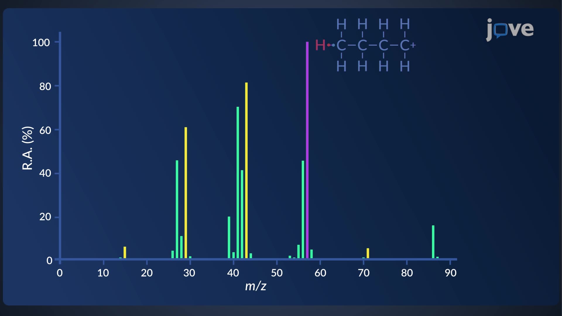
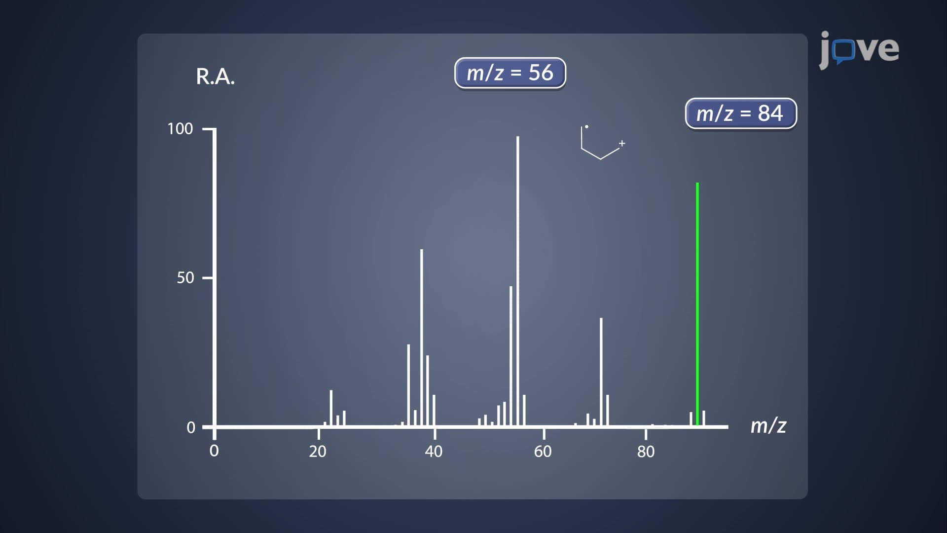
ESI spectra differ dramatically from traditional mass spectrometry results. Instead of a single molecular ion peak, ESI produces multiple peaks corresponding to different charge states of the same molecule. A protein with molecular weight 12,000 Da might appear as peaks at m/z 1,000 (12+ charge), m/z 1,200 (10+ charge), and m/z 1,500 (8+ charge). This charge distribution provides valuable information about molecular conformation and allows analysis of extremely large molecules that would exceed traditional mass spectrometer detection limits.
Students preparing for the MCAT or advanced placement chemistry exams should understand that peak interpretation requires calculating the original molecular weight from the observed m/z values and charge states. The presence of adduct ions-like the M+23 peak from sodium association-helps confirm molecular identity and provides insights into solution conditions.
Real-World Applications and Clinical Significance
ESI-MS has transformed pharmaceutical development, environmental monitoring, and clinical diagnostics across the United States. The FDA relies on ESI-MS for drug purity analysis, while hospitals use ESI-LC-MS systems to measure therapeutic drug concentrations in patient blood samples. Environmental protection agencies employ this technique to detect trace contaminants in drinking water supplies, and forensic laboratories use ESI-MS to identify unknown substances in criminal investigations.
Frequently Asked Questions
Electrospray ionization ESI mass spectrometry is a soft ionization technique that analyzes large biomolecules without fragmenting them. The process involves dissolving samples in polar solvents, spraying the solution through a high-voltage capillary into a vacuum chamber, and allowing solvent evaporation to create charged gaseous molecules for mass spectrometric detection.
MCAT questions typically focus on interpreting multiply charged ion spectra and calculating molecular weights from m/z ratios. College exams often test understanding of why ESI preserves molecular integrity compared to electron-impact ionization. Students should practice identifying charge states and explaining solvent selection rationale for different biomolecules.
AP Chemistry emphasizes ESI as a "soft" ionization method that doesn't fragment molecules, unlike electron-impact ionization. Test questions focus on explaining why polar solvents are necessary, how voltage creates charged droplets, and interpreting spectra with adduct ions like M+23 from sodium addition.
Major US pharmaceutical companies like Johnson & Johnson use ESI-MS for drug development and quality control, analyzing protein therapeutics and small molecule drugs. Hospitals employ ESI-LC-MS systems to monitor therapeutic drug levels in patients, detect drug metabolites, and identify unknown compounds in emergency toxicology cases.
Not at all-ESI builds on fundamental chemistry concepts you already know like polarity, charge, and phase changes. If you understand why oil doesn't mix with water and how evaporation works, you have the foundation for grasping ESI principles. The technique simply applies these concepts to create charged molecules for analysis.
Focus on practicing m/z calculations for multiply charged ions and understanding the relationship between charge state and peak position. Create concept maps connecting solvent polarity, voltage application, droplet formation, and ion detection. Practice interpreting spectra with multiple charge states and adduct ion formation.
ESI serves as a bridge to liquid chromatography-mass spectrometry (LC-MS), a powerful combination used throughout analytical labs. It also connects to protein biochemistry, pharmaceutical analysis, and environmental chemistry. Mastering ESI principles prepares you for advanced topics like tandem mass spectrometry and proteomics.
ESI-MS expertise opens doors to pharmaceutical research, clinical laboratory science, environmental consulting, food safety analysis, and forensic science careers. Major employers include the CDC, EPA, pharmaceutical companies, clinical reference laboratories, and university research institutions across the United States.
Related Micro-courses


19 Concepts