Loading subject page…


42,407 views

Video Summary: What Is Mass Spectrum
Ever wondered how forensic scientists identify unknown drugs in criminal investigations? Mass spectrum explained reveals how scientists break down molecules into charged fragments to create unique "molecular fingerprints." A mass spectrum shows the relative abundance of these fragments plotted against their mass-to-charge ratio, like how the FDA analyzes pharmaceutical compounds to ensure drug purity and safety. Understanding what a mass spectrum is becomes essential for chemistry students pursuing careers in medicine, forensics, or pharmaceutical research. Watch the full video on JoVE Coach to master this concept with expert-led visuals and step-by-step explanations.
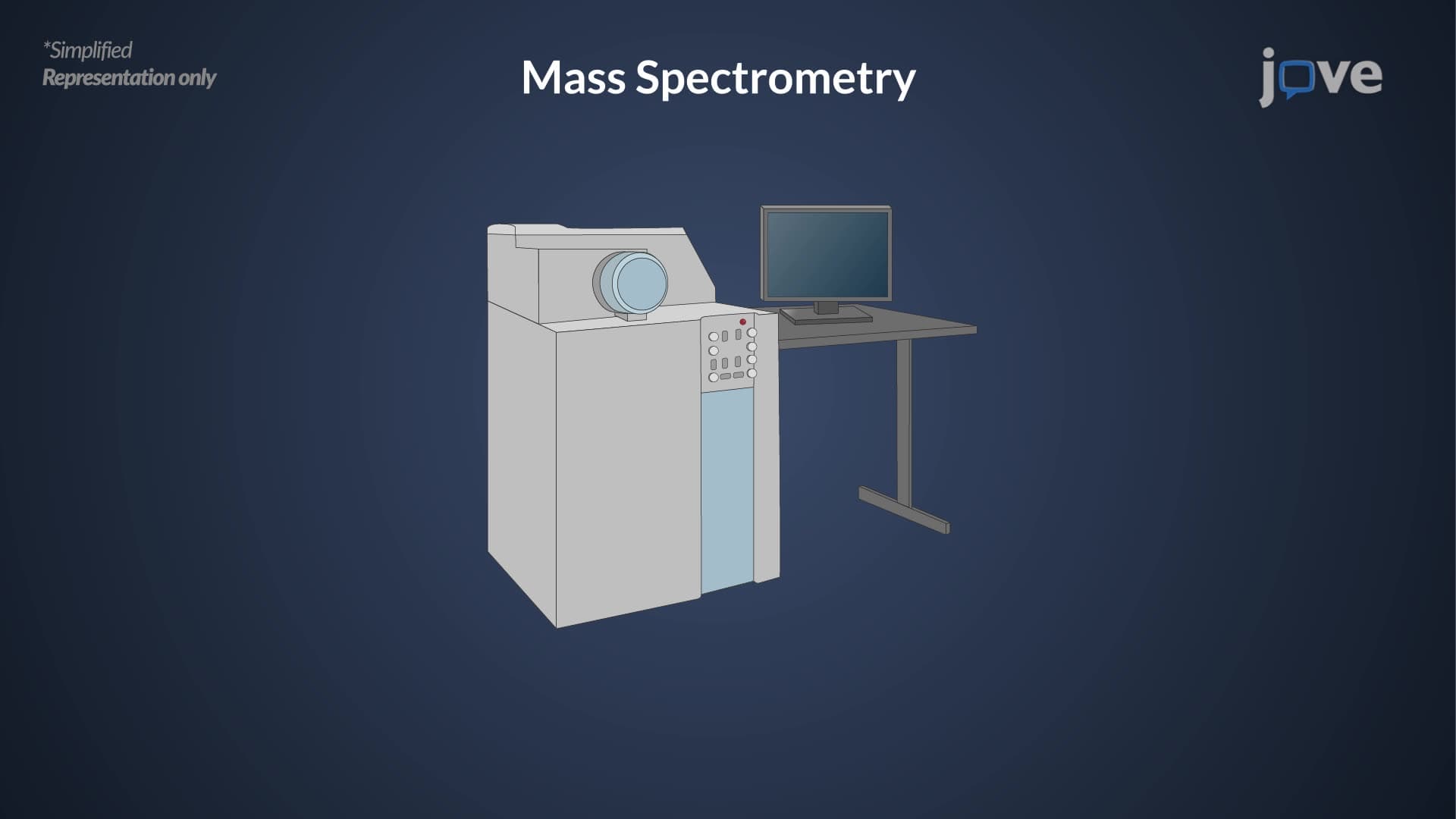
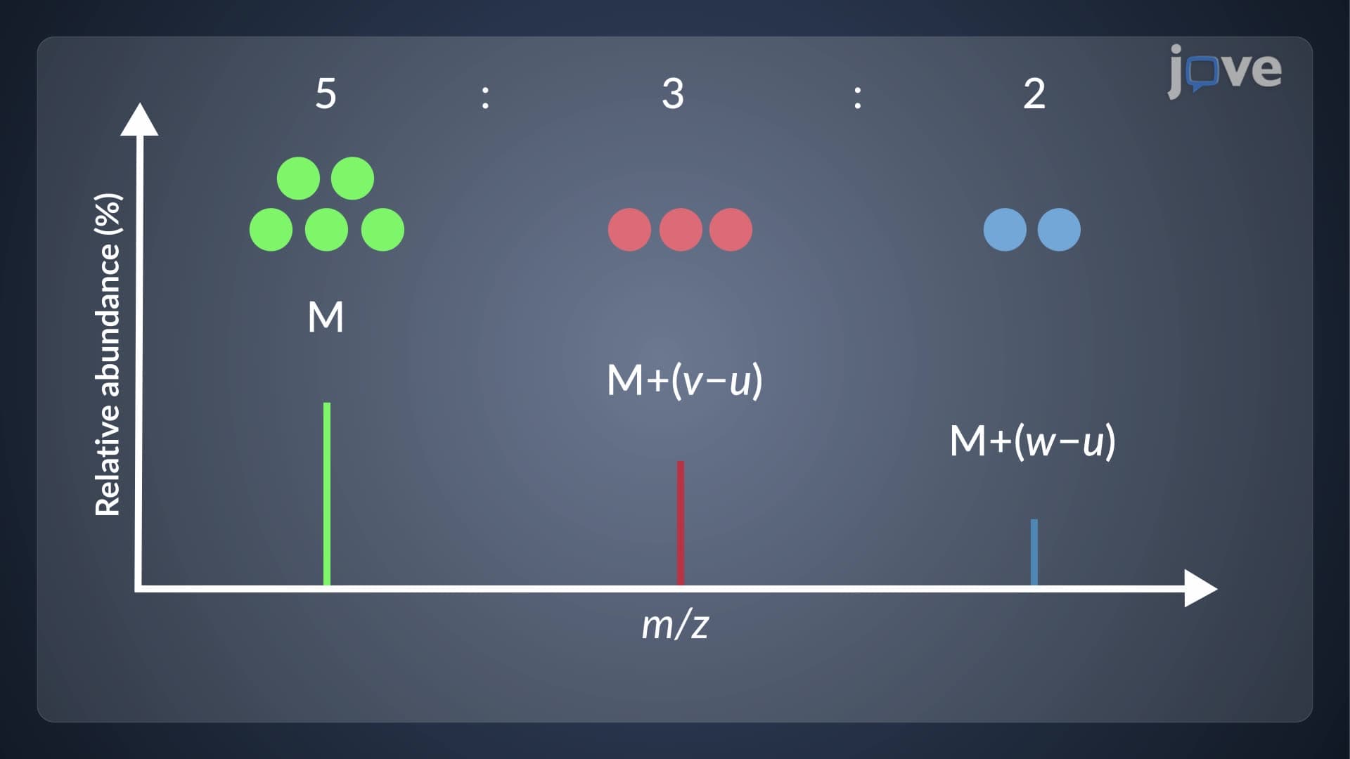
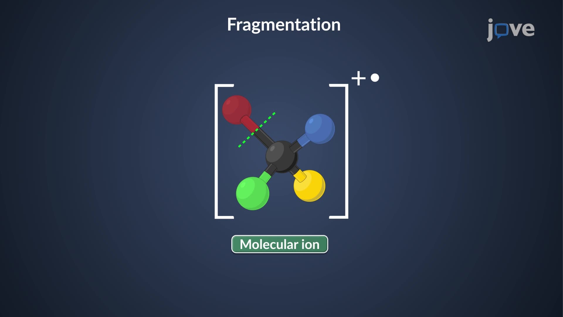
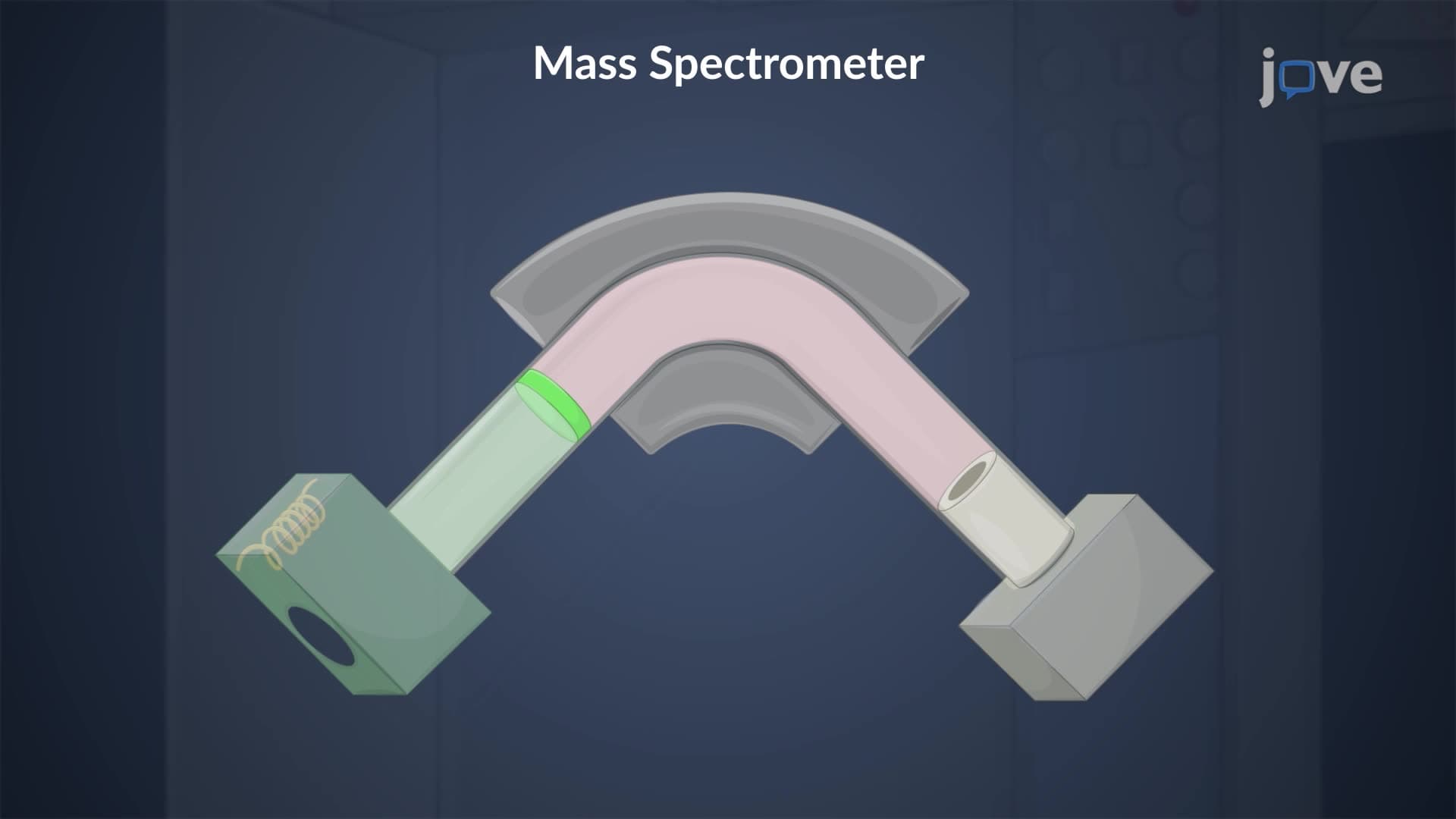
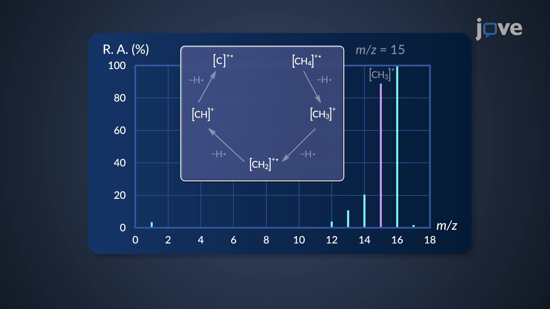
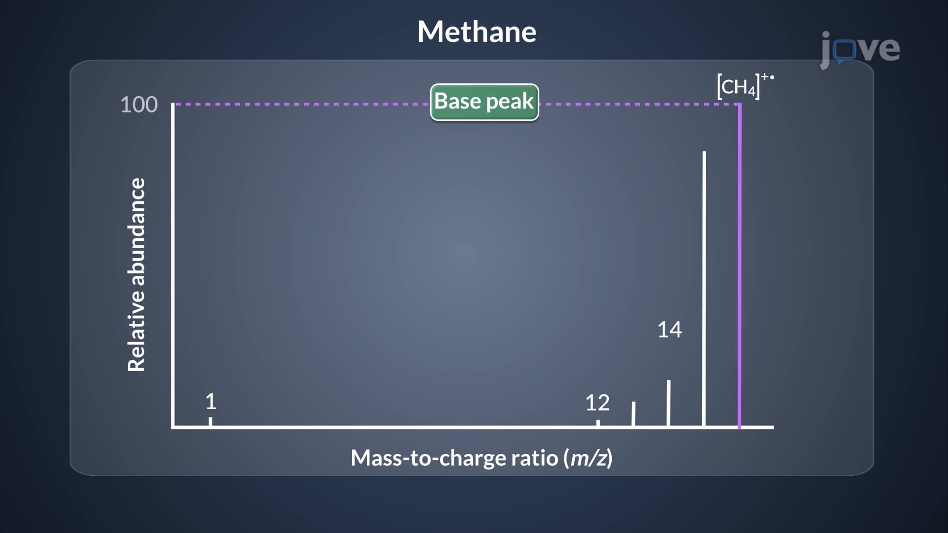
A mass spectrum functions as a unique molecular fingerprint, providing crucial information about a compound's identity and structure. When molecules are bombarded with high-energy electrons in a mass spectrometer, they lose electrons and break apart into charged fragments. The resulting mass spectrum displays these fragments as peaks, with the x-axis showing mass-to-charge ratio (m/z) and the y-axis indicating relative abundance.
This analytical technique proves invaluable across multiple fields. The DEA uses mass spectrometry to identify illegal drugs, while pharmaceutical companies like Pfizer employ it for quality control during drug manufacturing. Environmental agencies utilize mass spectra to detect pollutants in water supplies, and forensic laboratories analyze trace evidence from crime scenes.
The molecular ion peak represents the original molecule after losing just one electron, appearing at the highest m/z value in most spectra. This peak provides direct information about the compound's molecular weight. However, some molecules produce such unstable molecular ions that this peak may be weak or completely absent from the spectrum.
For students preparing for AP Chemistry or college organic chemistry courses, recognizing molecular ion peaks becomes crucial for solving structure determination problems. The molecular ion peak serves as the foundation for calculating molecular formulas and identifying unknown compounds in laboratory exercises.
The base peak represents the most abundant fragment in the spectrum, automatically set to 100% abundance. All other peaks are scaled relative to this maximum. Understanding fragmentation mass spectrum analysis helps predict which bonds will break most readily during ionization.
Common fragmentation patterns include loss of small neutral molecules (like water or methane) and formation of stable carbocations. Students studying for the MCAT will encounter these patterns when analyzing biological molecules, while those taking organic chemistry courses use fragmentation analysis to determine molecular structures.
Learning how to read a mass spectrum requires practice with real examples. Clinical laboratories use mass spectrometry to measure drug levels in patient blood samples, ensuring proper dosing for medications like warfarin. Research institutions employ this technique to identify metabolites in biochemical pathways, advancing our understanding of human metabolism and disease processes.
Related Micro-courses