Loading subject page…


99,205 views

Video Summary: Cranial Nerves Types Part I Explained
Ever wonder why closing your eyes to smell fresh coffee enhances the aroma? Cranial nerve types part one control this sensory magic through specialized neural pathways connecting your brain to sensory organs. From detecting the scent of apple pie (CN I) to reading street signs while driving (CN II), the first six cranial nerves orchestrate essential daily functions like vision, smell, and eye movement. Understanding Cranial Nerves Types Part I Explained reveals how neurological disorders can disrupt these basic abilities. Watch the full video on JoVE Coach to master this concept with expert-led visuals and step-by-step explanations.
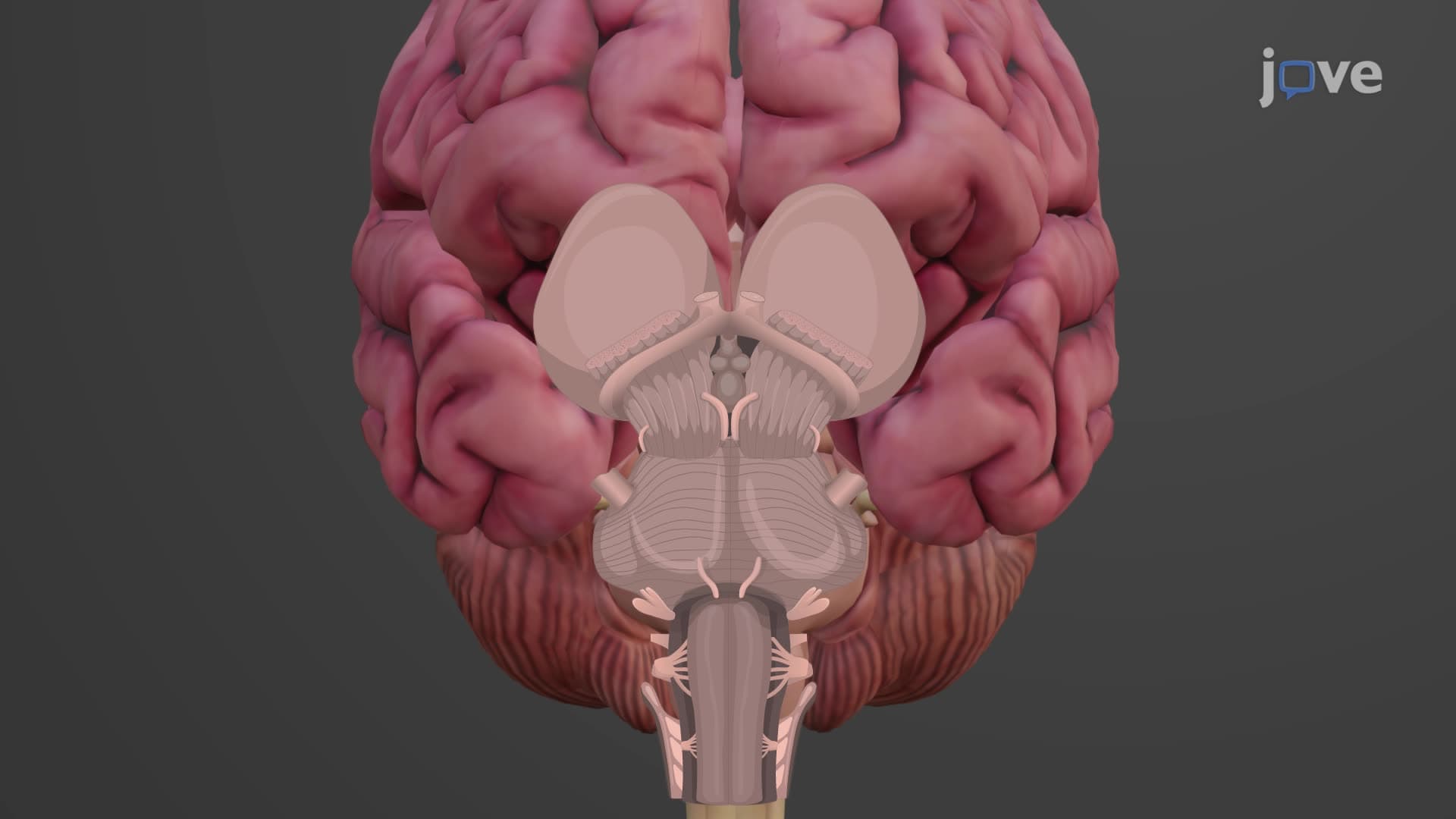
The first six cranial nerves represent a fascinating convergence of sensory perception and motor control that governs our most essential daily interactions. Unlike spinal nerves that follow predictable patterns, these cranial nerves demonstrate remarkable specialization, each emerging from distinct brainstem regions to serve unique physiological roles.
The olfactory nerve (CN I) stands apart as the only cranial nerve directly connected to the cerebral cortex, bypassing the thalamus entirely. This unique pathway explains why certain scents can trigger immediate emotional responses or vivid memories-a phenomenon crucial for understanding conditions like anosmia, which affects approximately 3% of Americans and gained attention during COVID-19. The optic nerve (CN II) processes over 10 million bits of visual information per second, making it essential for activities from reading standardized test questions to recognizing traffic signals.
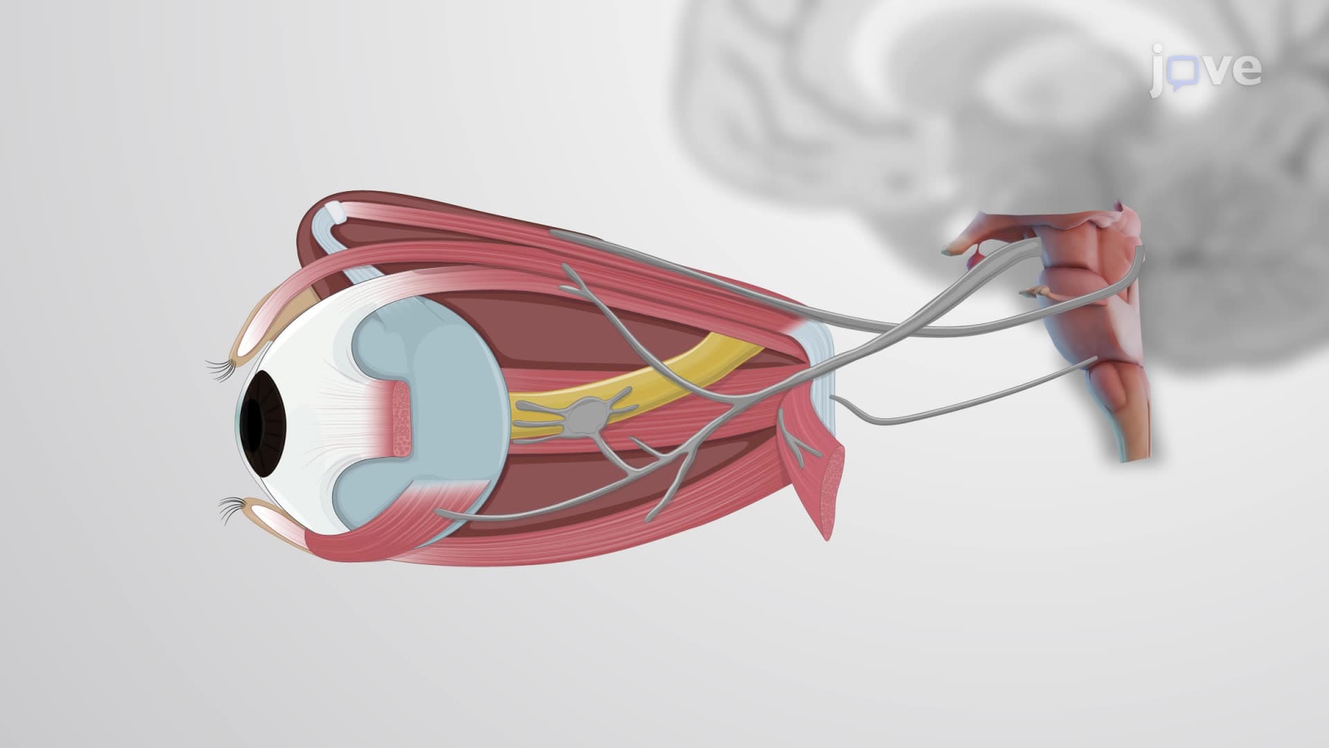
The oculomotor (CN III), trochlear (CN IV), and abducens (CN VI) nerves orchestrate the complex ballet of eye movements required for tracking moving objects, reading text, and maintaining visual focus. The oculomotor nerve controls most eye muscles plus pupil constriction-critical for understanding conditions like third nerve palsy, commonly tested in MCAT neuroscience sections. The trochlear nerve, despite being the smallest cranial nerve, enables the superior oblique muscle to rotate the eye downward and inward, essential for activities like walking downstairs safely.
The trigeminal nerve represents the largest cranial nerve complex, combining three distinct divisions that collectively enable facial sensation and mastication. Its ophthalmic division provides sensation to the forehead and cornea-explaining why eye injuries can trigger intense pain responses. The maxillary division innervates the upper jaw and nasal cavity, while the mandibular division uniquely combines motor control for chewing muscles with sensory innervation of the lower face. This complexity makes trigeminal neuralgia one of the most debilitating facial pain conditions, affecting over 150,000 Americans annually.
Understanding these cranial nerve types proves essential for AP Biology students tackling nervous system units and pre-med students preparing for MCAT passages on neuroanatomy and clinical correlations.
Related Micro-courses