Loading video…


 26 Concepts
 99.2K views

Video Summary: Diencephalon Thalamus and Information Relay Explained
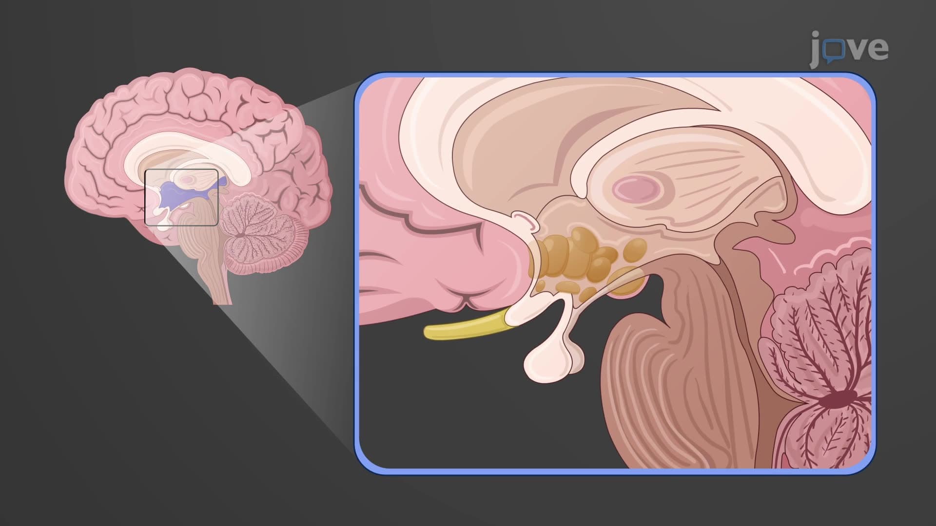
Ever wonder why damage to a brain region the size of a walnut can leave someone unable to feel touch or see properly? The thalamus information relay function serves as the brain's central switchboard, directing nearly all sensory and motor signals to the cerebral cortex. Consider how a stroke affecting the thalamus can instantly disrupt a patient's ability to process visual information at Johns Hopkins Hospital, demonstrating this structure's critical role. Understanding the Diencephalon Thalamus And Information Relay Explained reveals how this remarkable brain region coordinates consciousness, sensation, and movement through its specialized nuclei. Watch the full video on JoVE Coach to master this concept with expert-led visuals and step-by-step explanations.
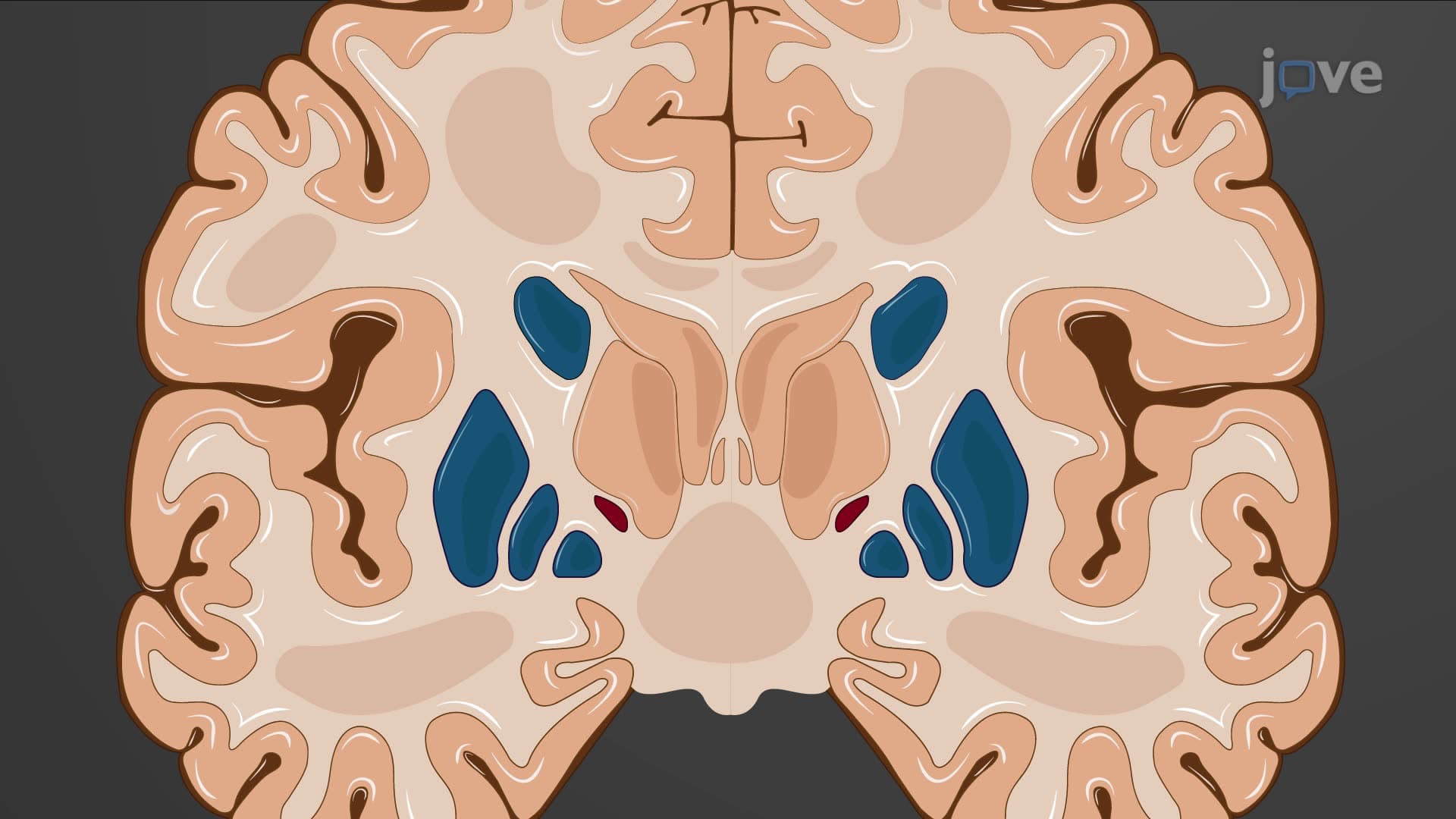
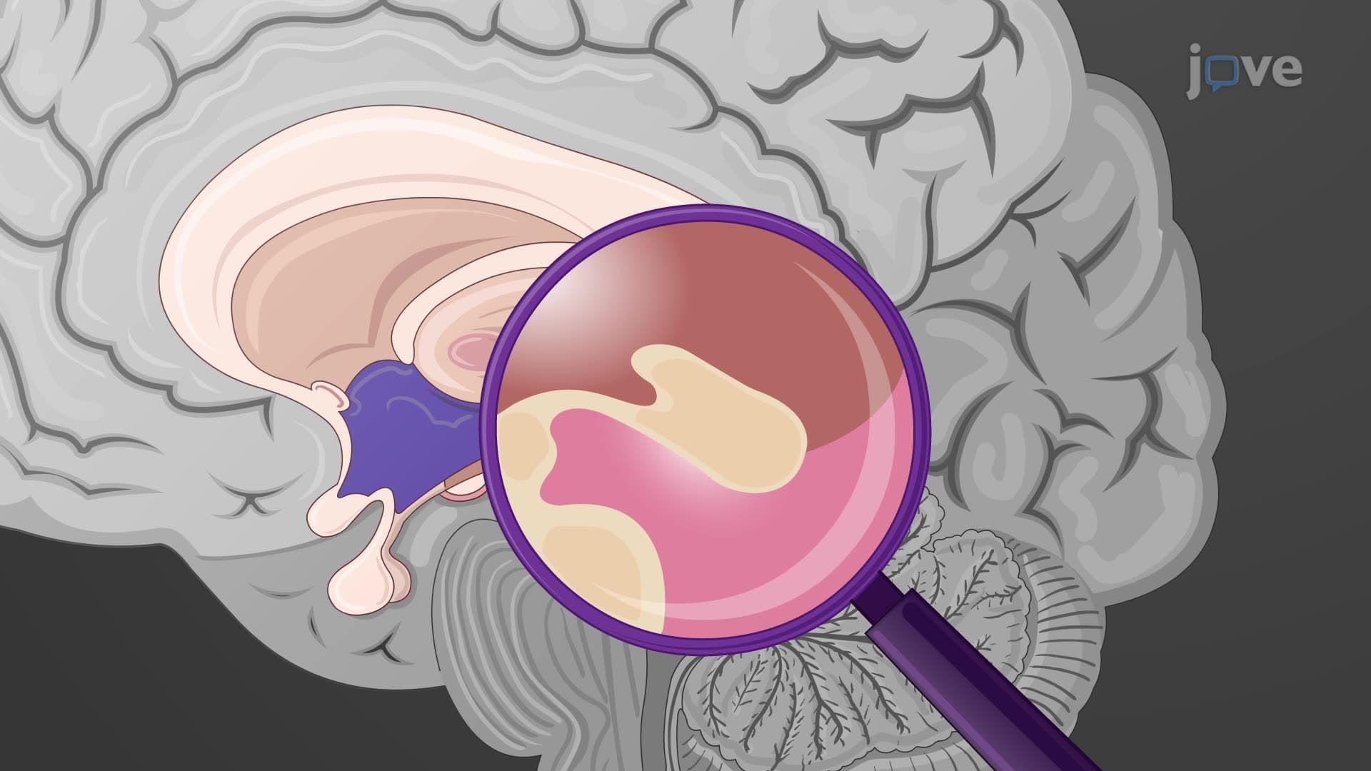
The thalamus represents one of neuroscience's most elegant examples of functional organization. This paired structure, located within the diencephalon, contains over 50 distinct nuclei that collectively process virtually every piece of information reaching the cerebral cortex. The thalamus information relay function operates like a sophisticated air traffic control system, ensuring that sensory data, motor commands, and emotional signals reach their proper cortical destinations.
The thalamic relay brain explained reveals a highly organized system where each nucleus group serves specific functions. The lateral geniculate nucleus exclusively handles visual information from the retina, channeling it to the primary visual cortex in the occipital lobe. This pathway becomes critically important when studying conditions like cortical blindness, where patients seen at UCLA Medical Center may retain some visual reflexes despite cortical damage.
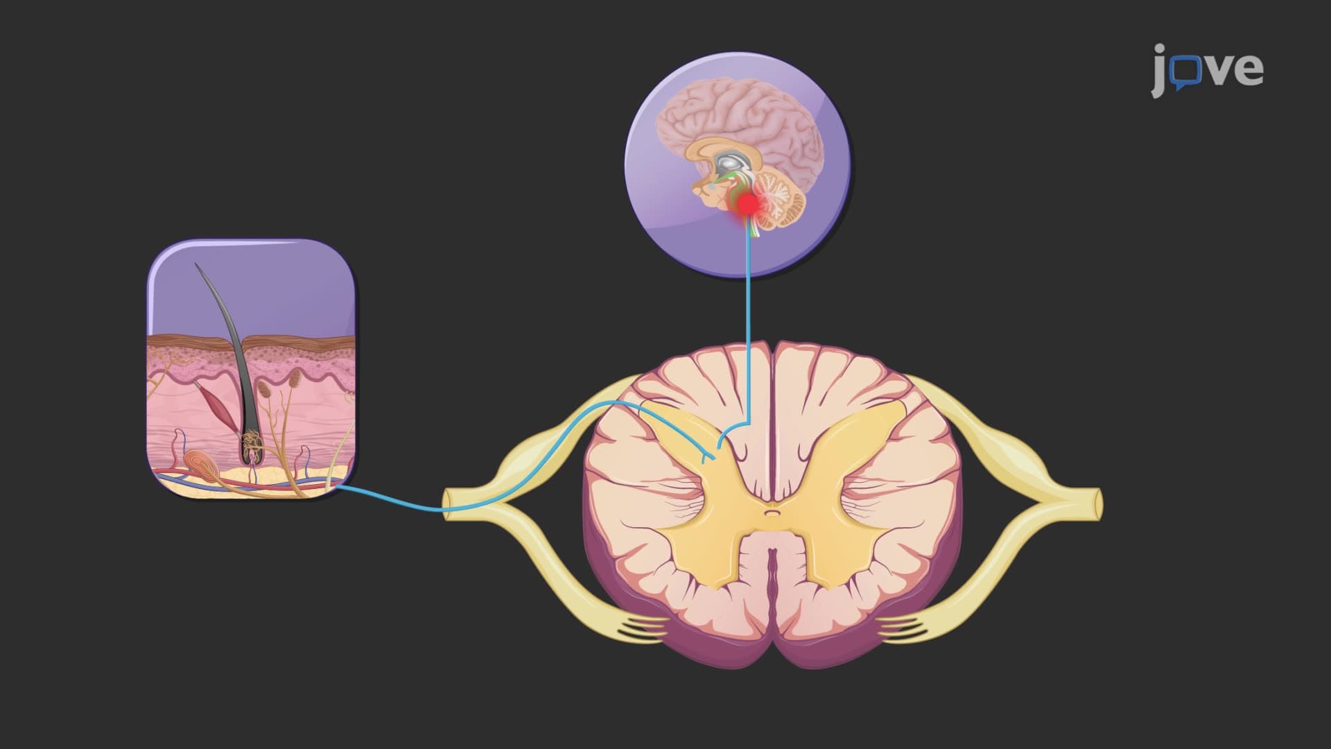
Similarly, the medial geniculate nucleus processes all auditory information, creating the foundation for our ability to interpret sounds, music, and speech. The ventral posterior nucleus manages somatosensory inputs-touch, pain, temperature, and proprioception-demonstrating how the thalamus relays information in the diencephalon through dedicated pathways.
Motor relay functions involve the ventral anterior and ventral lateral nuclei, which receive inputs from the basal ganglia and cerebellum respectively. These connections explain why thalamic strokes can produce both sensory deficits and movement disorders, a concept frequently tested on the MCAT and in neuroanatomy courses at institutions like Harvard Medical School.
Beyond simple relay functions, the thalamus plays crucial roles in consciousness and attention through its thalamic relay pathway connections with the reticular activating system. The intralaminar nuclei help maintain arousal and awareness, while the pulvinar nucleus integrates complex sensory information necessary for spatial attention.
Understanding these cortical thalamic projection patterns helps explain why thalamic lesions can produce such diverse symptoms. A patient at Mayo Clinic with a small thalamic hemorrhage might experience sensory loss, sleep disturbances, and attention deficits-all traceable to different nuclear groups within this compact structure.
This thalamic nuclei relay station organization frequently appears on AP Psychology exams, MCAT biological sciences sections, and undergraduate neuroscience courses. Students should focus on the functional relationships rather than memorizing every nuclear name, emphasizing how the thalamus enables sensory motor relay thalamus integration essential for conscious experience.
Related Micro-courses