Loading subject page…


99,205 views

Video Summary: Motor and Sensory Areas Cortex Explained
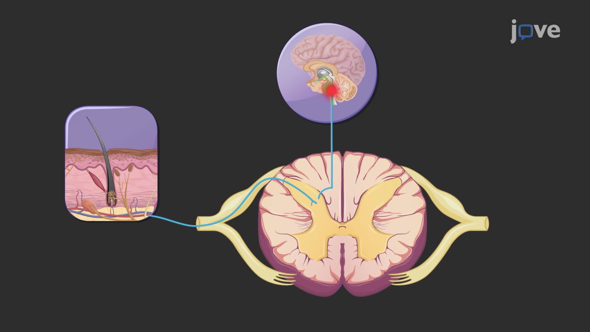
Ever wondered why touching a hot stove triggers an instant hand withdrawal? The motor sensory cortex areas work together like a sophisticated control center, with specialized regions processing touch sensations and coordinating movement responses. When a burn patient at Johns Hopkins receives treatment, medical teams rely on understanding these cortical regions to assess neurological damage and recovery potential. The Motor And Sensory Areas Cortex Explained reveals how your brain's frontal and parietal lobes create seamless communication between sensation and action. Watch the full video on JoVE Coach to master this concept with expert-led visuals and step-by-step explanations.
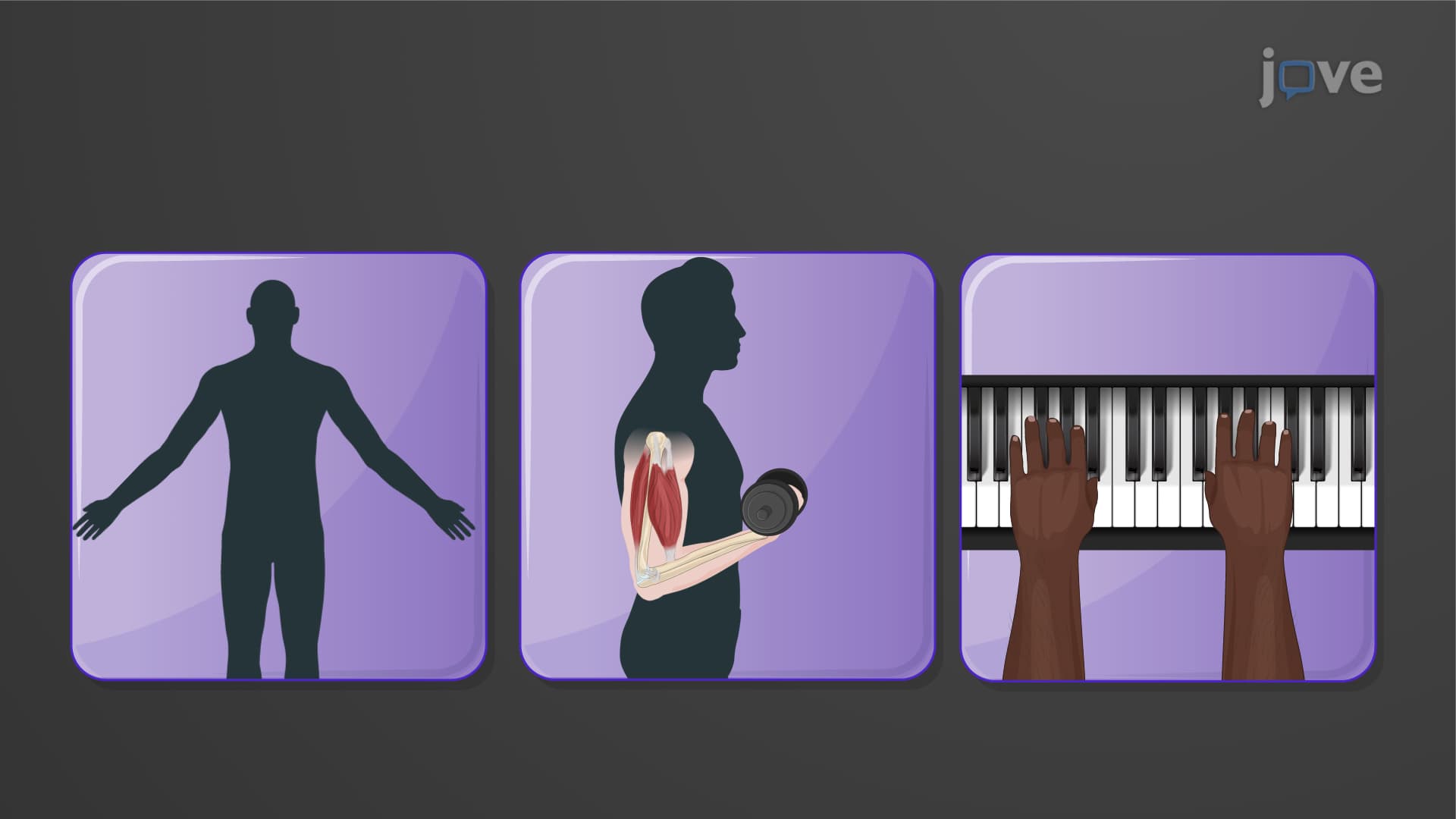
The human cerebral cortex represents one of evolution's most sophisticated information processing systems, with motor sensory cortex areas serving as the primary interface between our internal neural world and external environment. This intricate network spans multiple lobes, each specialized for distinct yet interconnected functions that enable everything from playing piano to recognizing a friend's voice.
The cortical motor sensory regions begin with the motor areas housed primarily in the frontal lobe. The primary motor cortex, located in the precentral gyrus (Brodmann area 4), functions as your brain's movement headquarters. This region exhibits remarkable organization through the motor homunculus cortex, a distorted body map where areas requiring fine motor control, like hands and face, occupy disproportionately large cortical territories.
Consider a violinist at the New York Philharmonic: their primary motor cortex has likely undergone extensive plasticity, with enlarged representations for finger movements. The premotor cortex, positioned anterior to the primary motor area, plans and sequences these complex movements before execution. Broca's area, a specialized region within the premotor cortex, coordinates the intricate muscle movements required for speech production, crucial for students mastering public speaking or preparing for college debate teams.
The somatosensory cortex area 3 (along with areas 1 and 2) in the postcentral gyrus processes touch, pressure, temperature, and proprioception. This sensory representation cortex mirrors the motor homunculus organization, with lips and fingertips claiming extensive neural real estate reflecting their sensory importance.
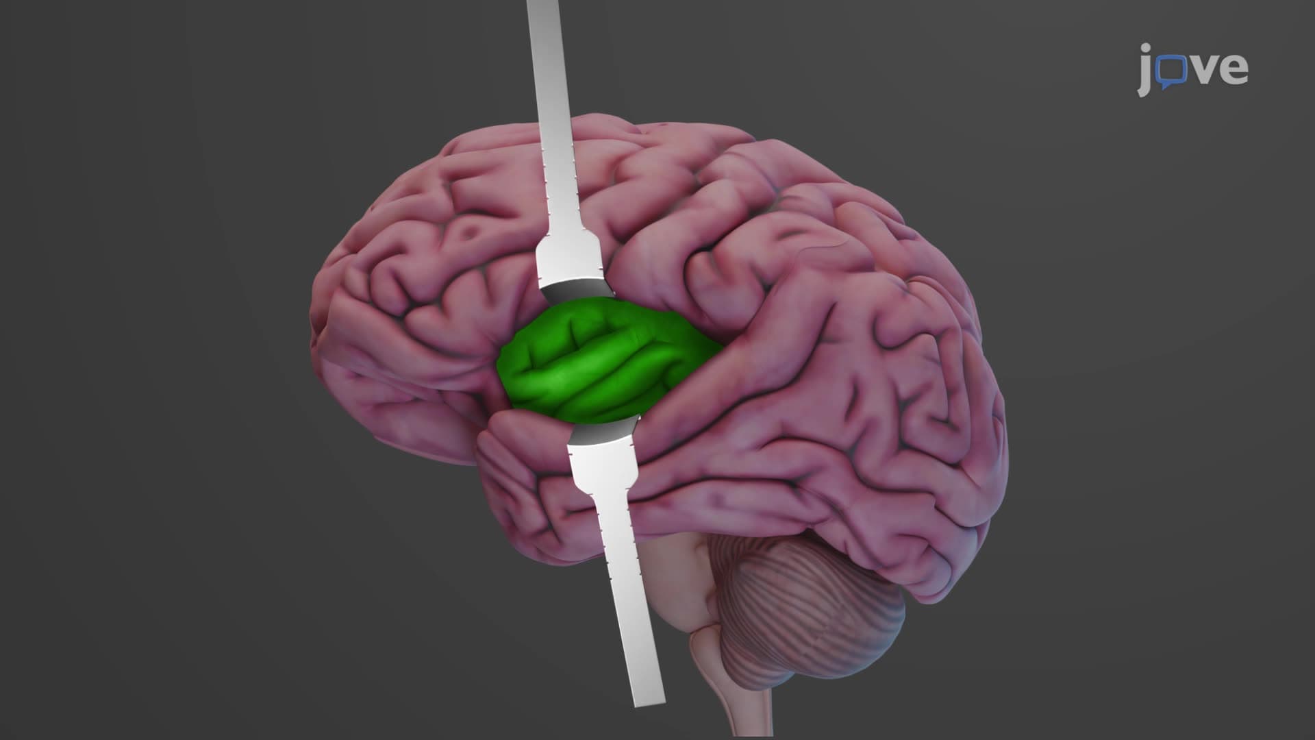
Beyond somatosensation, specialized sensory areas process distinct environmental inputs. The primary visual cortex in the occipital lobe processes visual information crucial for students studying visual perception in AP Psychology courses. The primary auditory cortex in the temporal lobe enables music appreciation and language comprehension, while the primary gustatory cortex in the insula processes taste, relevant for culinary arts students at institutions like the Culinary Institute of America.
Cortical area function extends beyond simple input-output relationships. Association areas integrate motor and sensory information, enabling complex cognitive functions tested on MCAT examinations. Understanding these relationships proves essential for pre-medical students studying neuroanatomy and for nursing students preparing for NCLEX examinations focused on neurological assessments.
Clinical applications abound in US healthcare settings. Stroke rehabilitation at facilities like the Mayo Clinic relies on cortical plasticity principles, where undamaged regions can partially compensate for injured areas. Physical therapists utilize this knowledge when designing recovery programs, understanding that repetitive, focused practice can strengthen cortical connections and improve patient outcomes.
Related Micro-courses