Loading video…


 26 Concepts
 99.2K views

Video Summary: What Is Anatomy Brain Ventricles
Did you know that your brain contains about 150 mL of fluid-roughly the amount in a small yogurt cup? This brain ventricles anatomy overview reveals how these fluid-filled chambers protect your central nervous system. Similar to how spinal fluid analysis helps diagnose conditions like meningitis at hospitals like Johns Hopkins, understanding what is anatomy brain ventricles is crucial for medical students and anyone studying neuroanatomy. Watch the full video on JoVE Coach to master this concept with expert-led visuals and step-by-step explanations.
The ventricular system brain explained represents one of the most sophisticated fluid management systems in human anatomy. These interconnected cavities form a complex network that maintains optimal brain function through precise cerebrospinal fluid (CSF) regulation. For students preparing for the MCAT or AP Biology exams, mastering ventricular anatomy is essential since questions frequently test the relationship between structure and function in the nervous system.
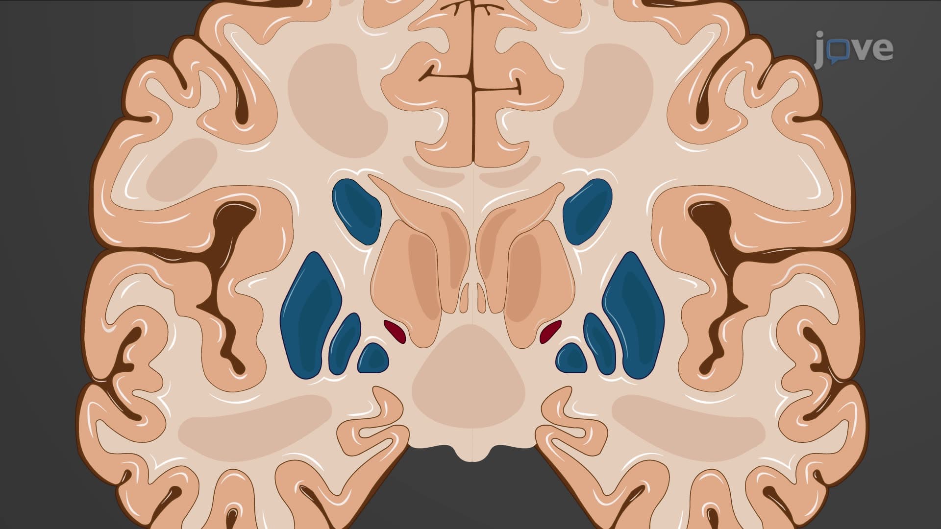
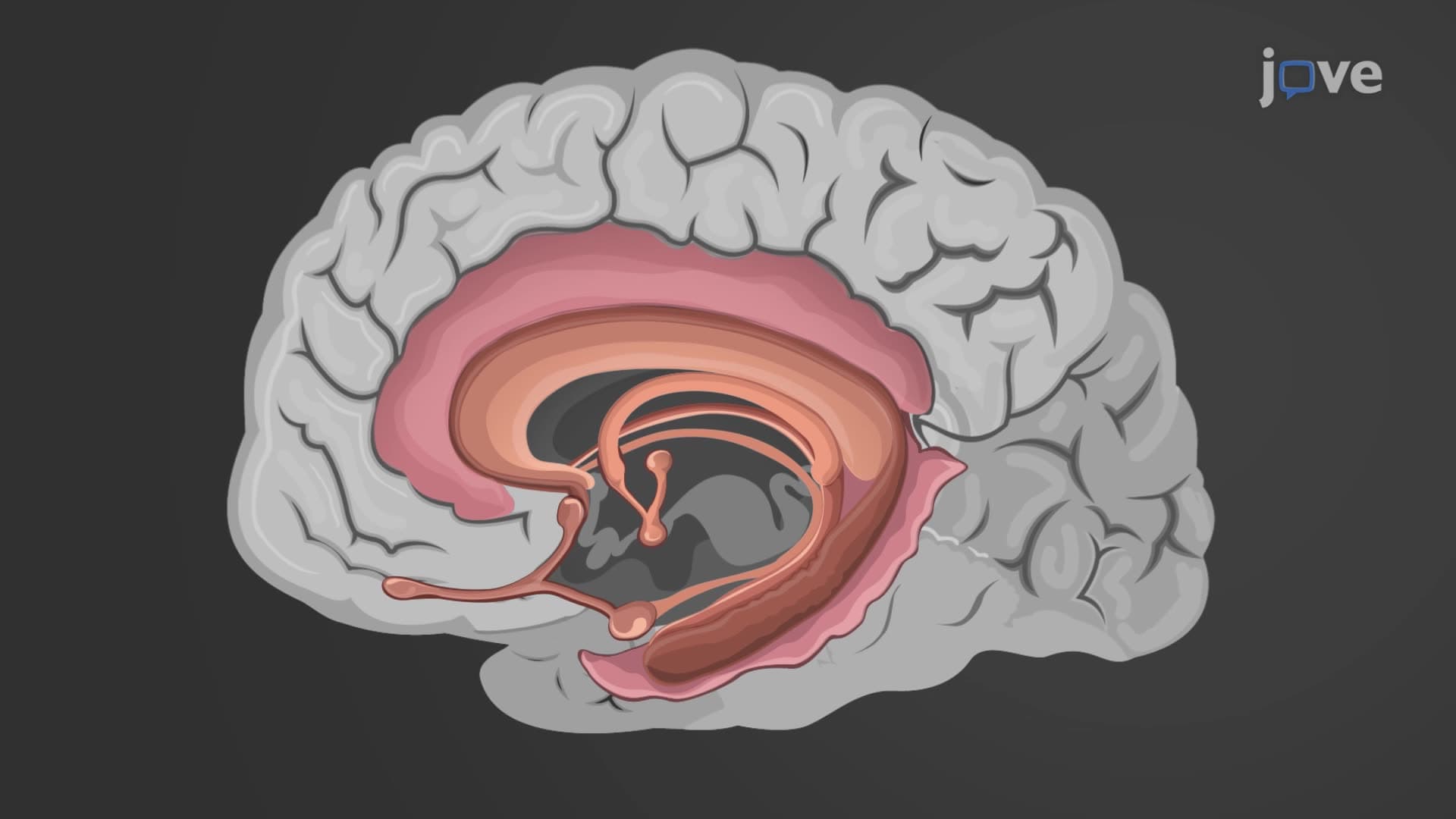
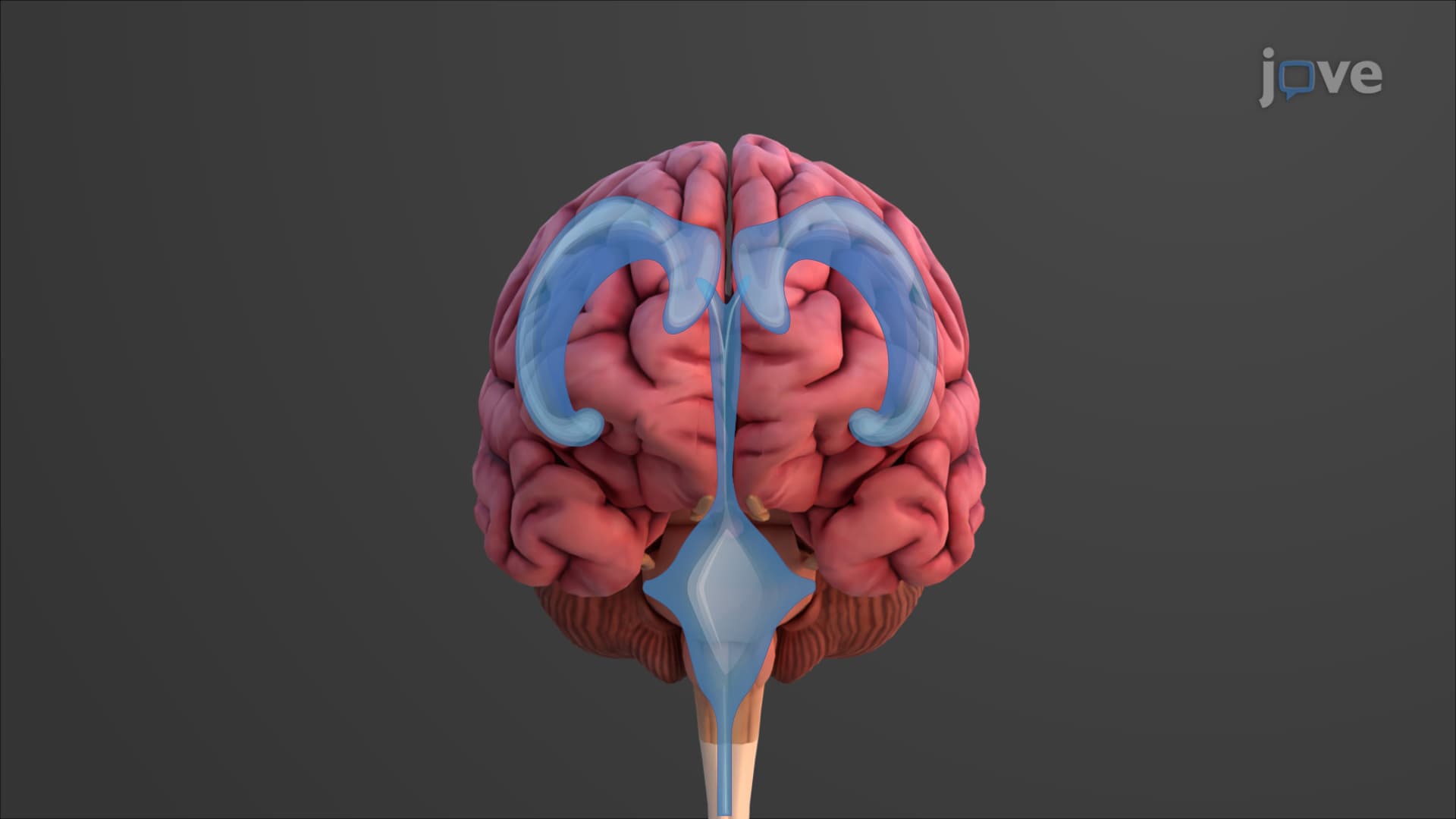
The lateral ventricle brain structures are the largest components of the ventricular system, with each cerebral hemisphere containing one. These C-shaped cavities extend into three distinct projections called horns. The anterior horn projects into the frontal lobe, the posterior horn extends into the occipital lobe, and the inferior horn curves into the temporal lobe. This complex shape reflects the ventricles' development alongside expanding cerebral hemispheres during embryonic growth. Medical students at institutions like Harvard Medical School study how lateral ventricle enlargement can indicate conditions such as Alzheimer's disease or normal pressure hydrocephalus.
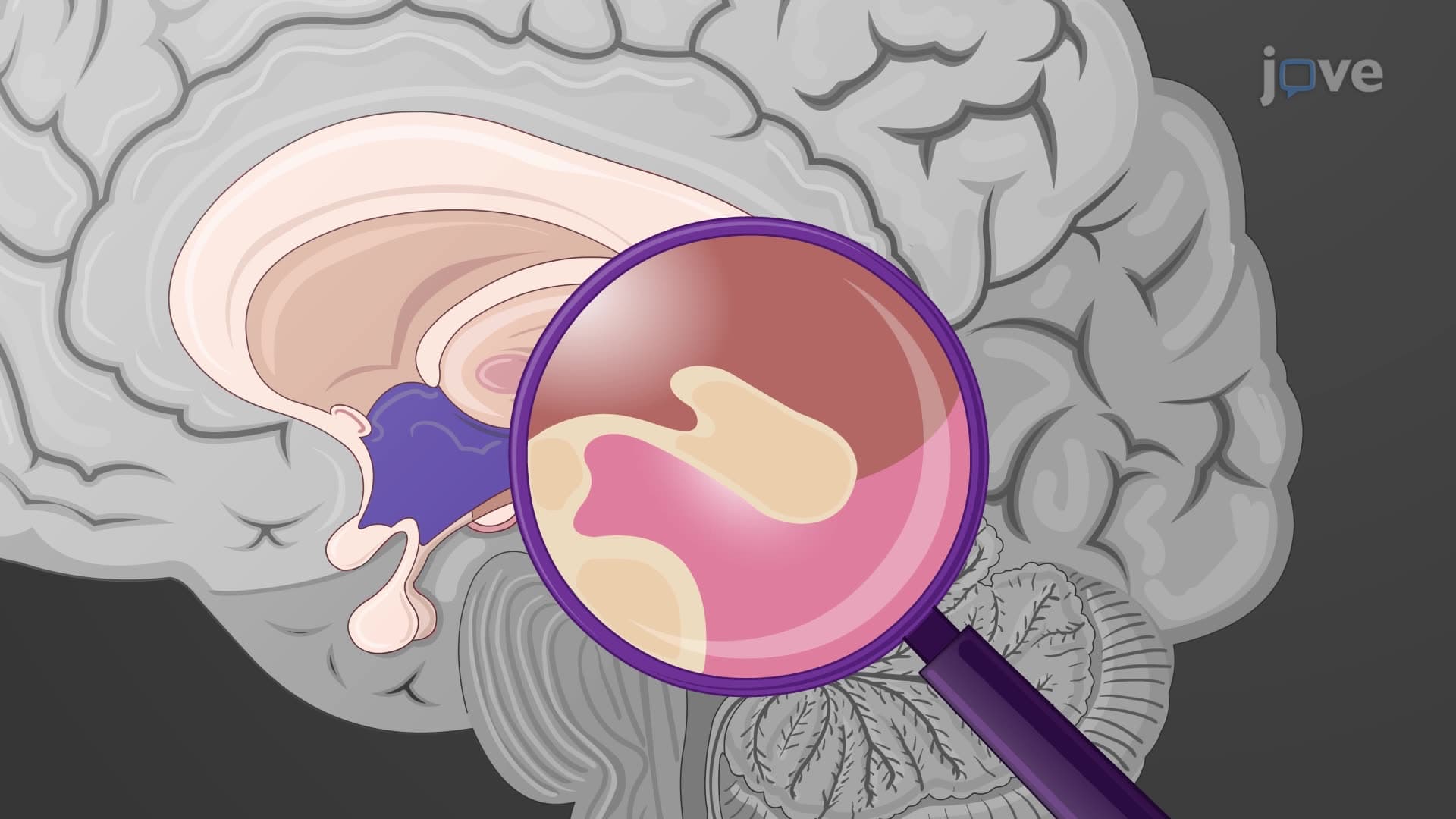
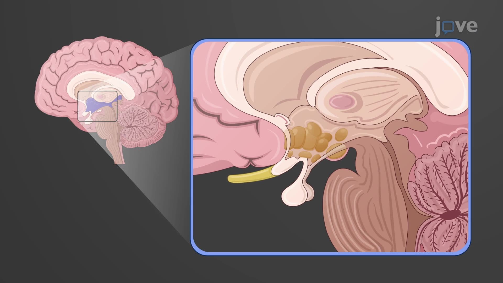
The third fourth ventricle system demonstrates remarkable anatomical precision in fluid circulation. The third ventricle, located within the diencephalon between the thalami, connects to lateral ventricles through interventricular foramina (also called foramina of Monro). The slender cerebral aqueduct (aqueduct of Sylvius) then channels CSF from the third to fourth ventricle. The fourth ventricle, positioned between the brainstem and cerebellum, serves as a crucial distribution point where CSF enters the central canal of the spinal cord and subarachnoid space. Understanding these connections helps explain why blockages at the aqueduct level can cause life-threatening hydrocephalus.
The choroid plexus CSF production system operates like a highly specialized filtration plant within each ventricle. These vascular structures contain networks of fenestrated capillaries surrounded by ependymal cells joined by tight junctions. This blood-CSF barrier selectively filters plasma while actively secreting CSF components. The choroid plexus produces approximately 500-600 mL of CSF daily, completely replacing the brain's CSF volume 3-4 times per day. This continuous production and circulation system maintains optimal intracranial pressure and removes metabolic waste products. College anatomy courses often emphasize how choroid plexus tumors can disrupt normal CSF production, leading to hydrocephalus in pediatric patients treated at children's hospitals like Boston Children's Hospital.
Related Micro-courses