Loading video…


 26 Concepts
 99.2K views

Video Summary: What Is Diencephalon Anatomical Regions
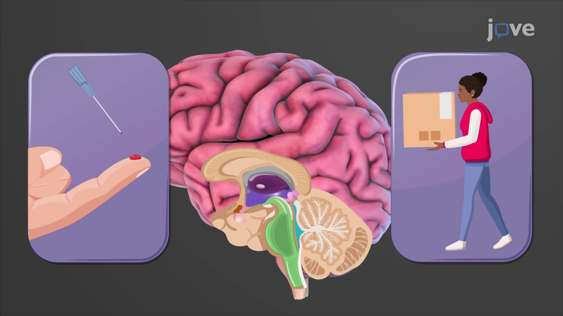
Ever wonder why you feel sleepy at night or how your brain processes the touch of a hot stove? The diencephalon anatomy regions control these vital functions through three key structures positioned between your brainstem and cerebral cortex. Students at Johns Hopkins University study how the thalamus, hypothalamus, and epithalamus work together as the brain's central relay station, processing everything from sensory input to hormone regulation. Understanding what are the anatomical regions of the diencephalon is crucial for mastering neuroanatomy concepts. Watch the full video on JoVE Coach to master this concept with expert-led visuals and step-by-step explanations.
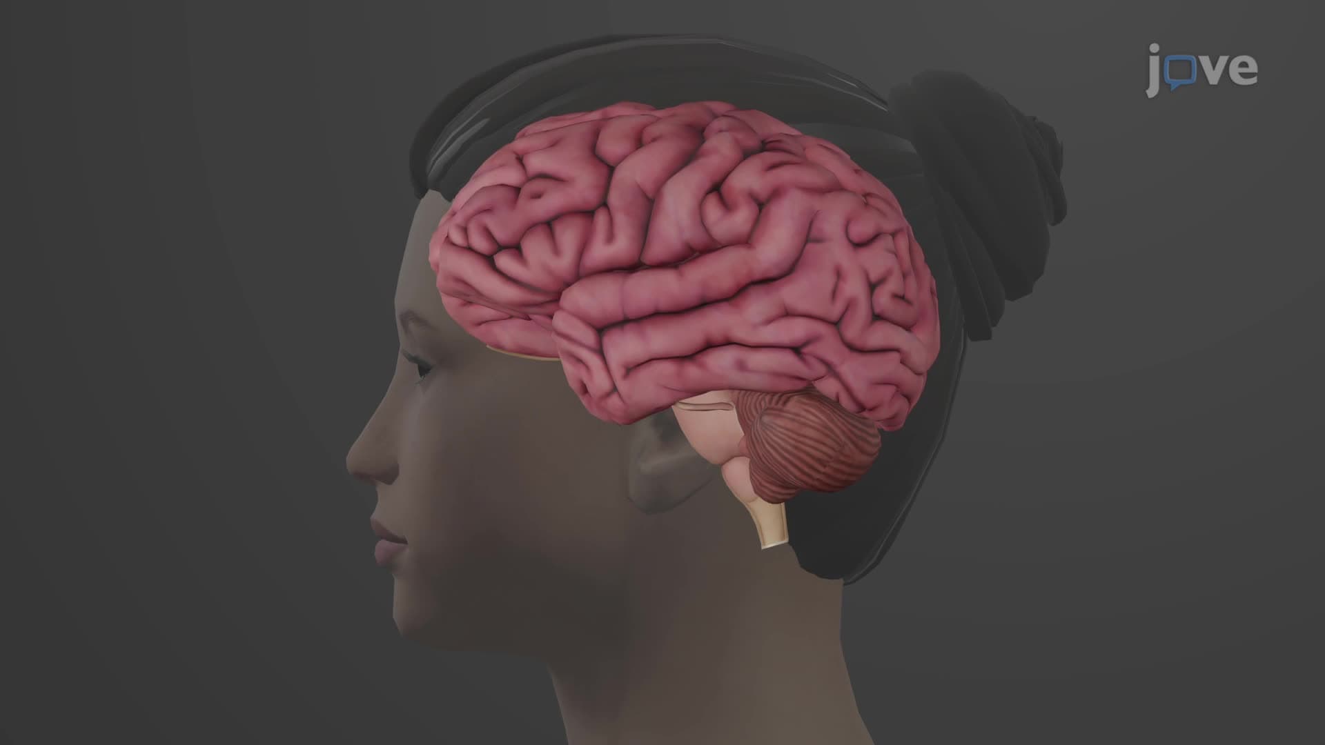
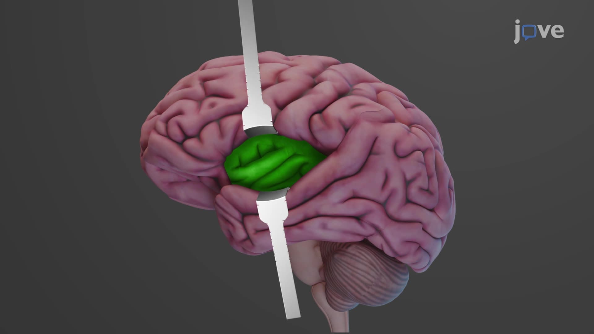
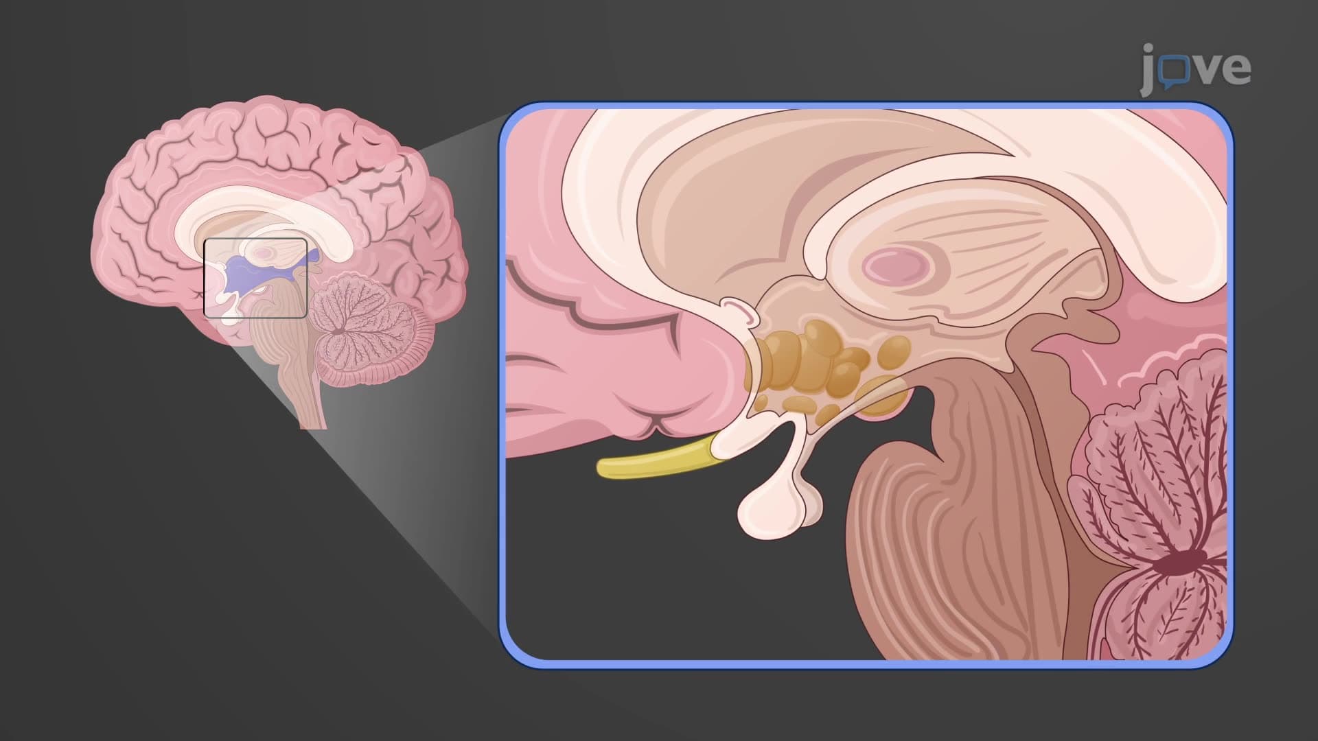
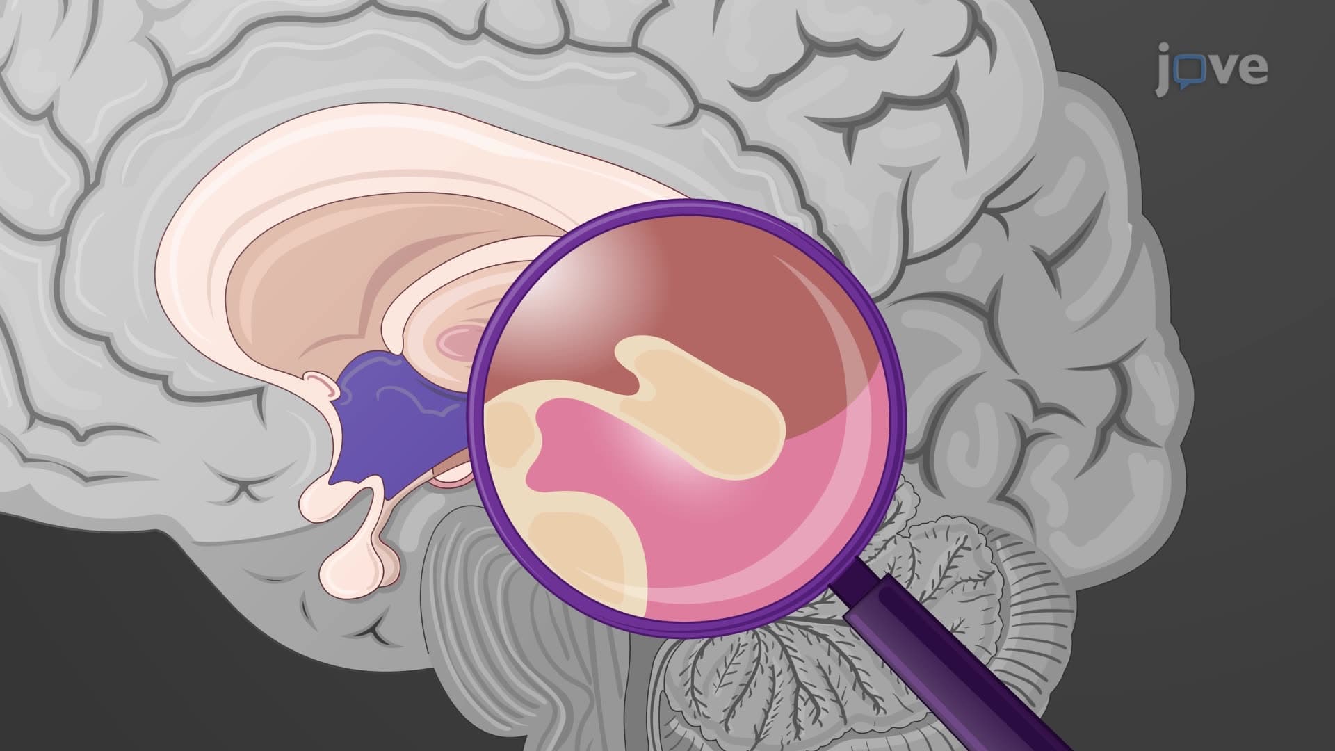
The diencephalon anatomy regions represent one of the most functionally diverse areas of the human brain, serving as a critical relay station between the brainstem and cerebral cortex. This complex structure, though hidden beneath the cerebral hemispheres, orchestrates essential processes that keep us alive and aware. For students preparing for the MCAT or AP Biology exams, mastering the diencephalon brain structure overview is fundamental to understanding neurophysiology.
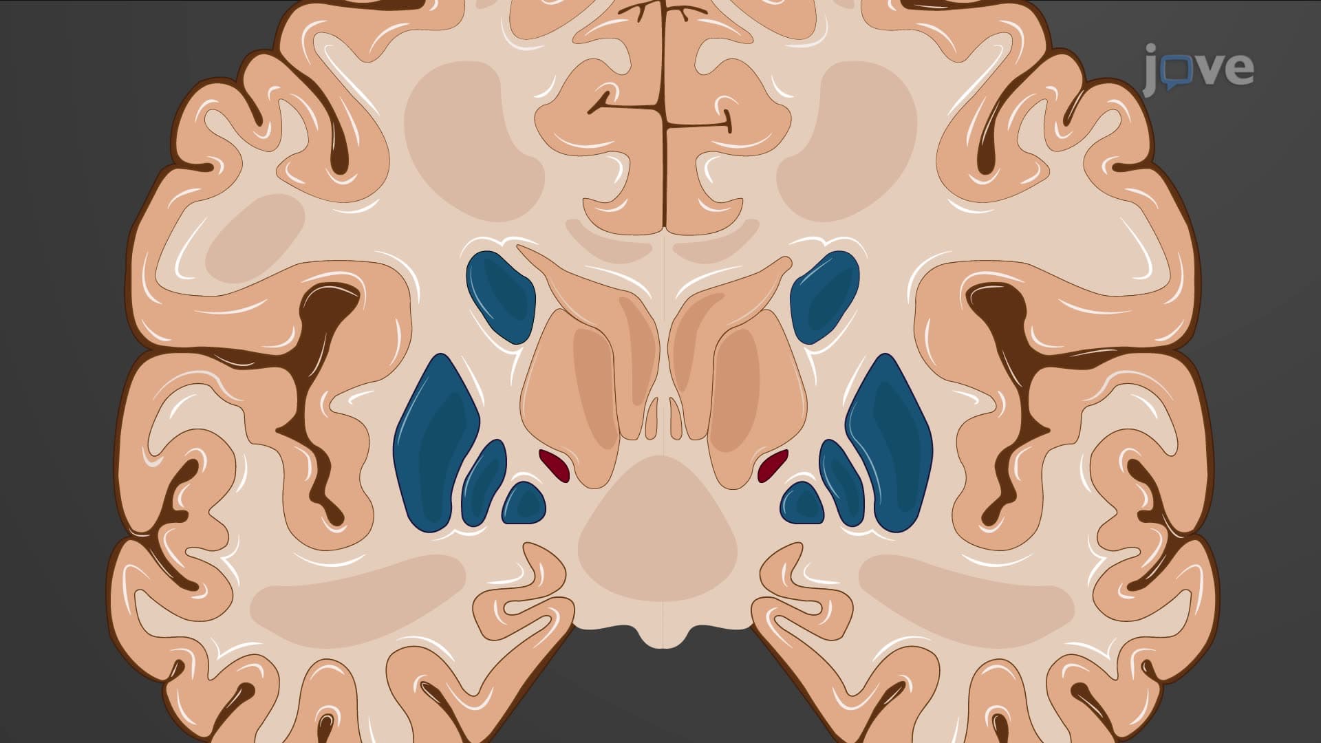
The thalamus functions as the brain's primary relay center, processing nearly all sensory information before it reaches the cerebral cortex. Its oval-shaped paired structures contain specialized nuclei organized into distinct groups: anterior, medial, lateral, ventral, intralaminar, and midline clusters. The reticular nuclei wrap around the lateral aspects, acting as gatekeepers for information flow.
Consider a patient at Massachusetts General Hospital experiencing a thalamic stroke. They might lose the ability to process touch sensations on one side of their body, demonstrating how the thalamus hypothalamus diencephalon network is essential for conscious perception. Medical students studying for the USMLE Step 1 exam frequently encounter questions about thalamic nuclei and their specific sensory pathways.
The hypothalamus, though small (roughly the size of an almond), controls numerous vital functions through its four distinct regions. The mammillary bodies process memory formation, while the tuberal region houses the arcuate nucleus that regulates appetite-explaining why patients with hypothalamic tumors often experience dramatic weight changes. The supraoptic region produces hormones like vasopressin and oxytocin, while the preoptic area controls body temperature.
Students at Stanford Medical School learn how hypothalamic dysfunction can cause conditions like diabetes insipidus or Kallmann syndrome, making this knowledge crucial for clinical practice.
The epithalamus pineal gland connection represents one of the most fascinating aspects of diencephalon region anatomy. This small, pine cone-shaped structure secretes melatonin in response to darkness, helping regulate our sleep-wake cycles. Jet lag experienced by travelers flying from New York to Los Angeles occurs partly because the pineal gland needs time to adjust melatonin production to the new time zone.
For college students preparing for neuroscience courses, understanding how the pineal gland responds to light signals from the retina through the sympathetic nervous system illustrates the interconnected nature of neural pathways-a concept frequently tested on AP Psychology and college-level examinations.
Related Micro-courses