Loading subject page…


99,206 views

Video Summary: What Is Spinal Nerves Plexus I
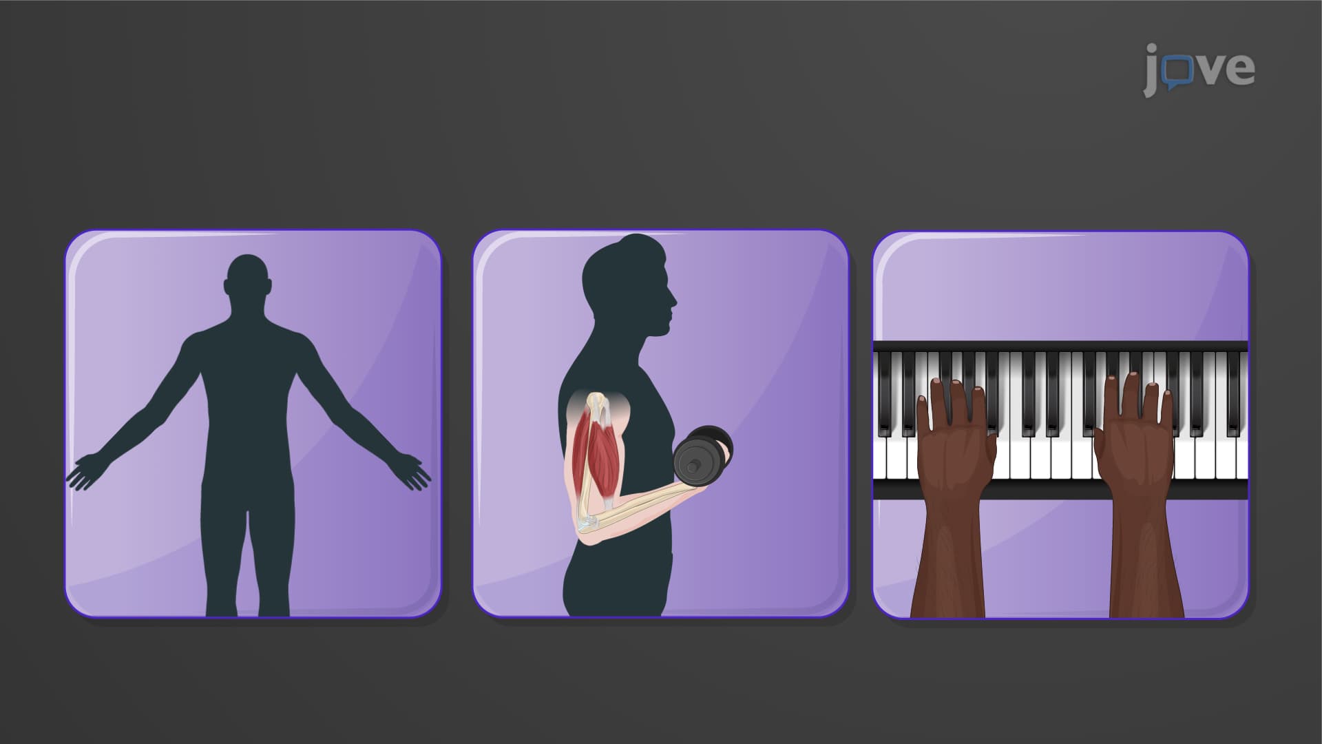
Did you know that when you take a deep breath, a single nerve from your neck controls your entire diaphragm? Spinal nerve plexus one anatomy reveals how cervical nerves C1-C4 form intricate networks that power essential functions like breathing and upper body movement. Consider how a pianist's finger dexterity depends on the brachial plexus coordinating five major nerves from C5-T1. Understanding What is Spinal Nerves Plexus I unlocks the foundation of peripheral nervous system organization. Watch the full video on JoVE Coach to master this concept with expert-led visuals and step-by-step explanations.
Spinal nerve plexus one anatomy represents a fundamental shift from the simple segmental pattern seen in thoracic nerves T2-T12. Unlike intercostal nerves that maintain individual pathways, cervical brachial plexus explained demonstrates how anterior rami from multiple spinal segments interconnect to form complex networks. This anatomical arrangement creates redundant innervation patterns that provide both functional advantages and clinical complexity.
The evolutionary benefit of plexus formation becomes apparent when considering upper limb mobility requirements. While thoracic intercostal nerves need only innervate specific rib segments, cervical and brachial regions must coordinate intricate movements spanning multiple joints and muscle groups. This complexity explains why what are the first spinal nerve plexuses in anatomy focuses on cervical (C1-C4) and brachial (C5-T1) networks.
Cervical plexus C1-C4 governs critical life-sustaining functions, making it essential knowledge for pre-med students preparing for the MCAT. The phrenic nerve, arising primarily from C4 with contributions from C3 and C5, represents the most clinically significant branch. High cervical spinal injuries above C4 often require mechanical ventilation because phrenic nerve damage eliminates diaphragmatic breathing control.
Motor components include the ansa cervicalis, which innervates infrahyoid muscles essential for swallowing and speech. AP Biology students studying human physiology should understand how these seemingly simple neck muscles coordinate with cranial nerves during deglutition. The four sensory branches-lesser occipital, great auricular, transverse cervical, and supraclavicular nerves-create overlapping sensory maps that emergency medicine physicians use for diagnostic nerve blocks.
Upper limb brachial plexus organization from C5-T1 creates the foundation for understanding orthopedic and neurological conditions affecting arm function. The five major terminal branches-axillary, musculocutaneous, radial, median, and ulnar nerves-each serve specific functional roles that physical therapy students must master for clinical practice.
College anatomy courses emphasize how brachial plexus injuries, such as Erb's palsy affecting C5-C6 roots, create predictable clinical patterns. The "waiter's tip" position results from specific muscle group paralysis, demonstrating how nerve plexus formation anatomy translates directly to clinical diagnosis. USMLE Step 1 preparation requires understanding these injury patterns alongside normal anatomical variations.
Medical school curricula integrate spinal nerve plexus anatomy with clinical correlations throughout multiple courses. First-year anatomy lab dissections reveal individual variations in plexus formation that explain why some patients experience atypical symptoms following nerve injuries. NCLEX-RN candidates must understand how cervical plexus blocks provide anesthesia for carotid endarterectomy procedures, while brachial plexus blocks enable upper extremity surgeries.
Related Micro-courses