Loading subject page…


99,207 views


Video Summary: What Is Spinal Nerves Plexus Ii
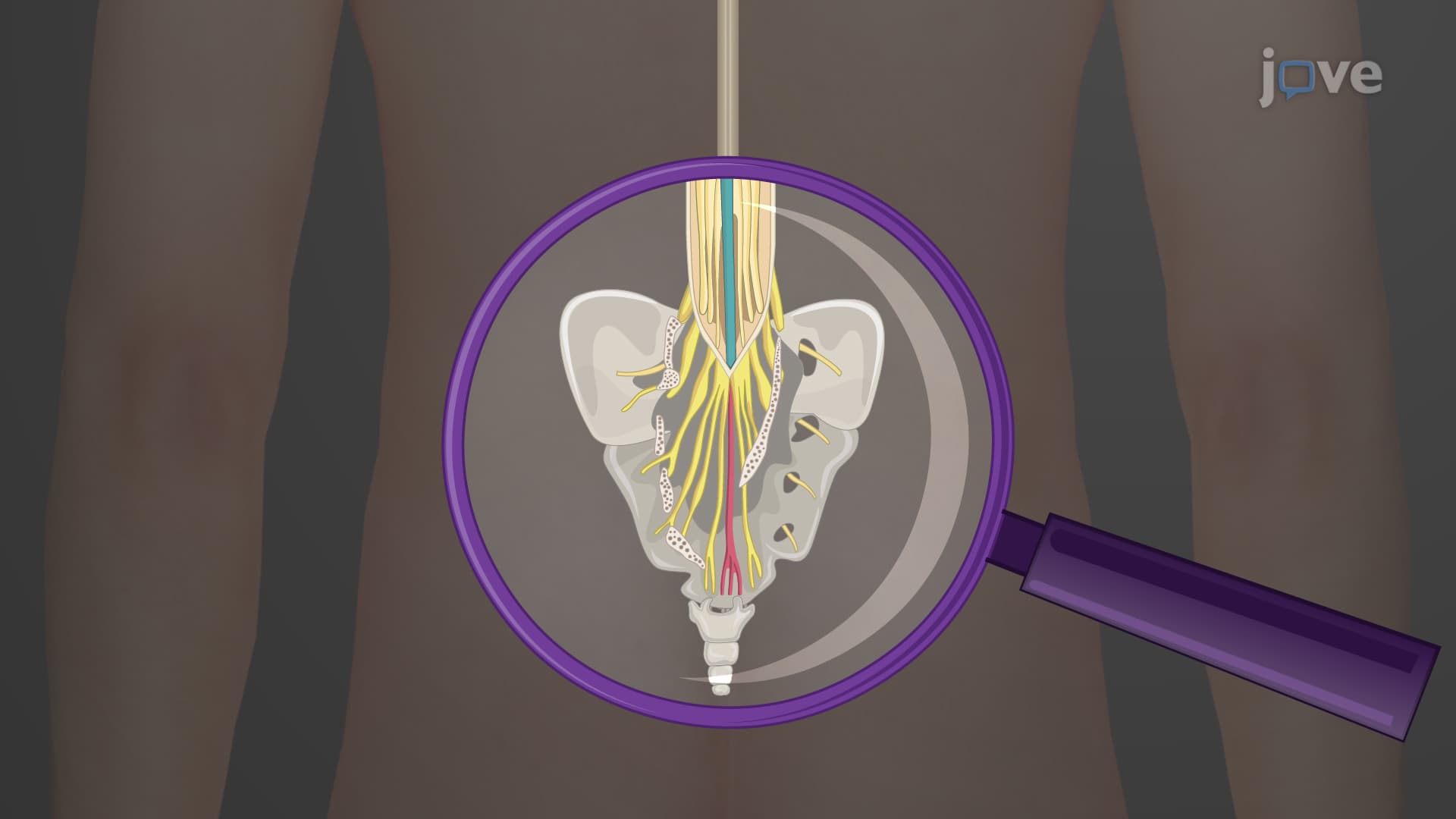
Ever wondered why a herniated disc in your lower back can cause shooting pain down your leg? The spinal nerve plexus two anatomy reveals how lumbar, sacral, and coccygeal nerve networks create this intricate connection. For instance, when a patient at Johns Hopkins experiences sciatica, doctors trace the pain pathway through the sacral plexus to the thick sciatic nerve. Watch the full video on JoVE Coach to master this concept with expert-led visuals and step-by-step explanations.
The lower body's neurological framework centers on three interconnected nerve networks that demonstrate remarkable organizational complexity. Unlike the upper extremity plexuses, these lower networks must coordinate both locomotion and vital pelvic functions, making their study essential for anyone pursuing healthcare careers.
The lumbar plexus L1-L4 operates as the primary control system for hip flexion and thigh sensation. Formed within the psoas major muscle, this network produces six major branches that medical students memorize using mnemonics like "I Like Getting Laid On Friday." The femoral nerve lumbar plexus represents the largest terminal branch, innervating the quadriceps muscles essential for knee extension and stair climbing. When physical therapists at Cleveland Clinic assess knee weakness, they often trace problems back to femoral nerve dysfunction originating in the lumbar plexus.
The obturator nerve, another crucial lumbar branch, controls thigh adduction-the movement that allows soccer players to bring their legs together for ball control. Medical boards frequently test knowledge of obturator nerve injuries, which can occur during difficult childbirth procedures at major medical centers like Mayo Clinic.
The sacral plexus L4-S3 forms the body's most robust nerve network, giving rise to the legendary sciatic nerve sacral plexus. This massive nerve, thicker than a pencil, carries both sensory and motor information for the entire posterior thigh, leg, and foot. MCAT questions often focus on sciatic nerve distributions because understanding this pathway explains why piriformis syndrome can mimic herniated disc symptoms.
Emergency room physicians at Johns Hopkins regularly encounter sciatic nerve injuries from motorcycle accidents, where damage to this critical pathway can result in foot drop-the inability to lift the front of the foot during walking. The superior and inferior gluteal nerves, also arising from the sacral plexus, control the powerful hip muscles that maintain pelvic stability during single-leg stance.
For AP Biology students, understanding what are the lumbar and sacral spinal nerve plexuses provides crucial foundation knowledge for advanced anatomy courses. The lumbosacral plexus integration demonstrates how embryological segmentation creates functional units that serve specific body regions.
NCLEX-RN examinations frequently test nursing students on nerve plexus anatomy when assessing patients with back injuries or surgical complications. Understanding that the coccygeal plexus serves only the tailbone region helps differentiate its limited clinical significance from the more complex lumbar and sacral networks.
Related Micro-courses