Loading subject page…


112,914 views

Video Summary: What are the Parathyroid Glands
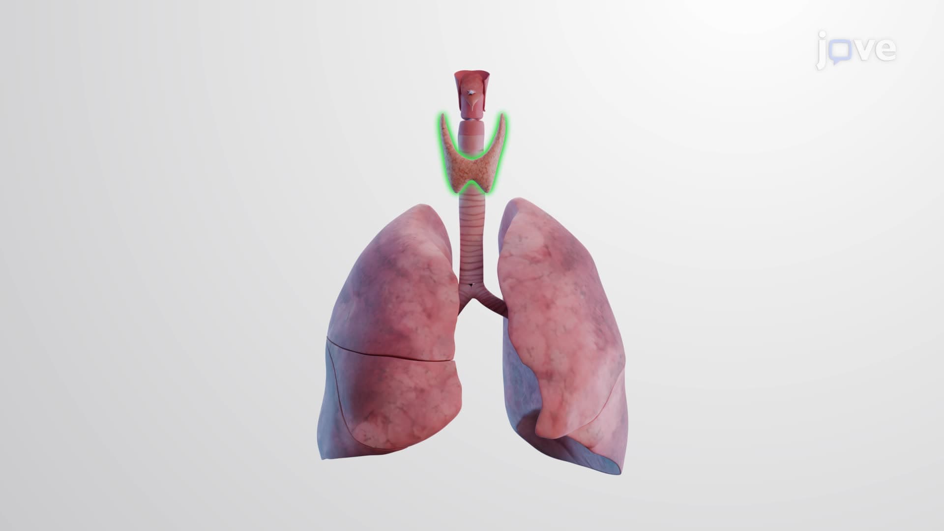
Did you know that four tiny glands smaller than rice grains control your body's calcium levels and bone strength? The parathyroid glands anatomy biology reveals how these embedded structures behind your thyroid prevent muscle cramps and maintain bone health-critical functions that become evident when patients at Johns Hopkins develop calcium imbalances after thyroid surgery. Understanding what are the parathyroid glands helps explain why calcium regulation is essential for everything from muscle contraction to nerve function. Watch the full video on JoVE Coach to master this concept with expert-led visuals and step-by-step explanations.
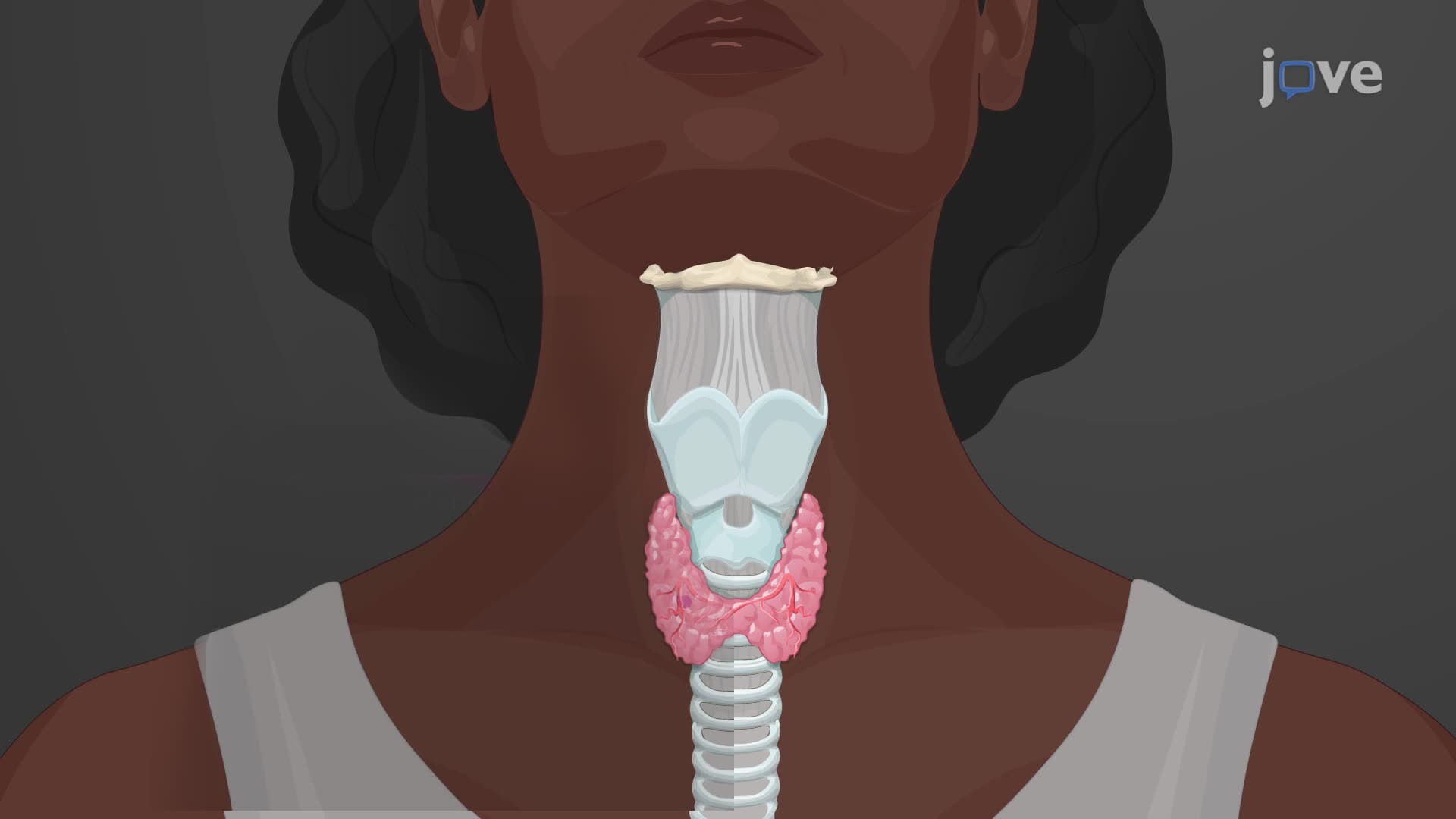
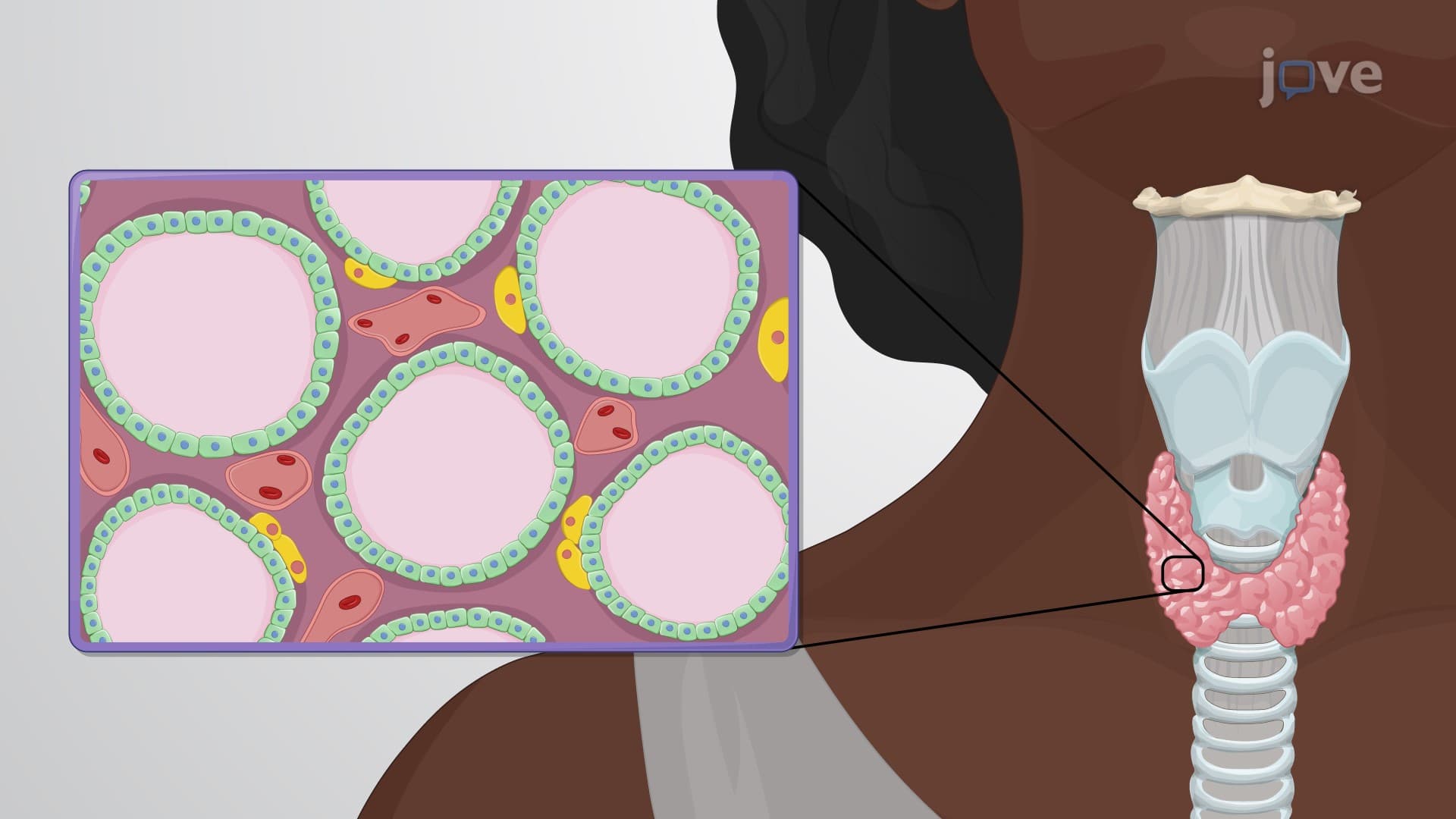
The parathyroid glands anatomy biology centers on four small, pea-sized endocrine glands embedded in the posterior surface of the thyroid gland. Unlike the thyroid itself, these glands are separated by a dense fibrous capsule that protects them during surgical procedures. Each gland measures only 3-8 millimeters in diameter, making them among the smallest endocrine organs in the human body. Their strategic location allows them to monitor blood calcium levels as blood flows through the highly vascularized neck region.
The parathyroid gland structure function depends on two primary cell populations. Principal cells (also called chief cells) constitute 80-85% of the gland's mass and serve as the primary calcium sensors. These cells contain calcium-sensing receptors that directly monitor blood calcium concentrations. Oxyphil cells appear during late adolescence but remain functionally mysterious-researchers at Mayo Clinic continue investigating their role, though they may represent aged principal cells or have metabolic functions.
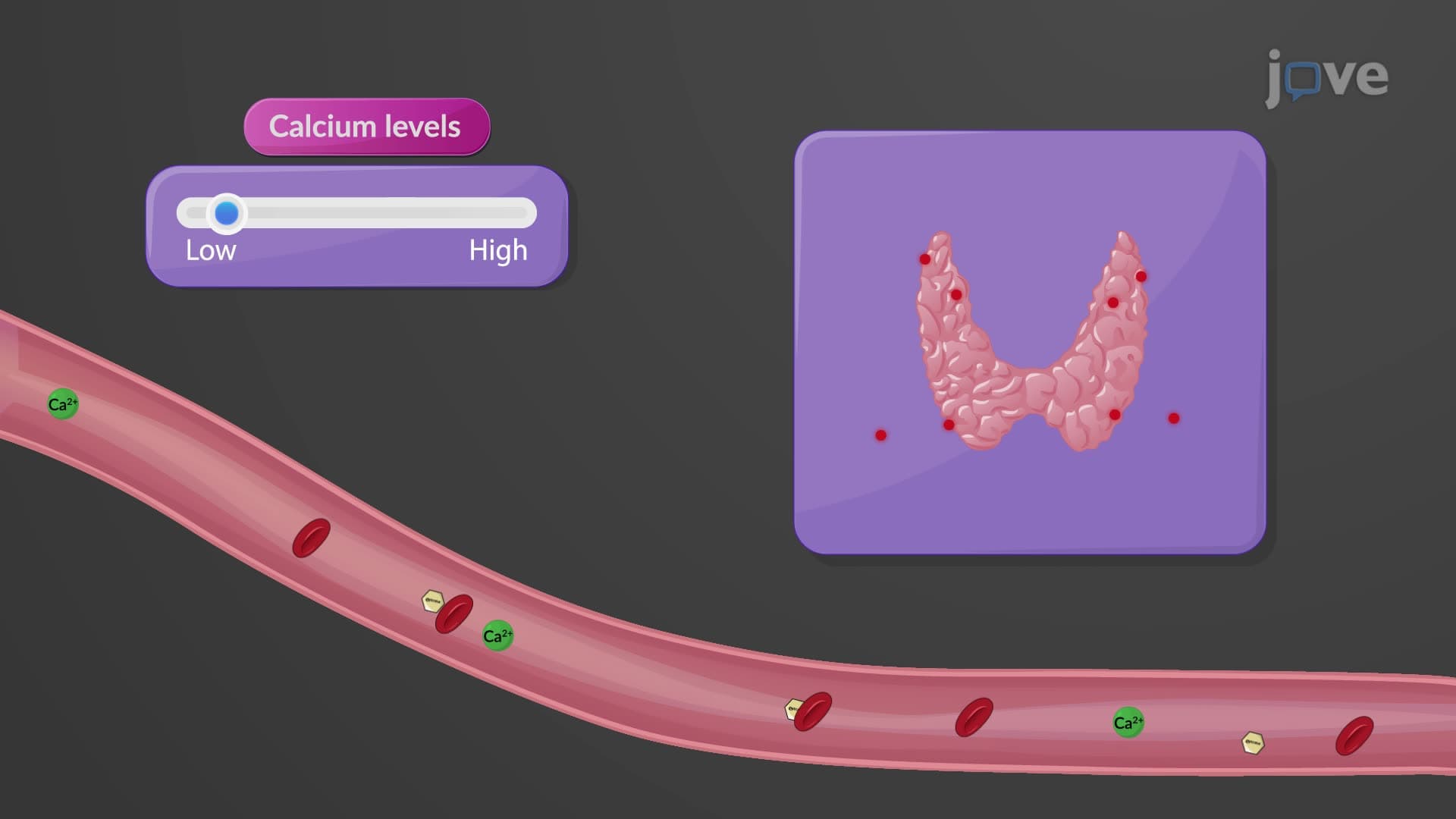
When blood calcium drops below 8.5 mg/dL, principal cells rapidly secrete parathyroid hormone (PTH), an 84-amino acid polypeptide. This parathyroid calcium sensor mechanism operates through sophisticated G-protein coupled receptors that detect even minute changes in ionized calcium. PTH acts on three target organs: bones (stimulating osteoclast activity to release stored calcium), kidneys (enhancing calcium reabsorption and reducing phosphate retention), and intestines (indirectly through vitamin D activation). Students preparing for the MCAT should understand this represents a classic negative feedback loop-as calcium levels normalize, PTH secretion decreases.
Understanding parathyroid anatomy function proves crucial for medical professionals. Primary hyperparathyroidism affects over 100,000 Americans annually, often requiring surgical intervention at specialized centers like the University of California San Francisco. Conversely, accidental removal during thyroid surgery can cause hypoparathyroidism, leading to dangerous hypocalcemia. For AP Biology students, this exemplifies how anatomical structure directly correlates with physiological function-the glands' posterior location protects them while maintaining proximity to major blood vessels for optimal calcium monitoring.
Related Micro-courses