Loading subject page…


112,914 views

Video Summary: What Is Anatomy Adrenal Glands
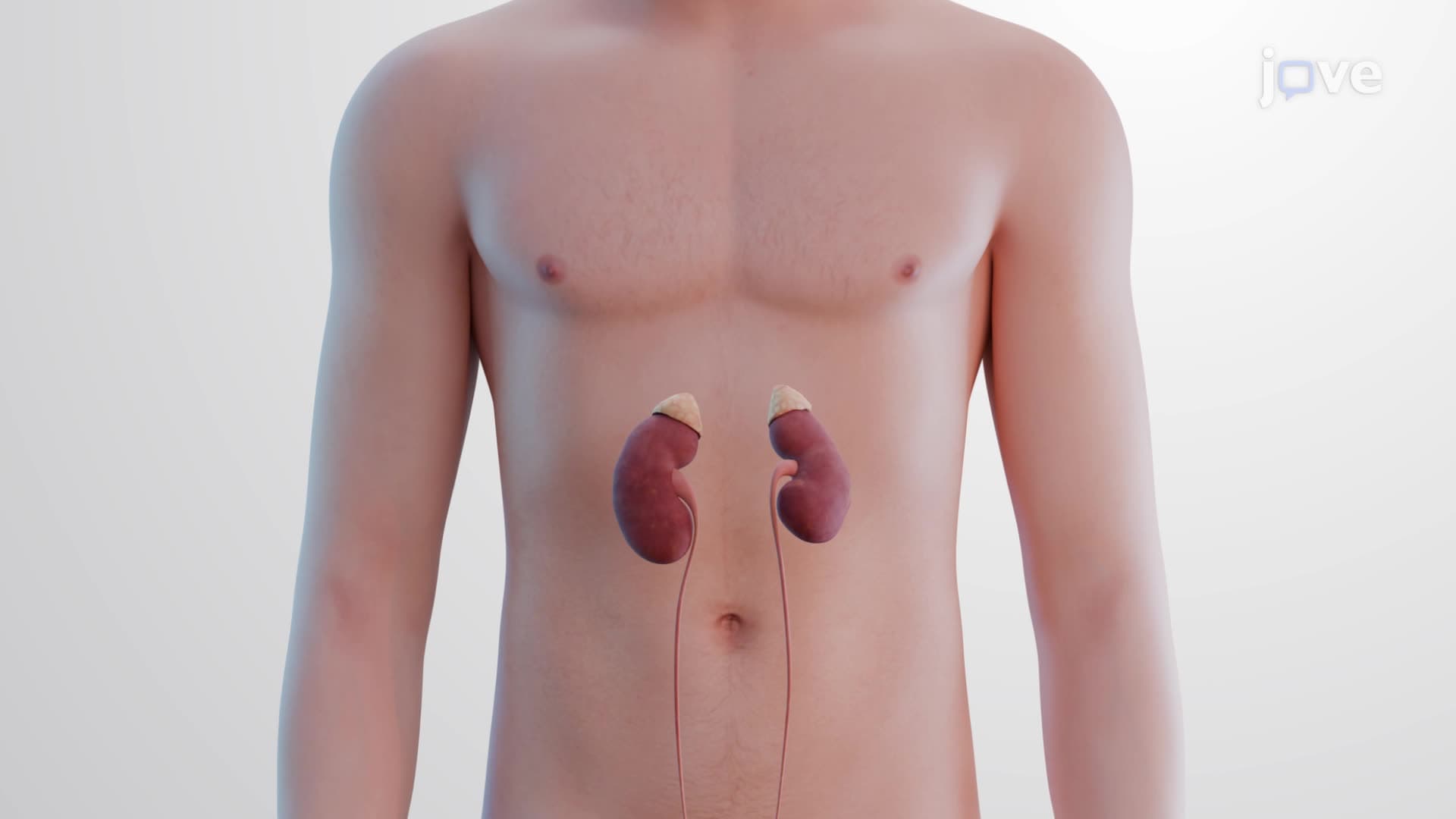
When you feel your heart racing during a stressful exam or emergency, your adrenal gland anatomy biology is working overtime to help you respond. These pyramid-shaped glands, positioned atop each kidney, contain two distinct regions that control everything from your fight-or-flight response to long-term stress management. For instance, when a student faces the SAT, their adrenal glands release both immediate stress hormones and sustained regulatory hormones. Understanding what is the anatomy of the adrenal glands reveals how these small but mighty organs coordinate your body's complex stress responses. Watch the full video on JoVE Coach to master this concept with expert-led visuals and step-by-step explanations.
The adrenal gland anatomy biology represents one of the most functionally diverse endocrine structures in the human body. These paired, pyramid-shaped organs sit atop each kidney in the retroperitoneal space, positioned at the level of the twelfth rib. The adrenal gland structure explained reveals a sophisticated two-part design that allows for both rapid emergency responses and sustained physiological regulation.
Each adrenal gland weighs approximately 4-5 grams and measures about 4-6 centimeters in length. The adrenal capsule structure consists of a tough fibrous outer layer surrounded by protective adipose tissue, providing both structural support and cushioning against physical trauma during movement or impact.
The adrenal cortex medulla anatomy begins with understanding the cortex as the outer, lipid-rich region comprising about 90% of the gland's mass. The cortex's yellow appearance results from stored cholesterol and fatty acids essential for steroid hormone synthesis. This region organizes into three distinct zones, each producing specific hormone classes.
The zona glomerulosa fasciculata reticularis represents the three-layer architecture from outer to inner. The zona glomerulosa produces mineralocorticoids like aldosterone, regulating sodium and potassium balance. The zona fasciculata, the thickest layer, synthesizes glucocorticoids such as cortisol for long-term stress management and glucose regulation. The innermost zona reticularis produces androgens and supports the body's response to chronic stress.
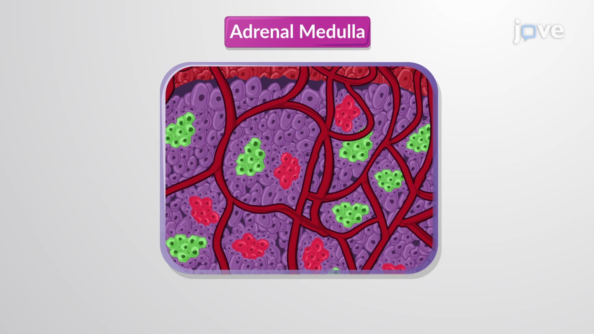
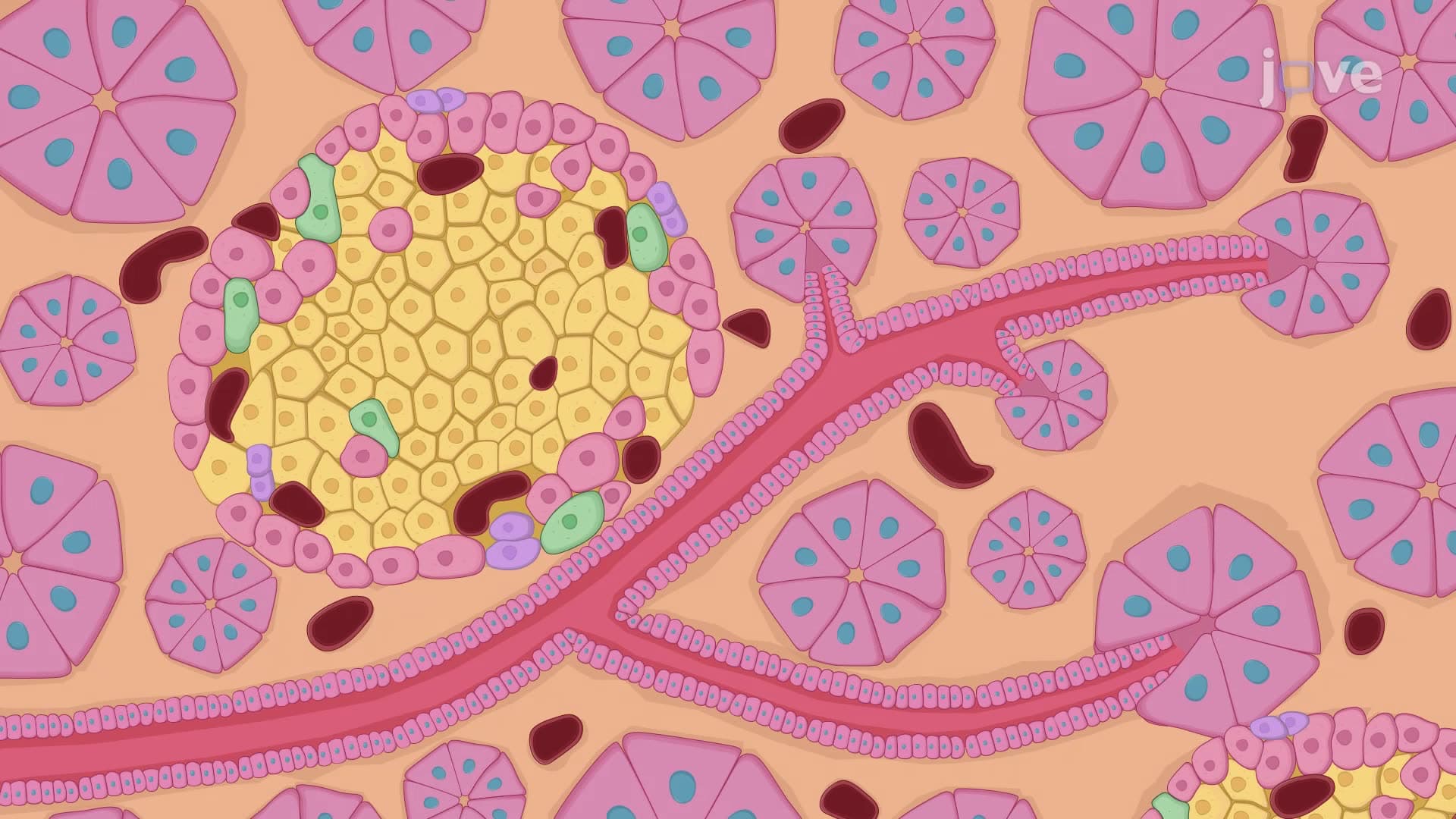
The adrenal medulla forms the inner core, composed entirely of nervous tissue derived from neural crest cells. This region contains specialized chromaffin cells arranged around extensive capillary networks. These cells synthesize and release catecholamines-primarily epinephrine (adrenaline) and norepinephrine-within seconds of sympathetic nervous system activation.
When students encounter high-stakes situations like AP Biology exams or MCAT testing, their adrenal medulla rapidly releases these hormones, increasing heart rate, blood pressure, and glucose availability for enhanced cognitive performance.
Understanding adrenal gland retroperitoneal positioning proves crucial for medical students preparing for USMLE Step 1, as adrenal pathology questions frequently appear on standardized exams. Conditions like Addison's disease (adrenal insufficiency) and Cushing's syndrome (excess cortisol) directly relate to specific anatomical regions within the gland structure.
Related Micro-courses