Loading subject page…


112,914 views


Video Summary: What Is the Pineal Gland
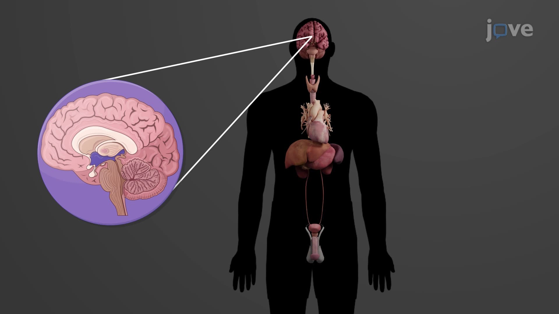
Ever wonder why you feel sleepy when the lights go out? The pineal gland biology anatomy reveals a tiny, pinecone-shaped structure in your brain that acts as your body's natural timekeeper. Located deep within the diencephalon, this remarkable endocrine gland produces melatonin-the hormone that makes millions of Americans reach for sleep aids each night. What is the pineal gland's secret to controlling our sleep-wake cycles? Watch the full video on JoVE Coach to master this concept with expert-led visuals and step-by-step explanations.
The pineal gland biology anatomy centers on a remarkably small but powerful endocrine structure measuring just 5-8 millimeters in adult humans. Often called the epiphysis cerebri pineal, this gland sits strategically positioned on the roof of the third ventricle, embedded within the diencephalon's epithalamus region. Its distinctive pinecone shape-from which it derives its name-makes it easily identifiable in neuroanatomy studies that frequently appear on AP Biology and college anatomy exams.
The gland's microscopic architecture reveals specialized secretory cells called pinealocytes, arranged in characteristic compact cords and clusters. These cells surround dense accumulations of calcium phosphate and carbonate deposits known as "brain sand" or corpora arenacea. These calcium deposits increase with age and serve as useful radiological landmarks for neurologists diagnosing brain abnormalities in US hospitals.
The pineal gland function explained revolves around its primary hormone, melatonin-a tryptophan-derived indoleamine synthesized from serotonin. This biochemical pathway, commonly tested on MCAT examinations, demonstrates how amino acid metabolism directly influences physiological processes. Pinealocytes convert serotonin to melatonin through a two-step enzymatic process involving arylalkylamine N-acetyltransferase (AANAT) and hydroxyindole-O-methyltransferase (HIOMT).
The pineal circadian rhythm regulation operates through a sophisticated neural pathway connecting the eyes to the pineal gland. Light detected by specialized retinal ganglion cells travels via the retinohypothalamic tract to the suprachiasmatic nucleus, then through sympathetic neurons to the pineal gland. This explains why exposure to bright screens before bedtime-a common concern among American teenagers-can suppress melatonin production and disrupt sleep patterns.
Understanding pineal gland light dark responses has practical implications for healthcare workers, students, and shift workers across America. Nurses working night shifts at hospitals like Mayo Clinic or Johns Hopkins often experience circadian rhythm disruption due to artificial lighting interfering with natural melatonin cycles. Similarly, students preparing for standardized tests may benefit from optimizing their sleep environment to support healthy pineal function.
The gland's influence extends beyond sleep regulation through its effects on reproductive physiology. Melatonin pineal secretion helps regulate seasonal breeding patterns in mammals and influences human puberty timing. Research conducted at Stanford University and Harvard Medical School has linked pineal dysfunction to delayed puberty, seasonal affective disorder, and certain sleep disorders commonly diagnosed in American adolescents.
Related Micro-courses