Loading subject page…

- Anatomy and Physiology
- The Integumentary System
- Changes in Skin Color: Clinical Perspectives
Micro-course: The Integumentary System
87,705 views
Learn key concepts and earn your certificate.




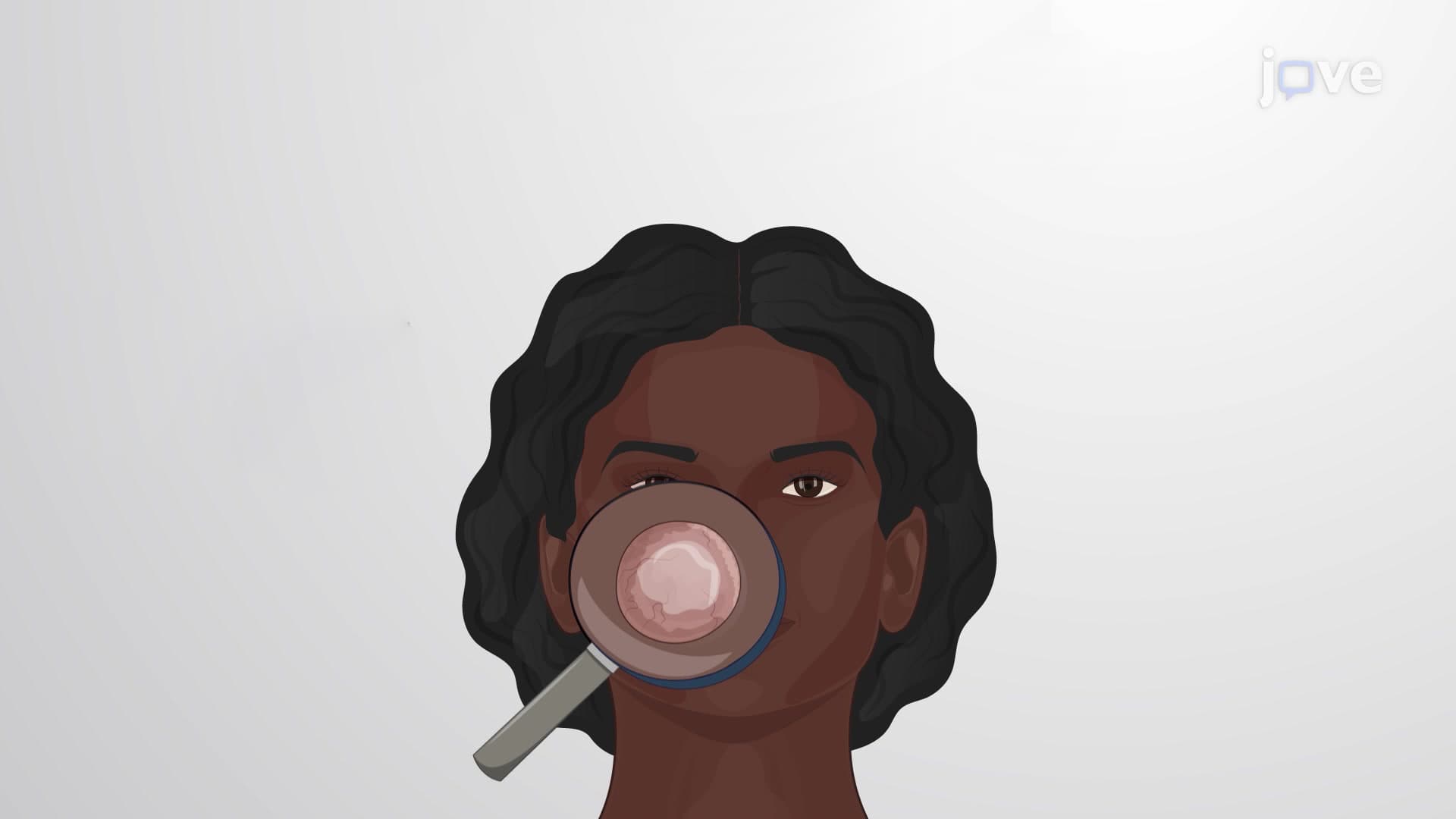
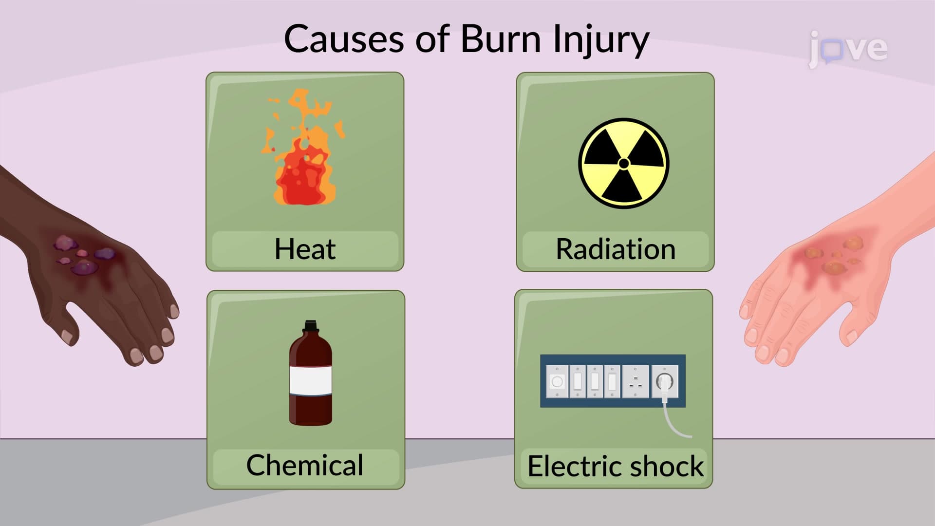
Changes in Skin Color: Clinical Perspectives

00:00
01:14
Video Summary: Changes in Skin Color Clinical Perspectives Explained
Did you know that your skin color can reveal critical health information that could save your life? Skin color changes clinical assessment is a fundamental diagnostic tool used in emergency rooms across America, from detecting oxygen deprivation in heart attack patients to identifying liver dysfunction in hepatitis cases. Understanding changes in skin color clinical perspectives explained empowers healthcare providers to make rapid, potentially life-saving diagnoses. Watch the full video on JoVE Coach to master this concept with expert-led visuals and step-by-step explanations.
- Understand the clinical significance of changes in skin color clinical perspectives explained and their role in medical diagnosis
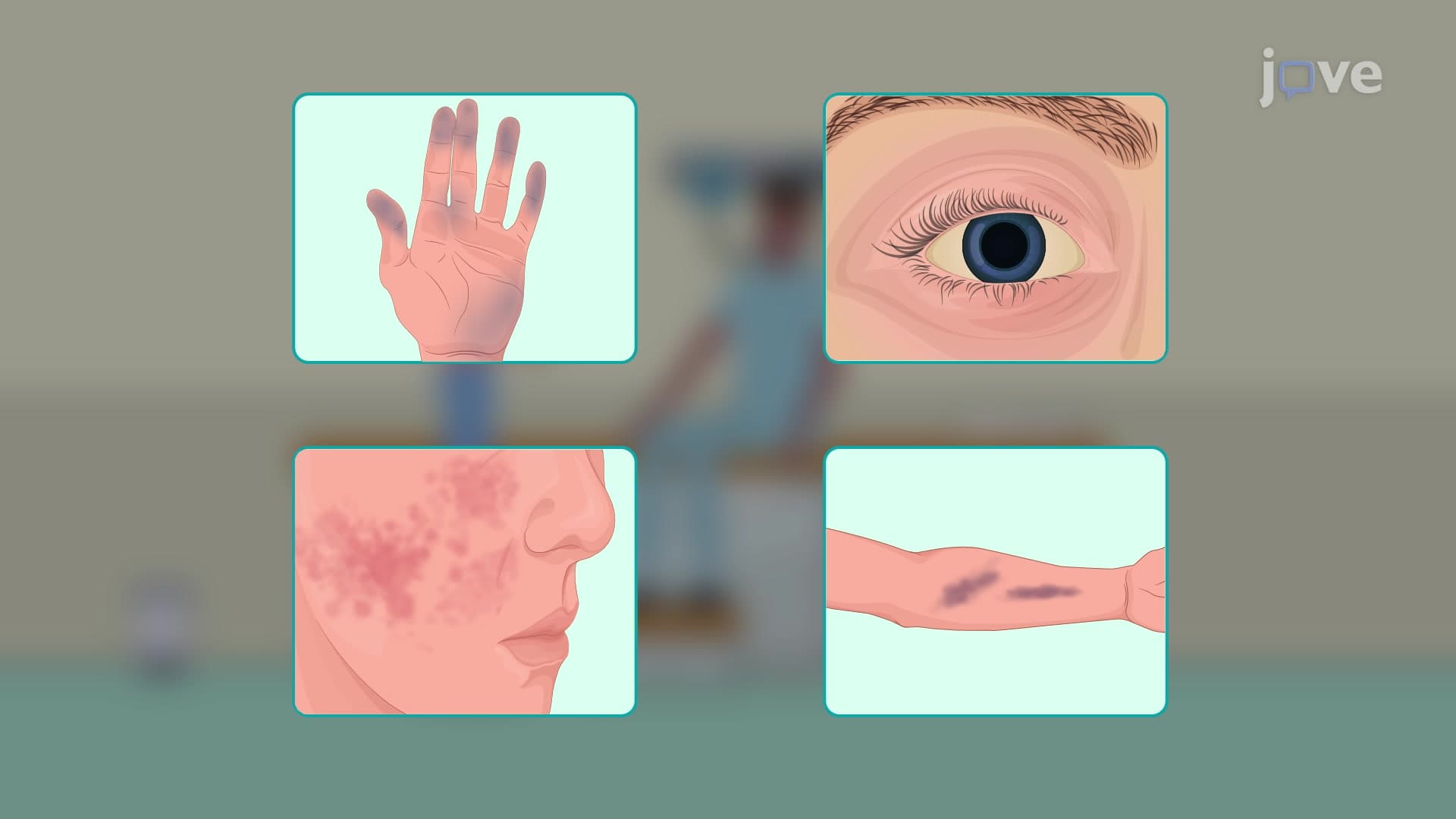
- Identify four major pathological skin color changes: cyanosis, jaundice, ecchymoses, and erythema
- Analyze the underlying physiological mechanisms causing abnormal skin pigmentation in clinical settings
- Compare different types of skin color disorders and their associated medical conditions
- Evaluate the diagnostic value of integumentary assessment in emergency and routine clinical practice
- Apply knowledge of skin color changes to recognize potential cardiovascular, respiratory, and hepatic disorders
- Recognize the difference between normal physiological color variations and pathological changes
- Explore how clinical skin color assessment integrates with other diagnostic tools in patient evaluation
Understanding Changes in Skin Color Clinical Perspectives Explained
Clinical assessment of skin color changes represents one of the most immediate and accessible diagnostic tools in modern healthcare. Healthcare providers in the United States routinely use clinical skin color changes explained as part of their initial patient assessment, particularly in emergency departments where rapid diagnosis can be life-saving. This integumentary assessment technique requires understanding both normal physiological responses and pathological conditions that manifest through altered pigmentation.
Cyanosis: The Blue Warning Sign
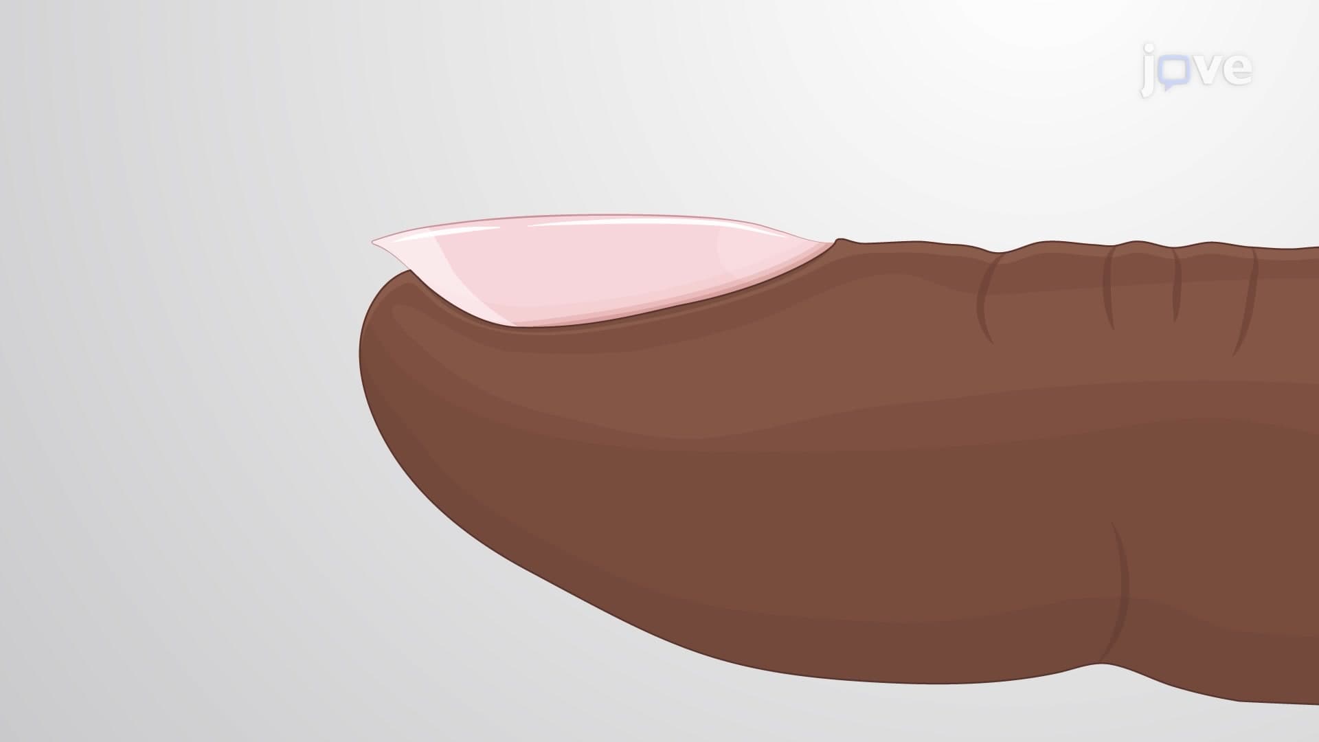
Pallor erythema cyanosis represents the most critical triad of color changes clinicians monitor. Cyanosis, characterized by a bluish discoloration of skin, mucous membranes, and nail beds, indicates insufficient oxygen saturation in the blood. In US emergency rooms, healthcare providers immediately assess for central cyanosis (affecting lips, tongue, and mucous membranes) versus peripheral cyanosis (limited to extremities). Central cyanosis often signals serious cardiac or pulmonary conditions requiring immediate intervention, while peripheral cyanosis may indicate circulatory compromise. This concept frequently appears on MCAT and nursing school examinations, particularly in cardiovascular and respiratory system assessments.
Jaundice and Hepatic Dysfunction
Jaundice skin color changes present as yellowing of the skin and sclera (whites of the eyes), indicating elevated bilirubin levels. In clinical practice across American hospitals, healthcare providers differentiate between pre-hepatic jaundice (excessive red blood cell breakdown), hepatic jaundice (liver dysfunction), and post-hepatic jaundice (bile duct obstruction). Understanding bilirubin metabolism proves essential for AP Biology students and pre-med undergraduates, as this topic integrates biochemistry with clinical medicine. The condition commonly appears in newborn assessments, where physiological jaundice affects approximately 60% of term infants in US hospitals.
Erythema and Ecchymoses: Inflammatory and Traumatic Responses
Skin color disorder clinical assessment includes recognizing erythema (redness from increased blood flow) and ecchymoses (bruising from subcutaneous bleeding). Erythema indicates localized inflammation, infection, or injury, commonly seen in conditions ranging from cellulitis to allergic reactions. Healthcare providers in urgent care centers frequently encounter erythema migrans, the characteristic bull's-eye rash of Lyme disease prevalent in northeastern US states. Ecchymoses, meanwhile, help clinicians assess trauma severity and bleeding disorders, particularly important in emergency medicine rotations for medical students.
Frequently Asked Questions
Changes in skin color clinical perspectives refer to the systematic assessment of abnormal skin pigmentation to diagnose medical conditions. These visual cues provide immediate diagnostic information about oxygen levels, liver function, circulation, and inflammatory processes. Healthcare providers use these observations as part of comprehensive patient assessment, often leading to rapid diagnosis and treatment decisions.
The MCAT frequently tests cyanosis in cardiovascular and respiratory passages, while nursing exams like NCLEX focus on recognizing jaundice in liver dysfunction scenarios. Expect questions linking skin color changes to underlying pathophysiology, particularly oxygen transport and bilirubin metabolism. These concepts integrate multiple body systems, making them popular for comprehensive assessment questions.
Focus on the relationship between hemoglobin oxygen saturation and skin color changes. Remember that central cyanosis indicates serious cardiopulmonary issues, while peripheral cyanosis may reflect poor circulation. Understand that cyanosis becomes visible when deoxygenated hemoglobin exceeds 5 grams per deciliter, and practice identifying this in case study questions.
Emergency physicians perform rapid visual assessment within seconds of patient arrival, checking for cyanosis (respiratory/cardiac emergency), jaundice (liver dysfunction), pallor (anemia/shock), or widespread erythema (sepsis/allergic reaction). This immediate assessment helps prioritize treatment and determine if patients need critical care resources. For example, central cyanosis in a chest pain patient immediately suggests serious cardiac or pulmonary pathology.
Basic understanding of oxygen transport and bilirubin metabolism suffices for introductory courses. Focus on the clinical significance rather than complex biochemical pathways initially. High school students should concentrate on recognizing color changes and their general causes, while college students can delve deeper into hemoglobin structure and liver function.
Practice with visual case studies and mnemonics linking colors to conditions: "Blue = Breathing problems, Yellow = Liver troubles, Red = Inflammation, Purple = Bleeding underneath." Review normal versus abnormal findings, and understand when each color change requires immediate intervention versus routine monitoring.
Skin color changes integrate with cardiovascular physiology, respiratory function, hepatic metabolism, and hematology. Consider studying oxygen transport mechanisms, liver function tests, inflammatory responses, and coagulation disorders. This foundation prepares you for advanced pathophysiology and clinical diagnosis courses.
Related Micro-courses

19 Concepts

11 Concepts
6 Concepts