Loading subject page…


87,705 views

Video Summary: What are Layers of the Epidermis
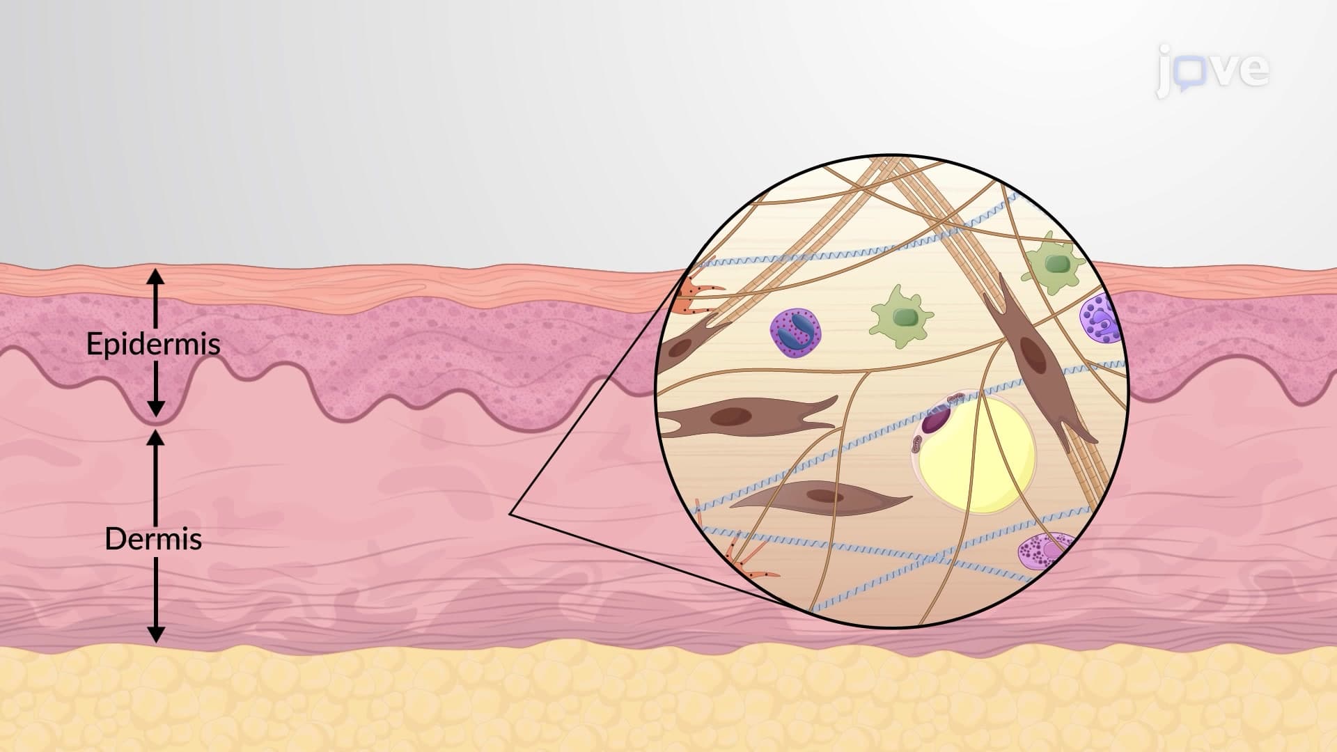
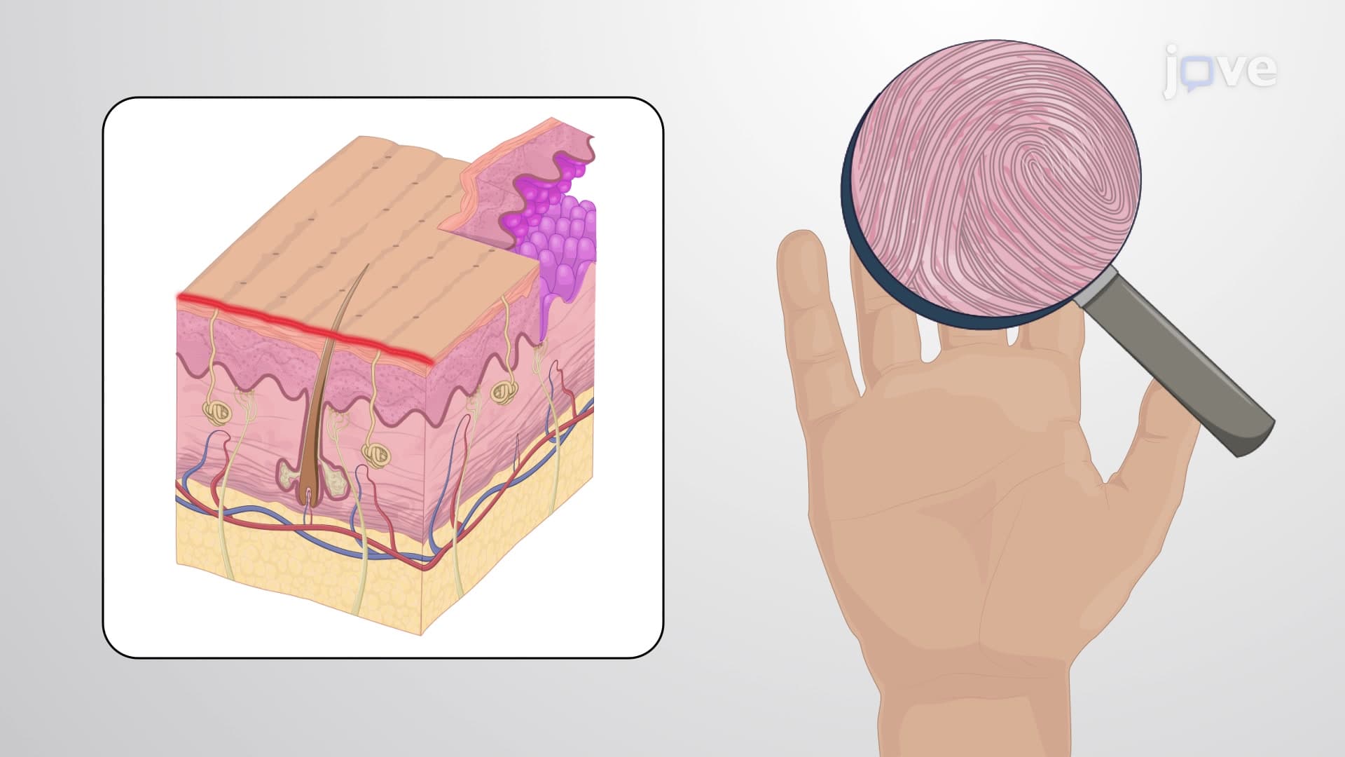
Your skin completely replaces itself every 28 days-but how does this remarkable process happen? The epidermis layers anatomy reveals a sophisticated cellular factory where millions of cells migrate upward through distinct zones. Consider how a dermatologist at Johns Hopkins examines a skin biopsy: they identify five precise layers, each with unique functions that protect your body from environmental damage. Understanding what are the layers of the epidermis provides crucial insight into wound healing, skin diseases, and protective mechanisms. Watch the full video on JoVE Coach to master this concept with expert-led visuals and step-by-step explanations.
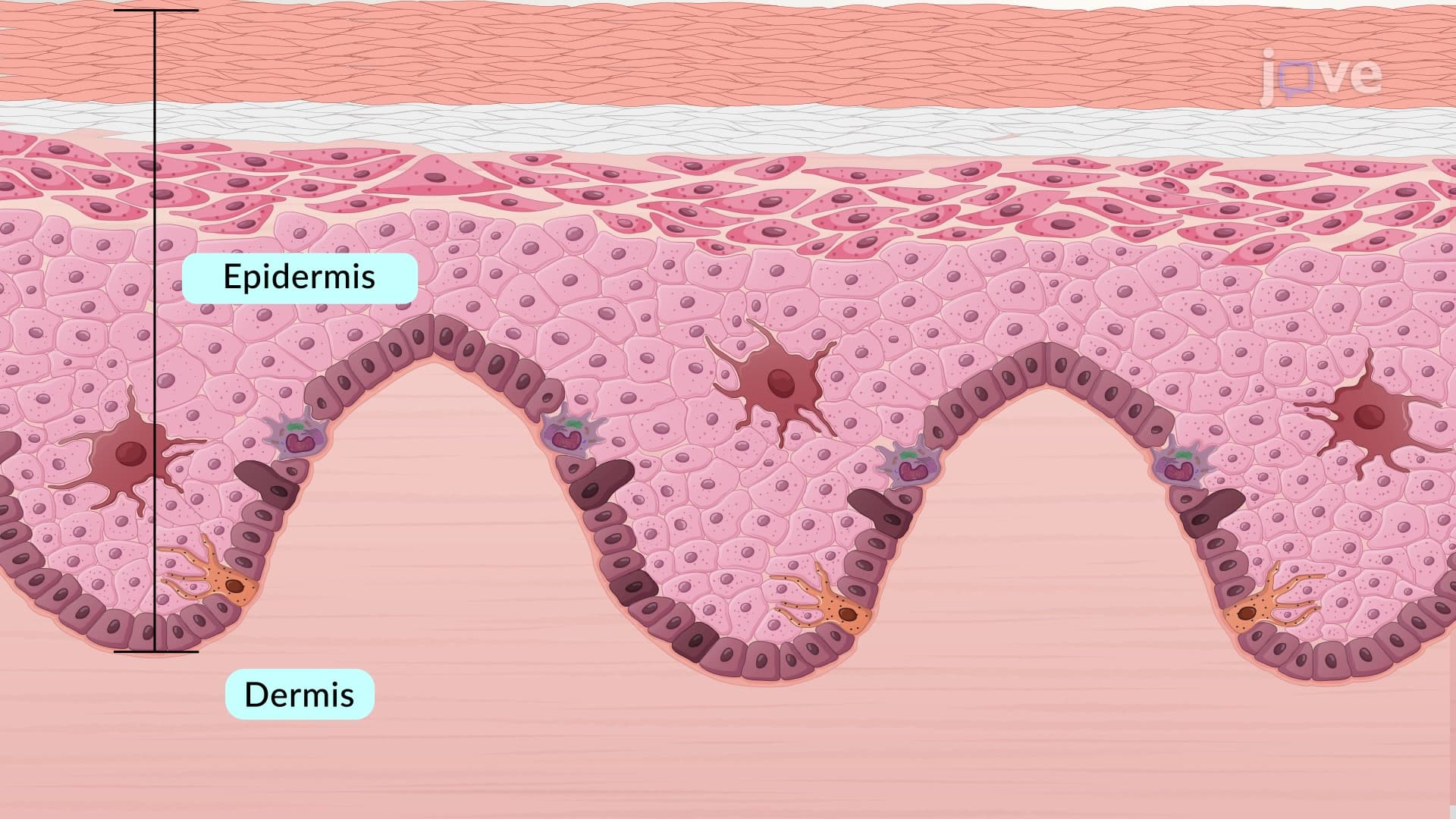
The epidermis layers anatomy represents one of biology's most elegant examples of cellular organization and renewal. Unlike other tissues, the epidermis maintains constant cellular turnover while providing an impermeable barrier against pathogens, chemicals, and water loss. Medical students at Harvard Medical School study these layers extensively because understanding skin epidermis layers explained is fundamental to dermatology, pathology, and wound care.
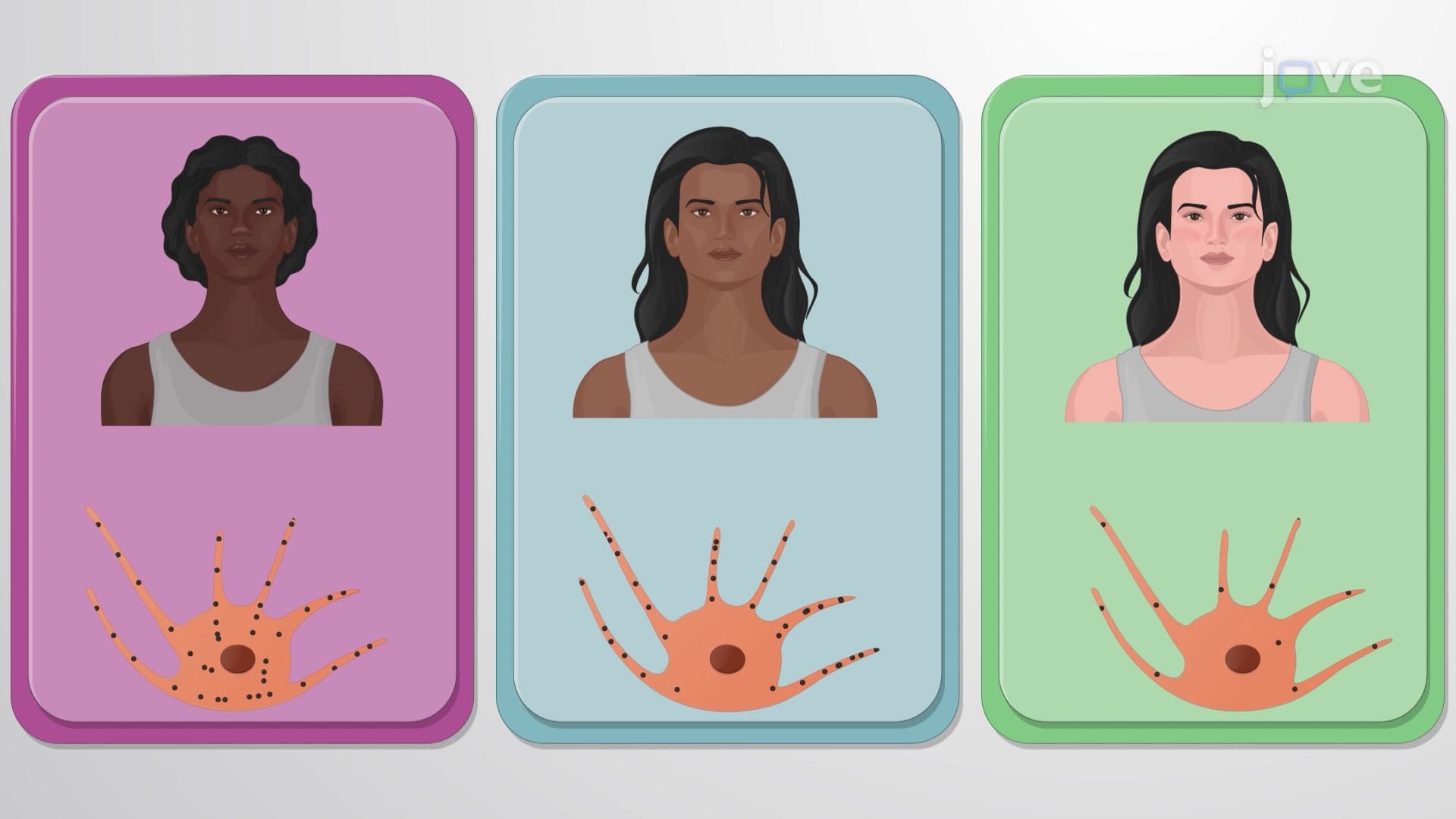
Stratum Basale: The Cellular Nursery The deepest layer, stratum basale, functions as the epidermis's stem cell reservoir. Keratinocytes here undergo continuous mitotic division, with daughter cells pushed upward into overlying layers. Melanocytes interspersed throughout this layer produce melanin, creating your skin's pigmentation. Merkel cells provide tactile sensation, while the basement membrane anchors this layer to the underlying dermis.
Stratum Spinosum and Granulosum: Processing Centers As keratinocytes migrate into the stratum spinosum granulosum region, they undergo dramatic structural changes. The spinous layer's characteristic "spiny" appearance under microscopy results from shrinkage artifacts around desmosomes-protein complexes that weld cells together. The granular layer marks where keratinization epidermal layer processes accelerate, with keratohyalin granules promoting protein cross-linking and cellular dehydration.
Stratum Lucidum and Corneum: The Protective Shield The translucent lucidum layer appears only in thick skin areas experiencing mechanical stress-palms and soles. The outermost stratum corneum basale layer consists entirely of dead, flattened corneocytes embedded in lipid matrix. This arrangement creates an effective barrier that prevents water loss while blocking harmful substances.
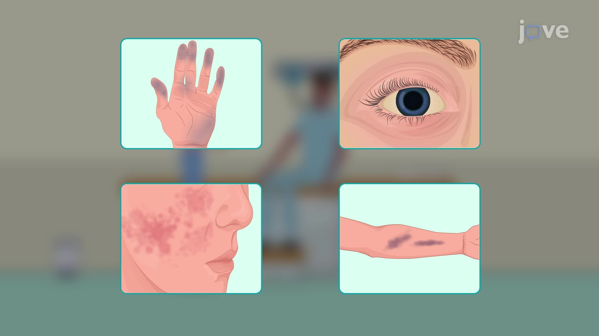
Understanding epidermal strata anatomy proves essential for MCAT preparation, particularly in biological sciences sections covering tissue organization. AP Biology students encounter epidermal layers when studying animal form and function. Pre-med students must recognize how disruption of normal skin layer arrangement contributes to conditions like eczema, psoriasis, and skin cancers.
Dermatologists at institutions like Mayo Clinic rely on epidermal layer knowledge when interpreting skin biopsies, planning treatments, and explaining conditions to patients. For example, understanding that the stratum corneum provides primary barrier function helps explain why topical medications require specific formulations to penetrate effectively.
Related Micro-courses