Loading subject page…

- Anatomy and Physiology
- The Integumentary System
- Accessory Structures of the Skin: Nails
Micro-course: The Integumentary System
87,705 views
Learn key concepts and earn your certificate.




Accessory Structures of the Skin: Nails

00:00
01:05
Video Summary: What Is Accessory Structures Skin Nails
Ever wondered why your nails turn white when you press them, then pink again when you release? This fascinating color change reveals the complex skin nail structure anatomy at work beneath your fingertips. From emergency room assessments of circulation to forensic investigations, medical professionals at hospitals like Mayo Clinic rely on understanding nail accessory structure skin components to diagnose everything from heart disease to nutritional deficiencies. What is Accessory Structures Skin Nails encompasses the intricate protective plates that shield our fingertips and toes. Watch the full video on JoVE Coach to master this concept with expert-led visuals and step-by-step explanations.
- Understand What is Accessory Structures Skin Nails and identify the major anatomical components including nail plate, nail bed, and nail matrix
- Recognize the functional relationship between nail bed capillaries and the characteristic pink coloration of healthy nails in clinical assessments
- Identify specialized structures like the lunula, hyponychium, and eponychium and explain their roles in nail protection and growth
- Analyze the keratinization process that transforms living cells in the nail matrix into the hard, protective nail plate structure
- Compare the structural differences between the free edge and nail body regions and their clinical significance
- Apply knowledge of nail anatomy to understand common nail disorders and injuries encountered in healthcare settings
- Evaluate how nail growth patterns and matrix activity contribute to nail regeneration after trauma or surgical procedures
- Explore the evolutionary advantages of nail structures as specialized accessory organs of the integumentary system
Understanding What is Accessory Structures Skin Nails: The Protective Shield System
Nail accessory structure skin represents one of nature's most ingenious protective mechanisms. These specialized keratinized plates serve as both shields and tools, protecting the sensitive fingertip pulp while enhancing our ability to manipulate small objects. In medical settings across the United States, from Johns Hopkins to UCLA Medical Center, healthcare professionals routinely examine nails as windows into overall health status.
The Nail Plate: Engineering Marvel of Dead Cells
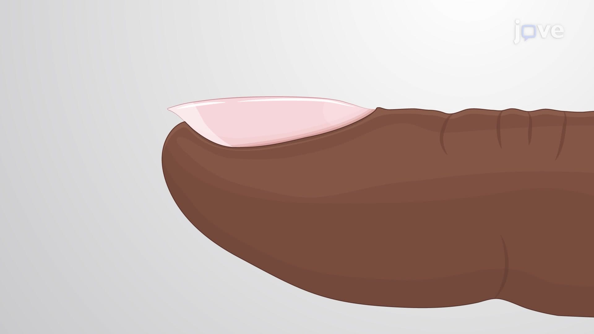
The visible nail plate free edge consists of densely packed, dead keratinocytes arranged in layers like microscopic roof shingles. This seemingly simple structure demonstrates remarkable engineering-strong enough to scratch surfaces yet flexible enough to bend without breaking. The transparency of healthy nails allows the pink nail bed beneath to show through, creating the characteristic coloration that medical students learn to assess during physical examinations on the MCAT and in clinical rotations.
Nail Bed and Hyponychium: The Living Foundation
Beneath the nail plate lies the nail bed hyponychium complex, where living tissue provides both nourishment and structural support. The nail bed's rich capillary network not only creates the pink coloration but serves as a crucial diagnostic tool. Emergency medicine physicians at trauma centers like Harborview Medical Center use capillary refill tests-pressing the nail to assess circulation-as rapid indicators of cardiovascular status.
Nail Matrix and Growth: The Cellular Factory
The nail matrix keratinization process represents a continuous cellular assembly line. Located beneath the skin at the nail's base, this region contains rapidly dividing stem cells that push older cells forward, gradually keratinizing them into the hard nail plate. Understanding this process proves essential for AP Biology students studying cellular differentiation and for pre-med students preparing for anatomy courses. The visible lunula-that whitish half-moon near the nail base-actually represents the distal edge of this active matrix region.
Cuticle and Nail Fold: Protective Barriers
The cuticle eponychium nail structure and surrounding nail fold anatomy structure create a sealed environment protecting the nail matrix from bacterial invasion. These tissues form natural barriers that prevent pathogens from reaching the growth center, explaining why proper nail care emphasizes gentle cuticle maintenance rather than aggressive removal.
Frequently Asked Questions
Nails are specialized protective plates made of dead keratin cells that cover and shield the tips of fingers and toes. They're considered accessory structures of the integumentary system because they develop from the same embryonic tissues as skin and hair. Think of them as natural armor plating that also enhances your ability to pick up small objects and scratch surfaces.
MCAT questions often focus on nail structure in relation to integumentary system disorders and diagnostic techniques. AP Biology exams typically include nails when discussing epithelial tissues, keratinization processes, or comparative anatomy. You'll need to identify structures like nail matrix, nail bed, and eponychium, plus explain how keratinocyte differentiation creates the nail plate.
Focus on nail assessment techniques, normal vs. abnormal nail coloration, and basic nail anatomy including nail bed, matrix, and cuticle. HESI A2 and TEAS exams often include questions about capillary refill testing and how nail appearance indicates circulation problems. Understanding clubbing, cyanosis, and other nail changes helps with clinical reasoning sections.
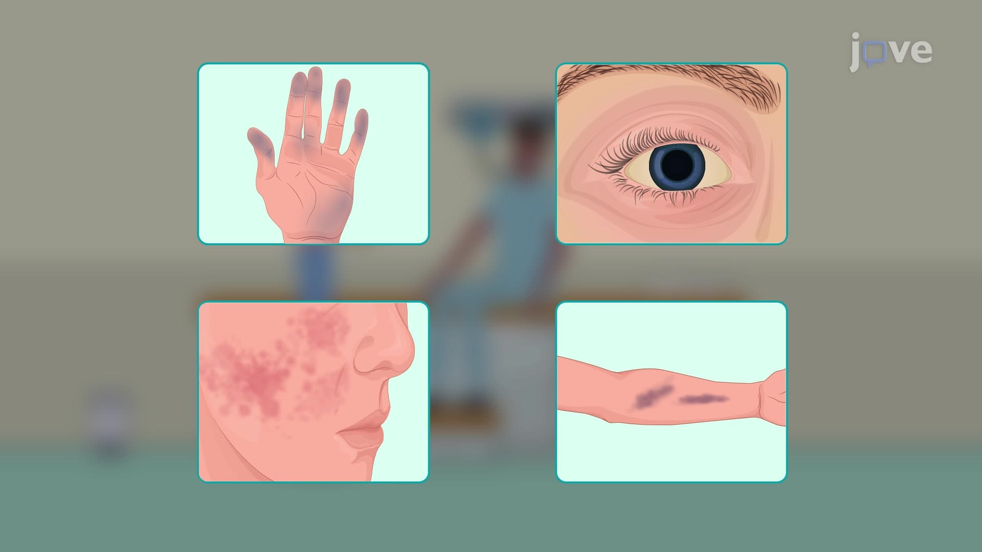
Nails provide non-invasive windows into systemic health conditions affecting circulation, nutrition, and metabolism. Medical students at schools like Harvard and Stanford learn to identify signs of heart disease, liver problems, and nutritional deficiencies through nail changes. This makes nail anatomy clinically relevant beyond just memorizing structural components.
When trauma damages nails, understanding matrix location becomes crucial for treatment decisions at hospitals nationwide. If the nail matrix remains intact after injury, complete regeneration typically occurs over 3-6 months. Plastic surgeons and emergency physicians use this knowledge when treating fingertip injuries to preserve long-term nail function.
Not at all-nail structure actually provides an excellent introduction to more complex anatomical systems. The basic components (nail plate, bed, matrix, cuticle) are straightforward, and the visible nature of nails makes the anatomy concrete rather than abstract. Most students find nail anatomy easier than other integumentary topics because they can observe the structures on themselves.
Create labeled diagrams showing nail cross-sections and practice identifying structures on actual nails (your own or volunteers'). Use mnemonics like "My Bed Needs Proper Care" for Matrix-Bed-Nail plate-Plate-Cuticle. Focus on functional relationships-how matrix damage affects growth, how nail bed circulation creates coloration-rather than just memorizing isolated facts.
Progress to hair follicle structure and the hair growth cycle, which shares many similarities with nail development. Then explore sebaceous and sweat glands, followed by skin layer organization (epidermis, dermis, hypodermis). This sequence builds naturally from simple keratinized structures to more complex glandular systems within the integumentary framework.
Related Micro-courses

19 Concepts

11 Concepts
6 Concepts