Loading subject page…


87,705 views


Video Summary: What Is Cells of the Epidermis
Ever wonder why your skin completely renews itself every month? The epidermis cell types work together like a biological factory, with four distinct cellular workers performing specialized functions. From keratinocytes producing protective proteins to melanocytes creating the pigment that gives your skin its color, these cells found in epidermis explained reveal how your body's largest organ stays healthy and functional. Consider how sunscreen protects these cellular processes when you visit California beaches during summer UV exposure. Watch the full video on JoVE Coach to master this concept with expert-led visuals and step-by-step explanations.
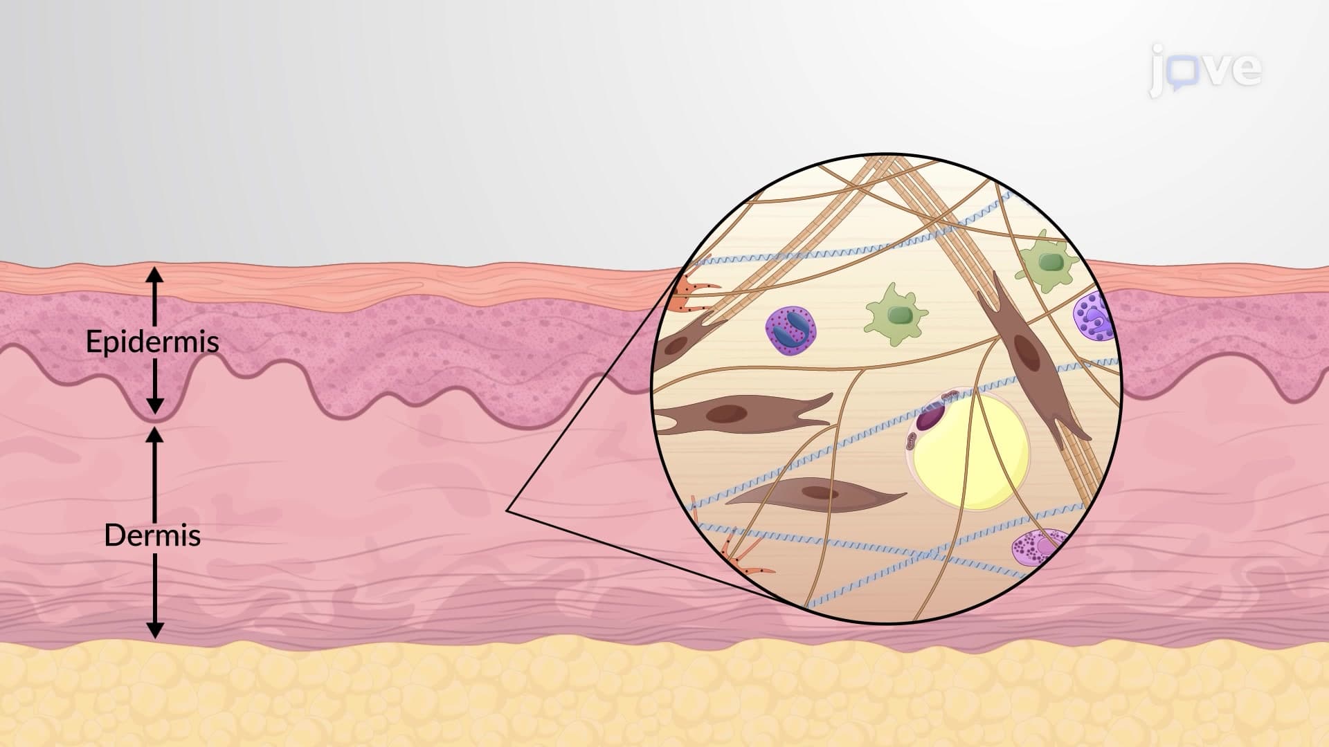
The epidermis cell types form a sophisticated biological system that protects your body from external threats while maintaining essential physiological functions. This outermost skin layer contains four primary cellular populations, each contributing unique properties that ensure skin integrity and health. Understanding these cellular components is crucial for students preparing for AP Biology exams and college-level anatomy courses.
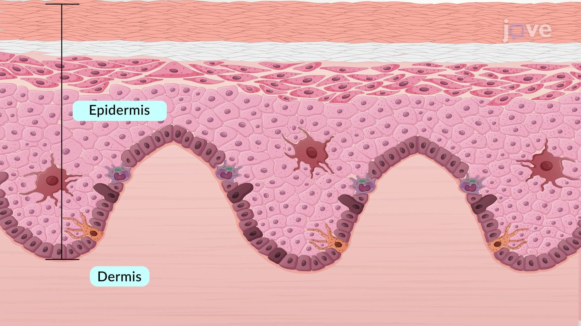
Keratinocyte epidermal cell populations constitute approximately 95% of all epidermal cells, making them the predominant cellular component. These cells originate in the basal layer and undergo a remarkable 25-45 day journey to the skin surface. During this migration, keratinocytes synthesize keratin, a fibrous protein that provides mechanical strength and waterproofing properties. This process, called keratinization, is essential for maintaining the skin barrier function that prevents water loss and pathogen entry.
Clinical applications of keratinocyte biology appear frequently in MCAT questions, particularly regarding wound healing and skin cancer development. For example, basal cell carcinoma, the most common skin cancer in the United States, originates from keratinocyte populations in the basal epidermis.
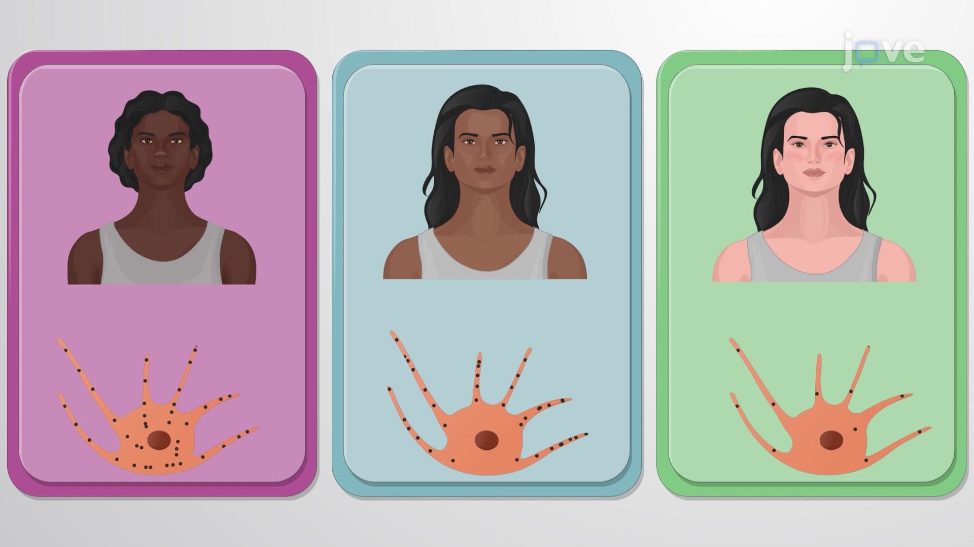
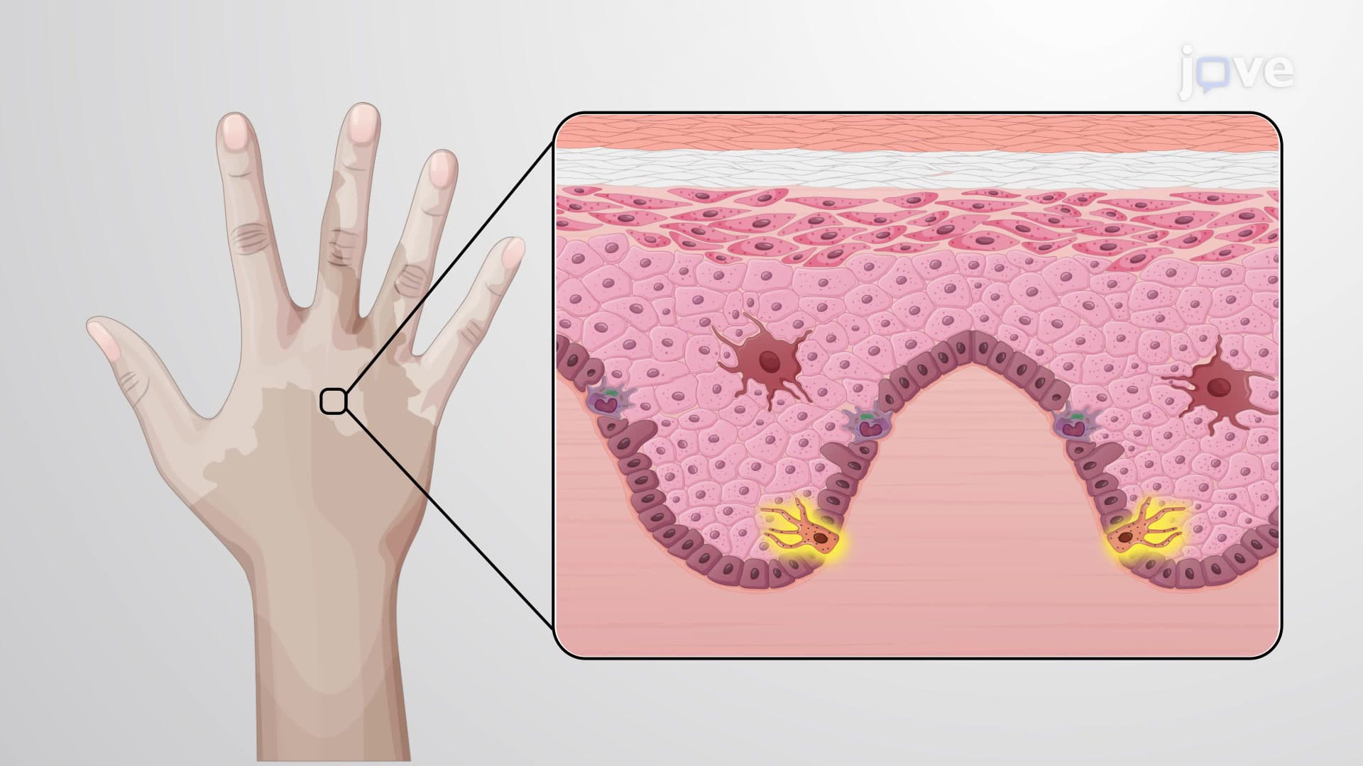
Melanocyte pigment cell networks extend throughout the epidermis with distinctive dendritic projections that contact surrounding keratinocytes. These cells synthesize melanin through complex biochemical pathways involving the enzyme tyrosinase. The melanin transfer process protects keratinocyte nuclei from DNA damage caused by ultraviolet radiation exposure.
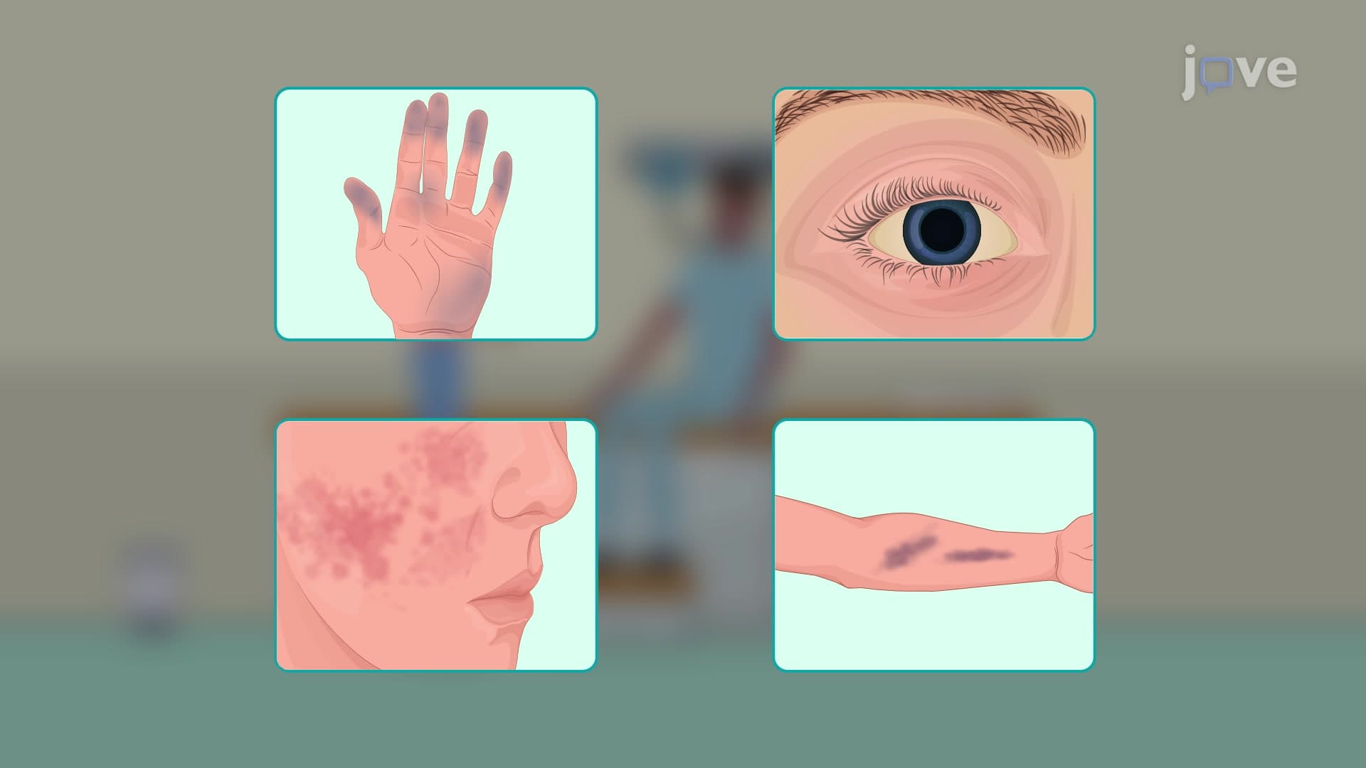
Melanocyte dysfunction underlies several important medical conditions frequently discussed in dermatology courses. Vitiligo, affecting approximately 1% of the US population, results from melanocyte destruction, while melanoma represents malignant transformation of these pigment-producing cells.
Langerhans immune epidermal cells serve as the skin's primary antigen-presenting cells, detecting foreign substances and initiating immune responses. These specialized dendritic cells process antigens and migrate to lymph nodes when activated, bridging innate and adaptive immunity. Merkel cell sensation receptors, located at the dermal-epidermal junction, contain specialized nerve endings that detect light touch and texture discrimination, contributing to tactile sensitivity.
Related Micro-courses