Loading subject page…

- Anatomy and Physiology
- The Integumentary System
- Reticular Dermis
Micro-course: The Integumentary System
87,705 views
Learn key concepts and earn your certificate.



Reticular Dermis


00:00
01:15
Video Summary: What Is Reticular Dermis
Ever wonder why surgical incisions heal better when made parallel to your skin's natural lines? The reticular dermis anatomy holds the answer, forming 80% of your skin's middle layer through thick collagen bundles that create these tension patterns. In U.S. plastic surgery, surgeons follow Langer's lines-mapped from reticular dermis structure-to minimize scarring in procedures from facelifts to reconstructive surgery. Watch the full video on JoVE Coach to master this concept with expert-led visuals and step-by-step explanations.
- Understand what is reticular dermis and its role as the skin's primary structural foundation
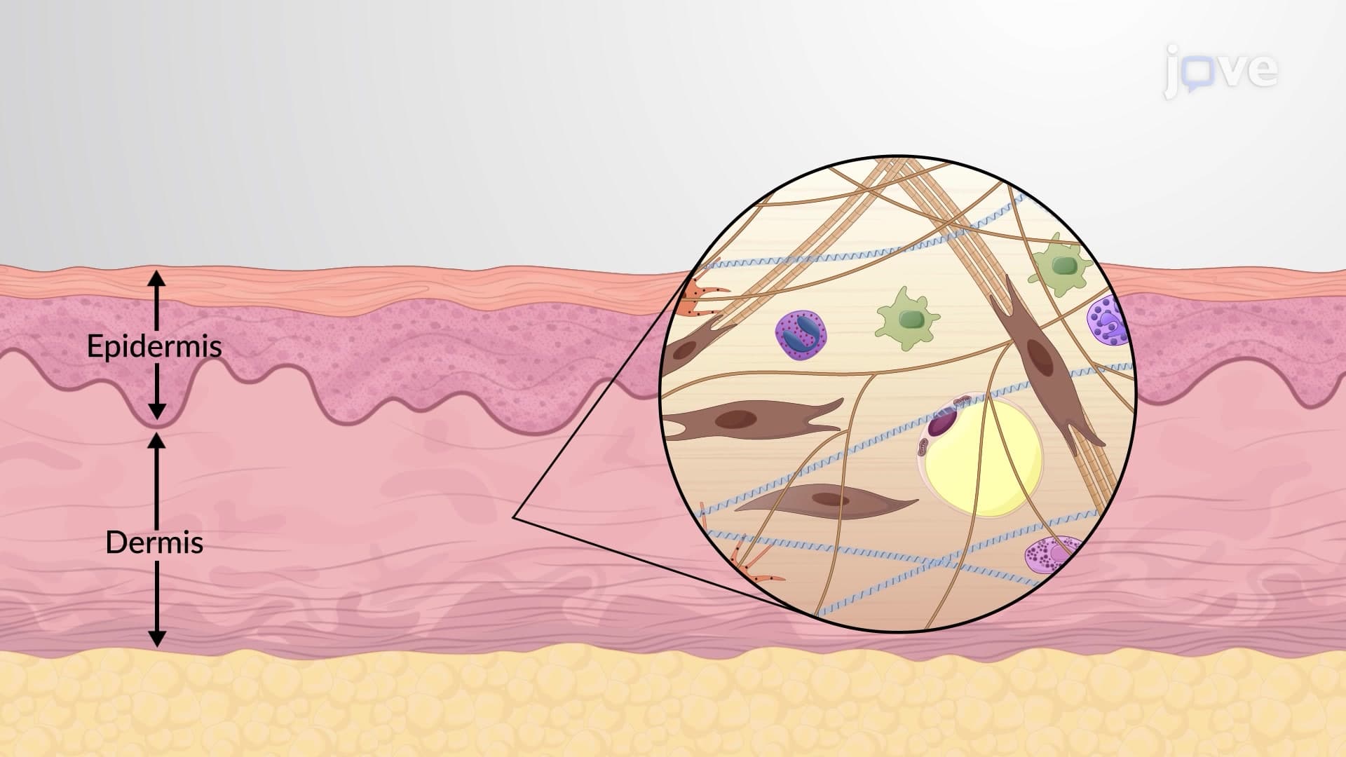
- Identify the cellular components including fibroblasts, stem cells, macrophages, and mast cells
- Analyze how the extracellular matrix creates skin strength through collagen and elastic fiber networks
- Compare reticular dermis layer skin thickness to other dermal components (80% vs 20% papillary)
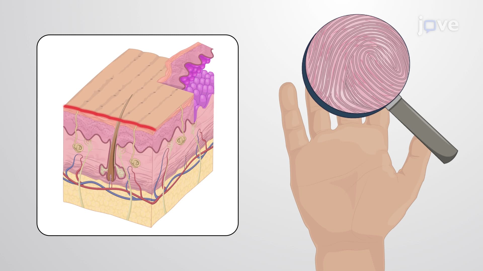
- Evaluate the vascular supply from cutaneous plexus and its support of skin appendages
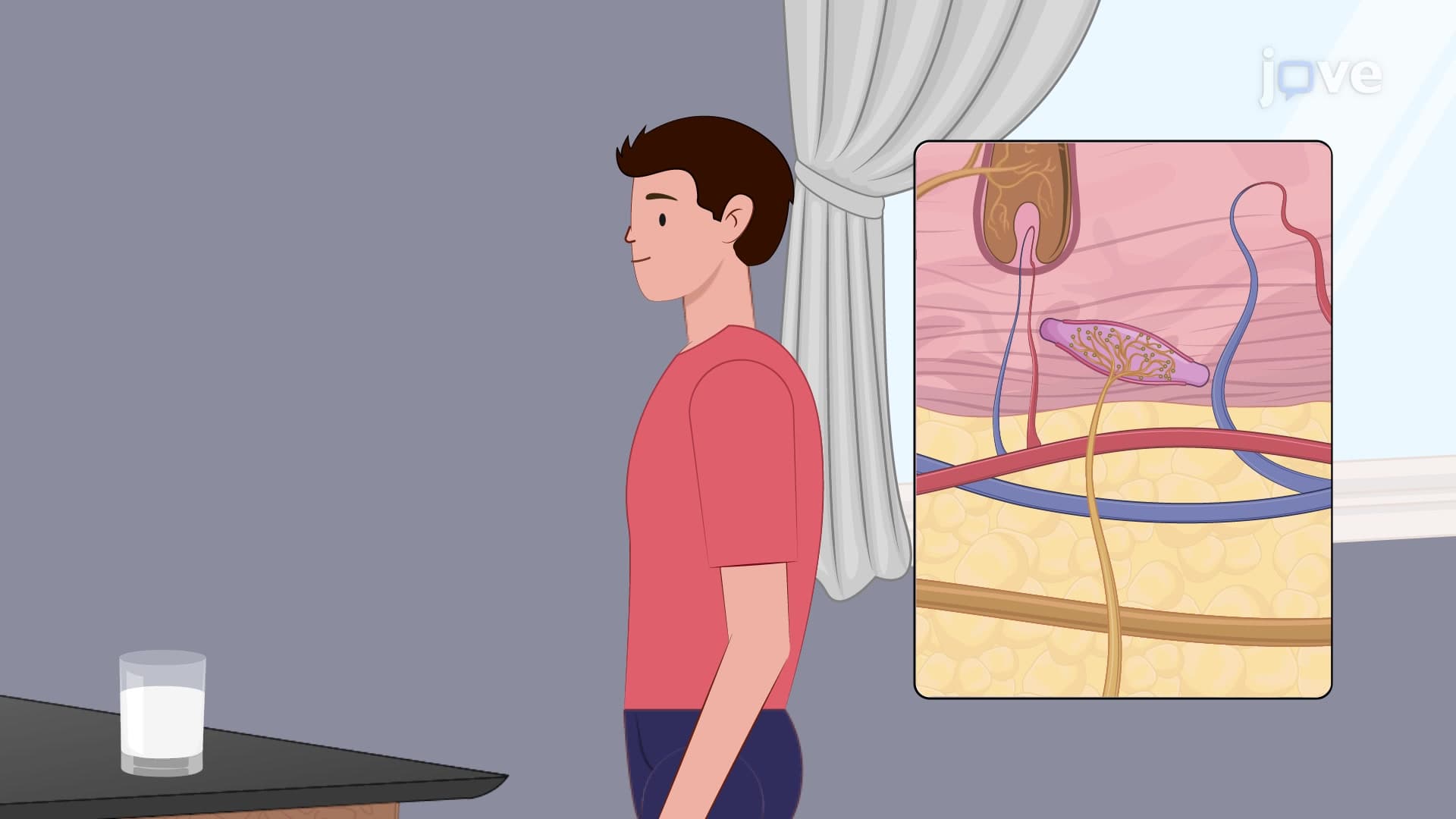
- Apply knowledge of sensory receptors like Pacinian and Ruffini corpuscles in pressure detection
- Recognize the clinical significance of cleavage lines in surgical procedures
- Explore the relationship between reticular structure and skin elasticity properties
Understanding Reticular Dermis Anatomy and Structure
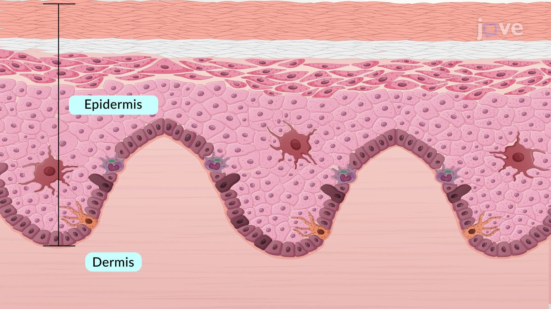
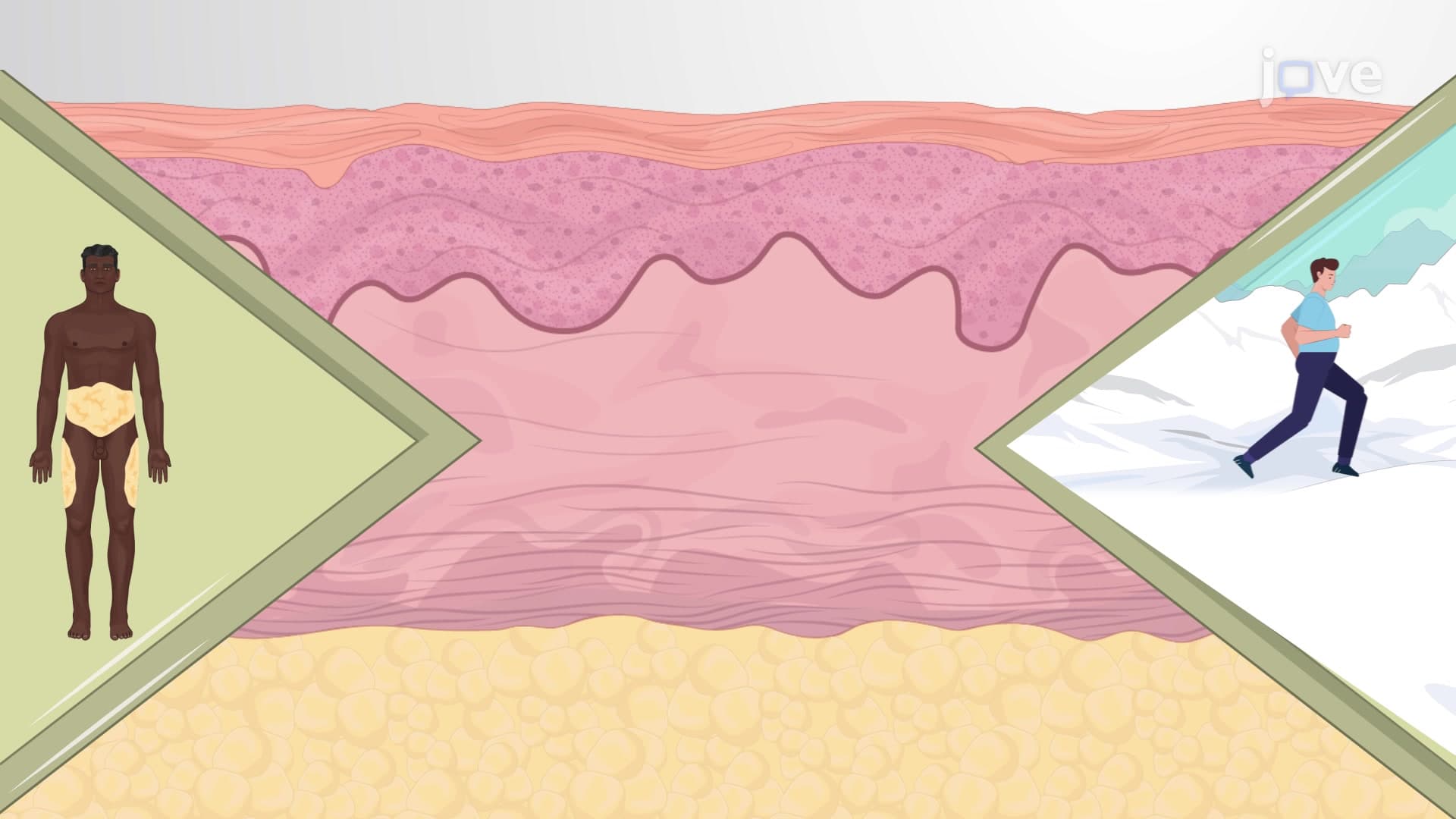
The reticular dermis anatomy represents the skin's engineering masterpiece, comprising the deepest and thickest portion of the dermis. Unlike the thin papillary layer above it, this reticular dermis layer skin forms approximately 80% of total dermal thickness, extending from just below the dermal-epidermal junction down to the subcutaneous fat layer. Medical students studying for the MCAT often compare this structure to reinforced concrete-the collagen provides tensile strength while elastic fibers allow flexibility.
Deep Dermis Glands Follicles and Cellular Architecture
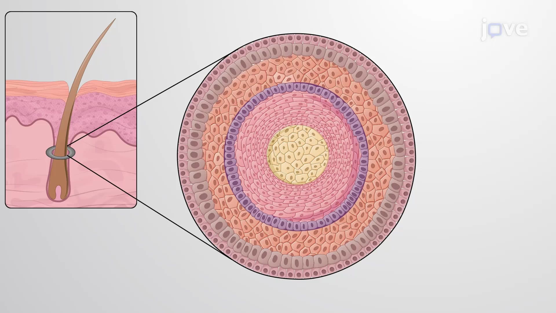
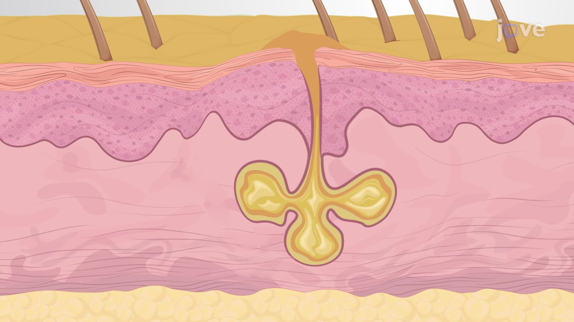
The reticular layer thickness houses critical skin appendages that define our daily physiology. Hair follicles extend deep into this layer, with the reticular dermis hair follicle relationship being particularly important for understanding conditions like androgenic alopecia, common in American adults. Sebaceous glands cluster around these follicles, while eccrine sweat glands-essential for thermoregulation in hot climates like Arizona or Florida-also call this layer home. Cellular residents include fibroblasts that continuously produce collagen (crucial for wound healing), macrophages providing immune surveillance, and mast cells involved in allergic responses.
Reticular Layer Collagen Elastic Matrix Organization
The hallmark of dermis reticular structure lies in its extracellular matrix composition. Thick collagen bundles, predominantly type I collagen, weave together in a basket-like pattern that provides exceptional tensile strength-up to 20-30 kg per square centimeter. Interspersed elastic fibers, composed mainly of elastin, allow the skin to return to its original shape after stretching. This combination explains why young skin bounces back quickly from pinching, while aged skin (with degraded elastic fibers) shows prolonged tenting-a clinical sign physicians use to assess dehydration.
Clinical Applications and Tension Lines
The practical implications of reticular dermis organization appear dramatically in surgical settings across American hospitals. Cleavage lines, also called Langer's lines, result from the predominant collagen fiber orientation within this layer. Plastic surgeons at institutions like Johns Hopkins or Mayo Clinic strategically align incisions parallel to these lines to minimize scarring and optimize healing. AP Biology students often encounter this concept when studying tissue organization, while pre-med students preparing for the MCAT must understand how collagen synthesis and degradation affect wound repair processes.
Frequently Asked Questions
The reticular dermis is the deep, thick layer of the dermis that makes up about 80% of the skin's middle section, located between the papillary dermis and subcutaneous tissue. Unlike the thin papillary dermis above it, the reticular layer contains thick collagen bundles and houses major skin structures like hair follicles and sweat glands. It provides the skin's primary structural strength and elasticity through its dense network of collagen and elastic fibers.
MCAT questions often focus on collagen synthesis, wound healing mechanisms, and the relationship between structure and function in skin elasticity. AP Biology exams typically test tissue organization, comparing reticular dermis thickness to other layers, and understanding how extracellular matrix components contribute to tissue properties. Students should know the 80/20 ratio between reticular and papillary dermis layers and be able to explain how collagen degradation affects skin aging.
Medical students studying dermatology must understand how skin conditions affect different dermal layers and how treatments penetrate to the reticular level. This knowledge is essential for USMLE Step 1 questions about skin pathophysiology and appears frequently in dermatology residency board exams. Understanding reticular dermis anatomy helps explain why certain laser treatments or injectables work at specific depths.
Plastic surgeons across American medical centers use reticular dermis anatomy to plan surgical incisions along natural tension lines, reducing scarring in procedures from breast reconstruction to facial surgery. Emergency medicine physicians assess skin turgor by understanding how reticular layer elasticity changes with age and dehydration, helping diagnose fluid status in patients at hospitals nationwide.
Not at all-reticular dermis concepts build naturally on basic biology knowledge about tissues and cells that students learn in freshman biology. The key is visualizing it like a woven fabric where collagen provides strength and elastic fibers add stretch, similar to materials in athletic clothing. Most students find the 80% thickness ratio easy to remember and can relate the concept to everyday experiences like skin stretching.
Focus on the "big three" components: cellular residents (fibroblasts, macrophages, mast cells), structural matrix (collagen and elastic fibers), and housed appendages (hair follicles, glands). Create comparison charts showing reticular versus papillary dermis differences, and practice drawing cross-sections labeling the 80/20 thickness ratio. Many students find success using tactile learning-feeling their own skin stretch helps reinforce the elastic fiber concept.
Students should dive deeper into collagen biochemistry and the aging process, exploring how sun damage affects elastic fibers in conditions like solar elastosis. Advanced topics include wound healing phases, scar formation mechanisms, and how various dermatological treatments target different dermal layers. Pre-health students benefit from studying connective tissue disorders that affect reticular dermis structure.
Related Micro-courses

19 Concepts

11 Concepts
6 Concepts