Loading subject page…


218,421 views

Video Summary: Lymphatic Vessels and Lymph Transport Explained
Did you know that your body has a second circulatory system that processes over 3 liters of fluid daily? Lymphatic vessels lymph transport creates an intricate network that returns filtered fluid from your tissues back to your bloodstream, much like how New York City's subway system moves millions of passengers through interconnected tunnels. Understanding Lymphatic Vessels And Lymph Transport Explained reveals how specialized vessels, valves, and ducts work together to maintain fluid balance and immune function. Watch the full video on JoVE Coach to master this concept with expert-led visuals and step-by-step explanations.
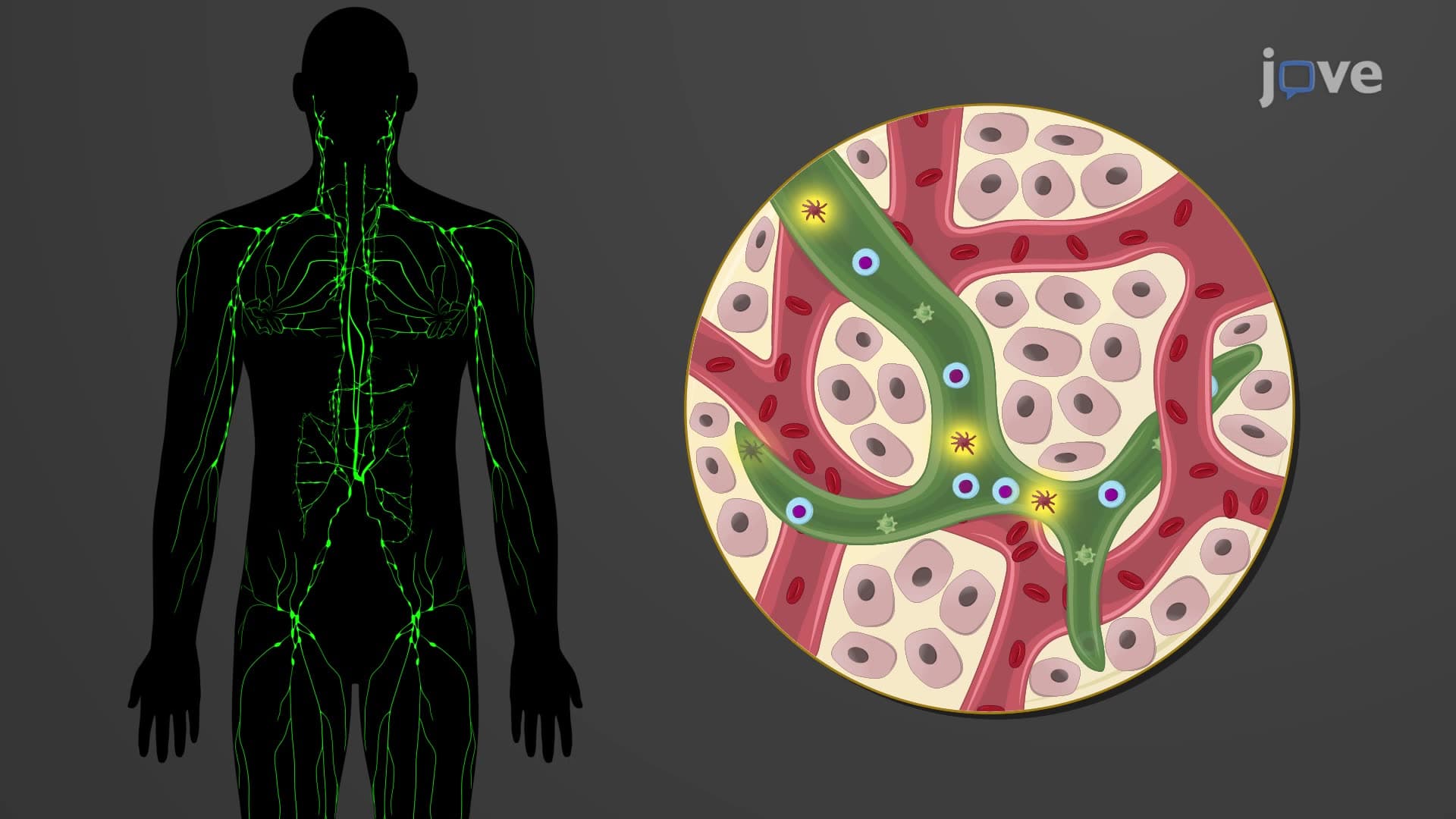
The lymphatic system operates as your body's sophisticated drainage network, collecting excess tissue fluid and returning it to circulation through a precisely organized transport system. Unlike blood vessels that form closed loops, lymphatic vessels lymph transport follows a one-way journey from peripheral tissues toward the heart, making this system fundamentally different from cardiovascular circulation.
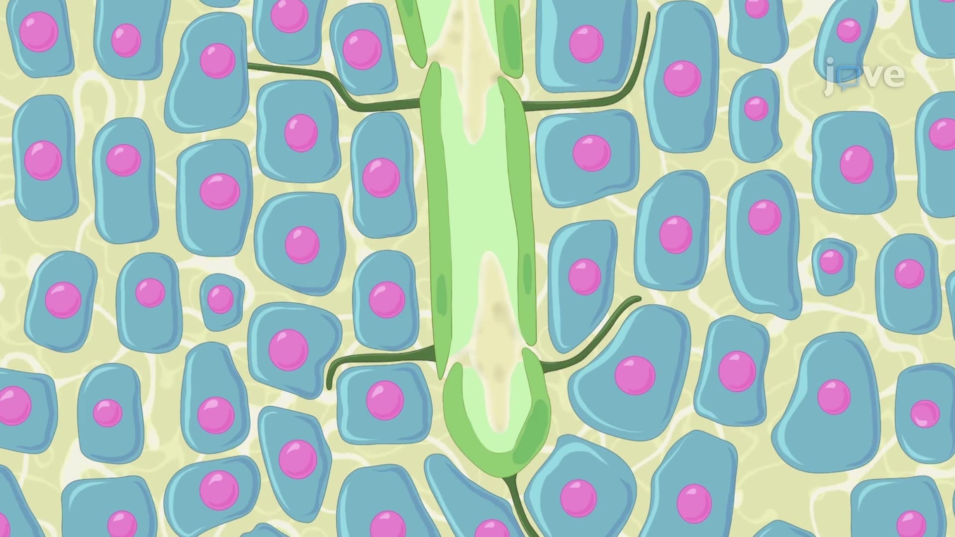
Lymph capillary collecting vessel networks begin as microscopic, blind-ended tubes interwoven throughout your tissues. These initial vessels feature overlapping endothelial cells that create one-way valves, allowing fluid entry but preventing backflow. When tissue pressure increases-such as during exercise or inflammation-these mini-valves open wider, enhancing lymph collection. The capillaries then drain into progressively larger collecting vessels equipped with smooth muscle walls and internal valves that propel lymph forward through rhythmic contractions.
Thoracic duct lymph drainage handles approximately 75% of your body's lymphatic return, collecting from everything below the diaphragm plus the left upper body. This largest lymphatic vessel begins as the cisterna chyli, a dilated sac located near your lower spine that receives lymph from intestinal and lumbar trunks. Medical students preparing for the MCAT or USMLE must understand that thoracic duct obstruction-such as from trauma during cardiac surgery-can cause chylothorax, where lymph accumulates in the chest cavity.
The right lymphatic duct manages lymph from the right upper quadrant of your body, including the right arm, right side of the head and neck, and right lung. This shorter duct forms where the right jugular, subclavian, and bronchomediastinal trunks converge. Understanding this asymmetrical drainage pattern proves crucial for AP Biology students studying immune responses and for pre-med students learning clinical correlations. For instance, breast cancer staging requires knowledge of lymphatic drainage patterns to predict potential metastatic spread.
Both major ducts ultimately empty into the venous angle-the junction between internal jugular and subclavian veins-where lymph flow valve vessel mechanisms prevent blood from backing up into the lymphatic system. This anatomical arrangement ensures efficient fluid return while maintaining pressure differentials essential for proper circulation.
Related Micro-courses