Loading subject page…


218,422 views

Video Summary: What are Secondary Lymphoid Organs
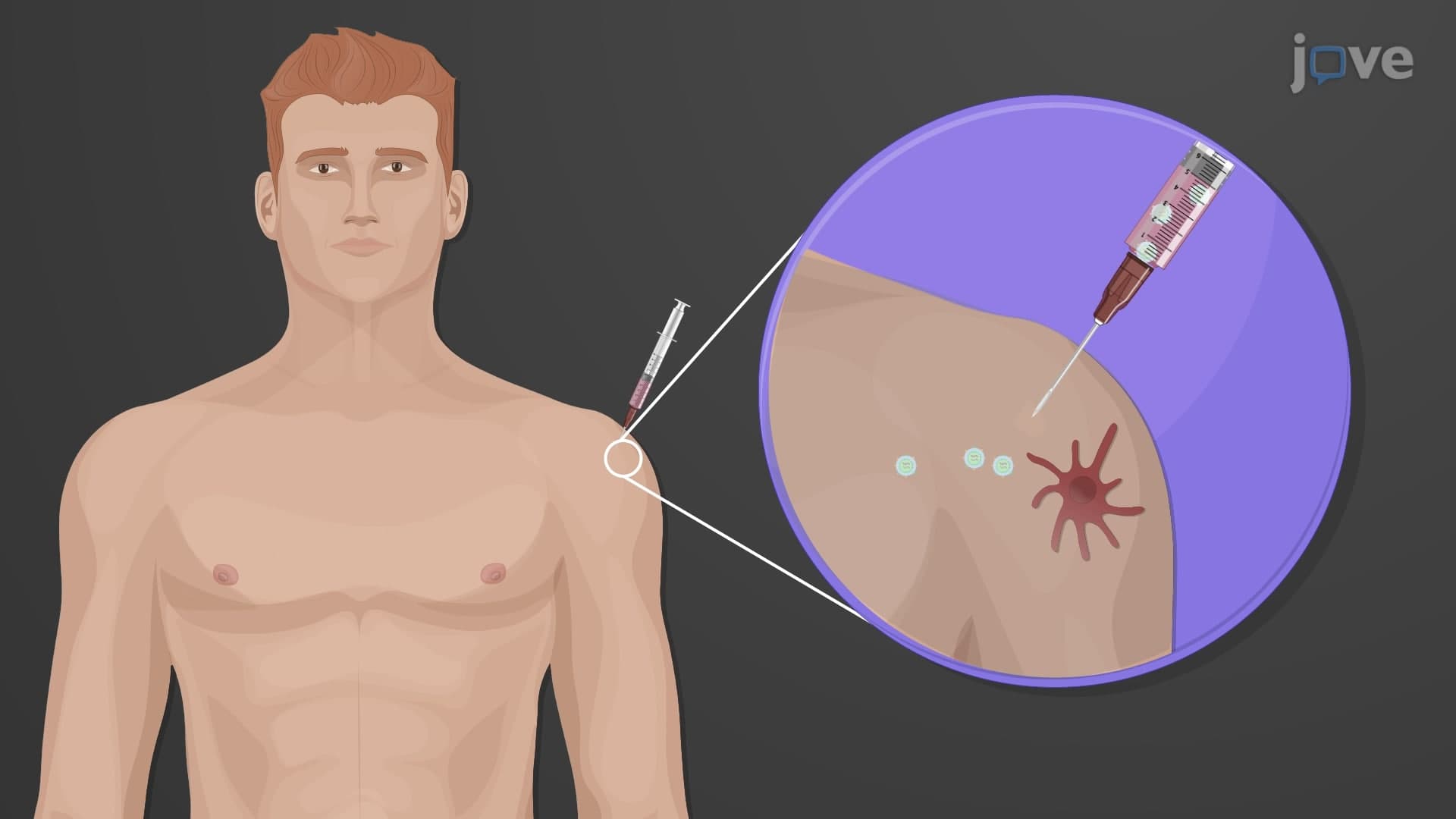
Did you know that every time you get a vaccine at your doctor's office, specialized structures throughout your body are working to create lasting immunity? Secondary lymphoid organs biology involves three critical components-the spleen, lymph nodes, and mucosa-associated lymphoid tissues (MALT)-that serve as meeting points where immune cells first encounter and respond to foreign invaders. For instance, when you receive a flu shot at a US clinic, your lymph nodes swell slightly as they activate immune responses. Understanding what are secondary lymphoid organs reveals how your body builds defense systems against diseases. Watch the full video on JoVE Coach to master this concept with expert-led visuals and step-by-step explanations.
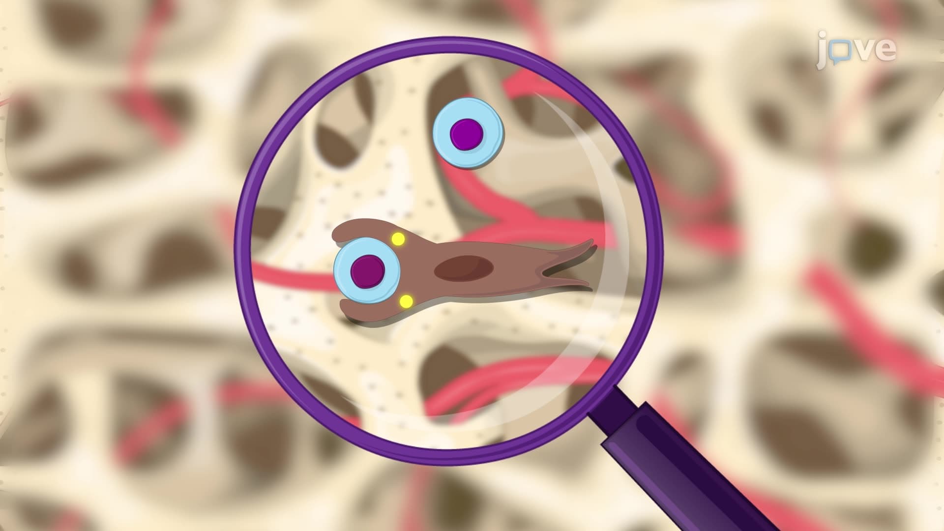
Secondary lymphoid organs biology forms the foundation of adaptive immunity, representing specialized anatomical sites where naive lymphocytes encounter antigens for the first time. Unlike primary lymphoid organs (bone marrow and thymus) where immune cells develop, secondary lymphoid structures facilitate mature immune cell activation and orchestrate targeted responses against specific pathogens.
The spleen functions as both a spleen immune secondary organ and blood filtration system. Located in the left upper abdomen beneath the rib cage, this fist-sized organ processes approximately 500 liters of blood daily. Its unique architecture divides into white pulp (immune function) and red pulp (blood filtration). The white pulp contains specialized zones where B cells form germinal centers during immune responses-a concept frequently tested on AP Biology exams and MCAT immunology sections. Clinically, patients with splenectomy (spleen removal) require additional vaccinations due to increased infection susceptibility, particularly from encapsulated bacteria like Streptococcus pneumoniae.
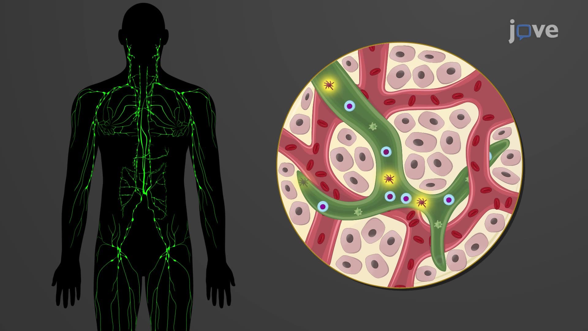
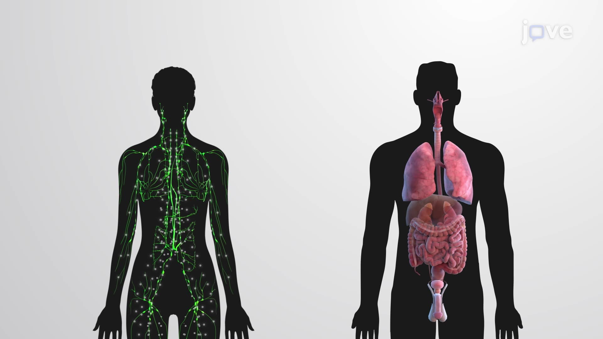
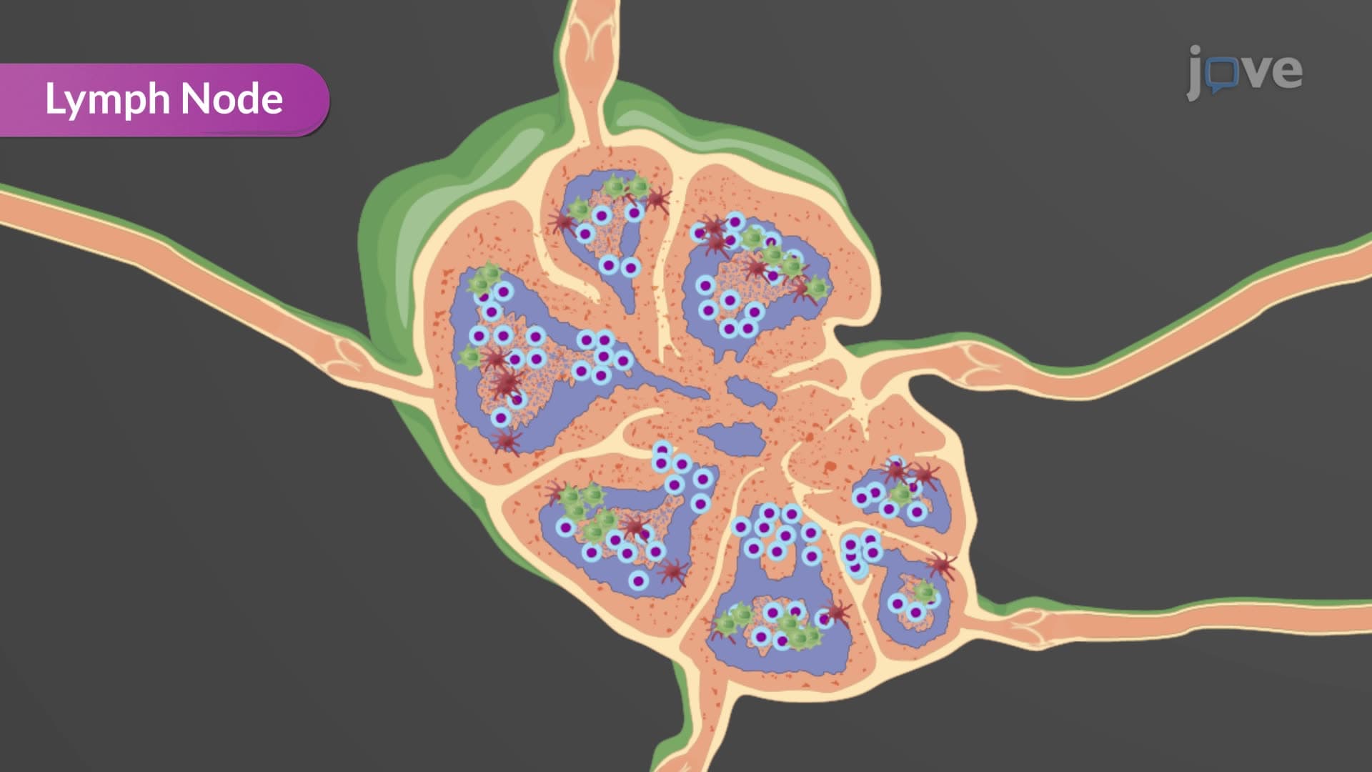
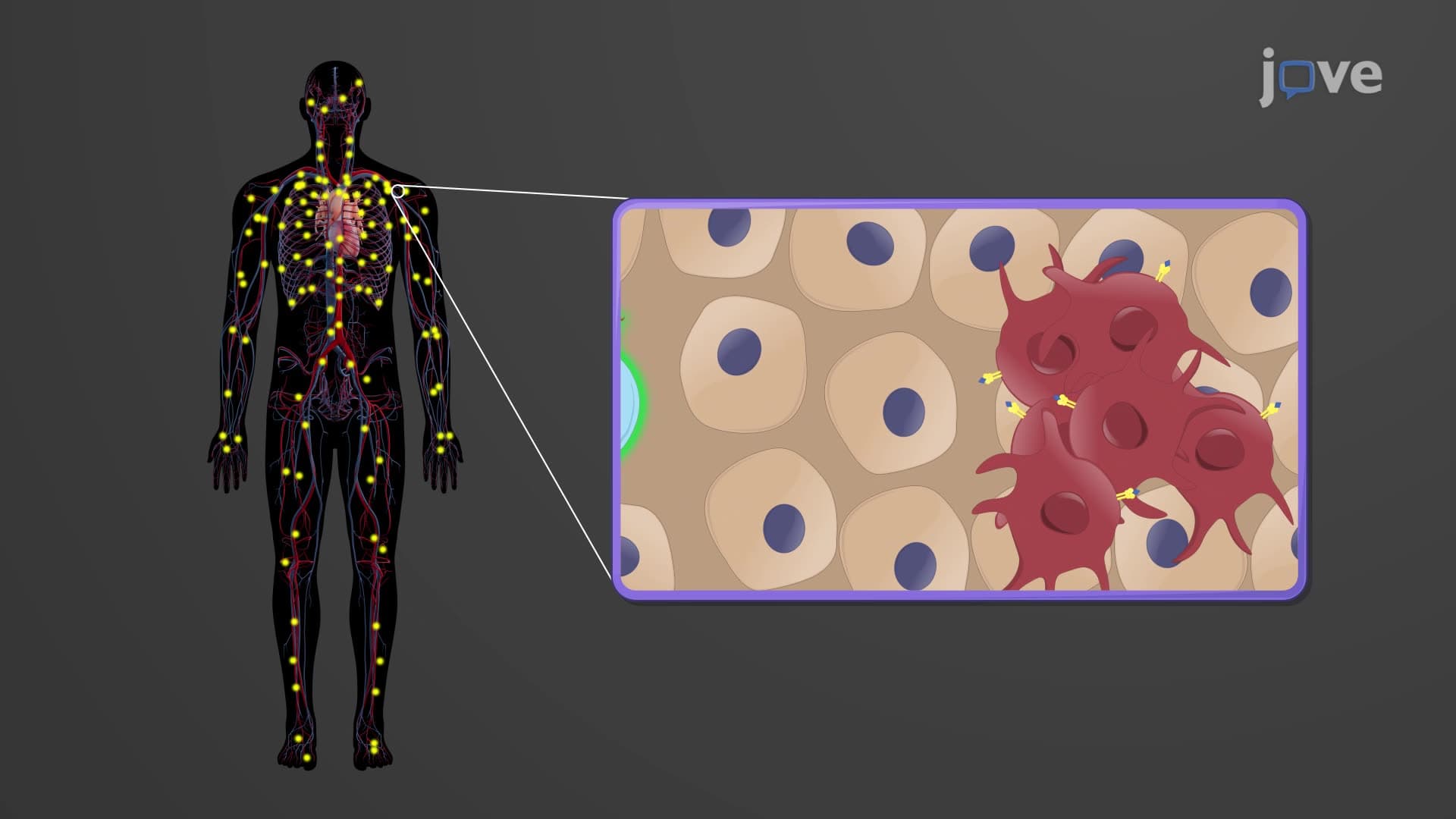
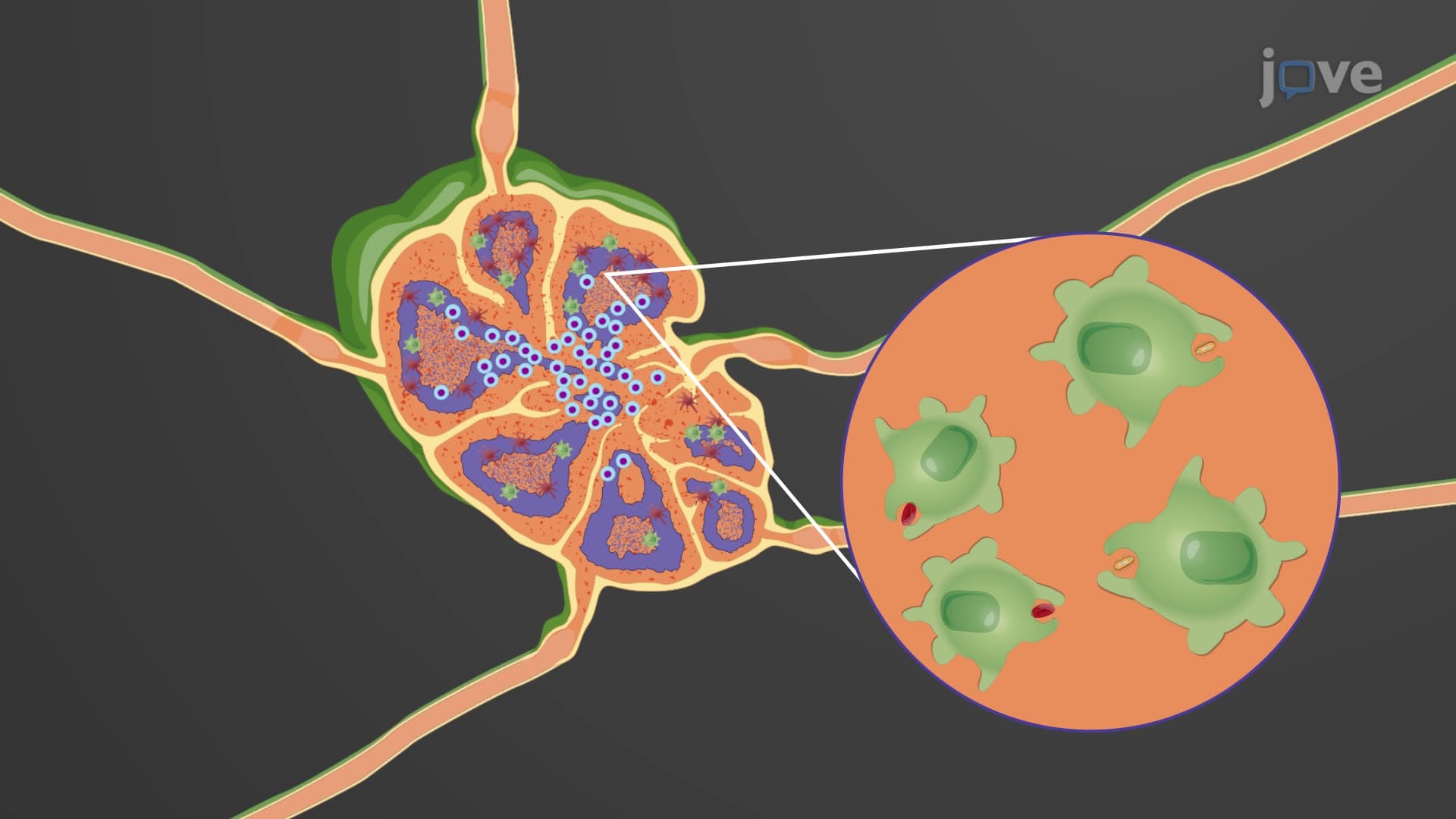
Lymph node spleen MALT systems work collaboratively, but lymph nodes serve unique filtering functions. These bean-shaped structures cluster in strategic locations-neck (cervical), armpits (axillary), and groin (inguinal) regions-intercepting lymphatic fluid before it returns to circulation. The germinal center lymph node formation represents active immune responses, where B cells undergo somatic hypermutation to produce high-affinity antibodies. Healthcare providers routinely palpate lymph nodes during physical examinations, as enlargement often indicates infection or malignancy. Students preparing for NCLEX or HESI A2 exams should understand that lymphadenopathy (enlarged lymph nodes) serves as a key diagnostic indicator.
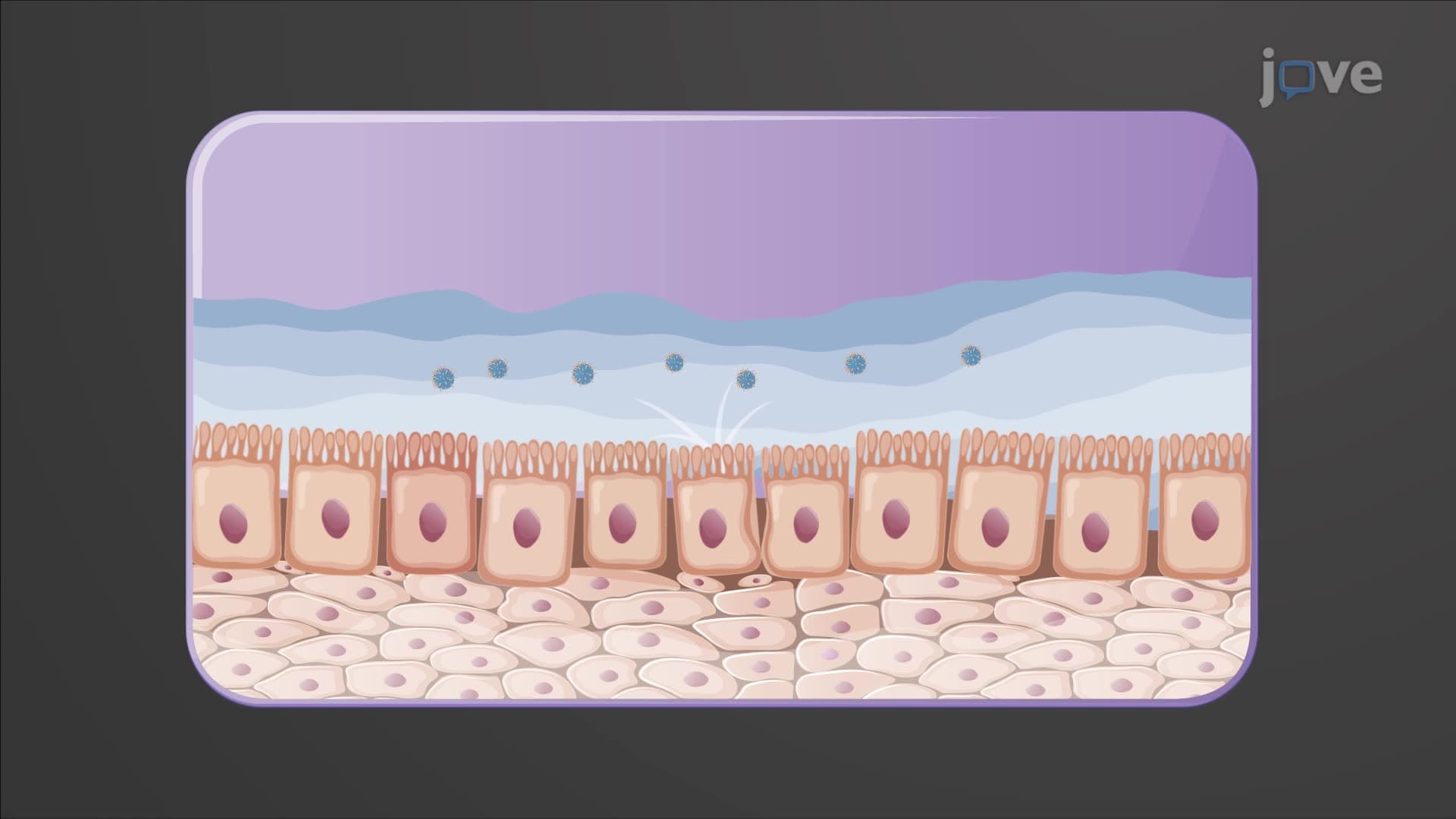
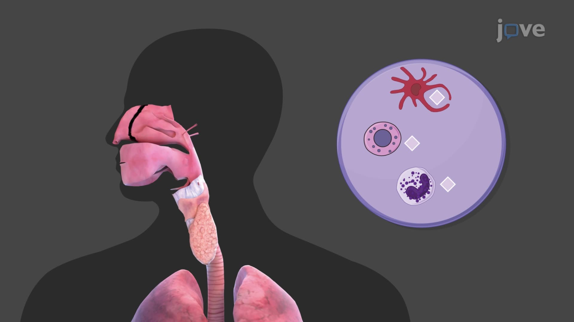
Mucosa-associated lymphoid tissues protect body surfaces exposed to environmental pathogens. Peyer's patches in the small intestine and palatine tonsils exemplify specialized MALT structures. These tissues lack capsules, allowing direct antigen sampling from mucosal surfaces. Antigen presentation secondary responses in MALT often produce secretory IgA antibodies, providing localized protection. Understanding MALT function helps explain why oral vaccines (like rotavirus vaccine administered to US infants) can provide both systemic and mucosal immunity.
Related Micro-courses