Loading subject page…
- Biology
- Membranes and Cellular Transport
- Introduction to Membrane Proteins
Micro-course: Membranes and Cellular Transport
2,101,768 views
Learn key concepts and earn your certificate.




Introduction to Membrane Proteins

00:00
01:16
Video Summary: Introduction to Membrane Proteins Structure and Function
Did you know that every insulin pump used by diabetic patients relies on membrane proteins to detect blood glucose levels? The introduction to membrane proteins: structure reveals how these molecular gatekeepers control what enters and exits our cells. From the sodium-potassium pumps maintaining nerve signals in your brain to the receptors that allow your taste buds to detect sweetness, membrane proteins are the unsung heroes of cellular function. Understanding their peripheral and integral classifications unlocks the mystery of how cells communicate and survive. Watch the full video on JoVE Coach to master this concept with expert-led visuals and step-by-step explanations.
- Understand the Introduction to Membrane Proteins: Structure and Function by distinguishing between peripheral and integral protein categories and their distinct cellular roles
- Identify the structural characteristics that differentiate peripheral proteins from integral proteins based on their membrane associations and hydrophobic properties
- Analyze how transmembrane proteins span cellular membranes using alpha helices or beta barrels to facilitate transport, enzymatic reactions, and signal reception
- Compare the attachment mechanisms of peripheral proteins (non-covalent interactions) versus integral proteins (hydrophobic embedding) and lipid-anchored proteins
- Evaluate the functional significance of amphipathic properties in integral proteins and how this relates to their orientation within phospholipid bilayers
- Apply knowledge of membrane protein structure to predict their roles as transporters, enzymes, receptors, and structural support elements
- Recognize how membrane protein dysfunction contributes to human diseases and therapeutic targets in modern medicine
- Explore the relationship between protein structure and cellular processes including signaling cascades and cytoskeletal organization
Understanding Introduction to Membrane Proteins: Structure and Function
Membrane proteins represent one of the most crucial yet complex topics in cell biology, forming the foundation for understanding how cells maintain their identity and communicate with their environment. These proteins serve as the cellular workforce, performing specialized tasks that keep organisms alive and functioning.
Peripheral Proteins: The Cellular Support Network
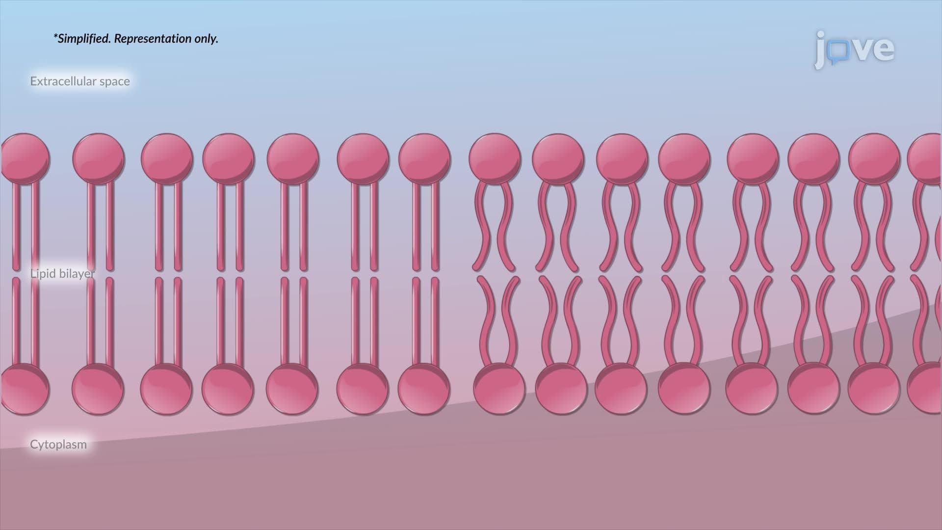
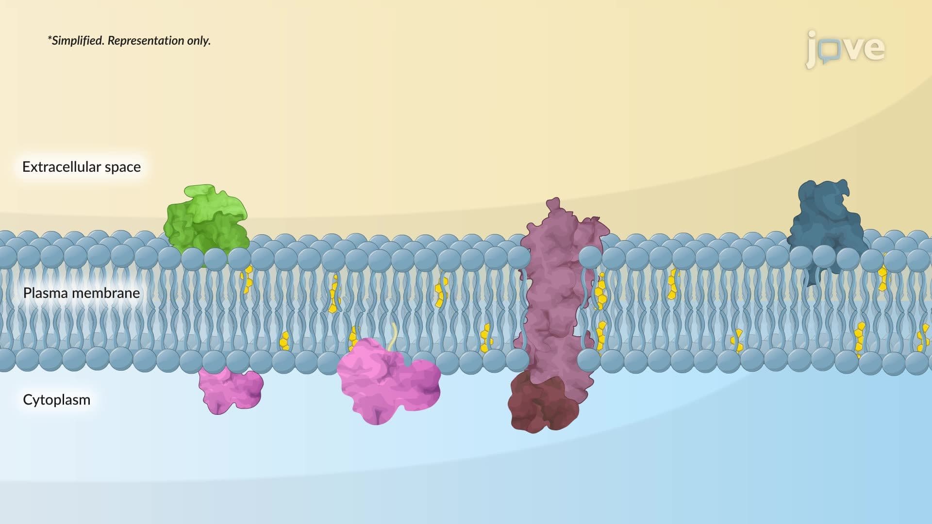
Peripheral proteins act like temporary workers, associating with membrane surfaces through weak, non-covalent interactions. Unlike their integral counterparts, these proteins can easily detach and reattach, making them perfect for dynamic cellular processes. Many peripheral proteins function in cell signaling cascades-think of them as cellular messengers that can quickly relocate when needed.
A prime example occurs in muscle contraction, where peripheral proteins like spectrin link the cell membrane to the underlying cytoskeleton in red blood cells. This connection provides the flexibility that allows these cells to squeeze through capillaries barely wider than the cells themselves. When this system fails, as in hereditary spherocytosis, patients experience anemia due to fragile red blood cells.
Integral Proteins: The Embedded Specialists
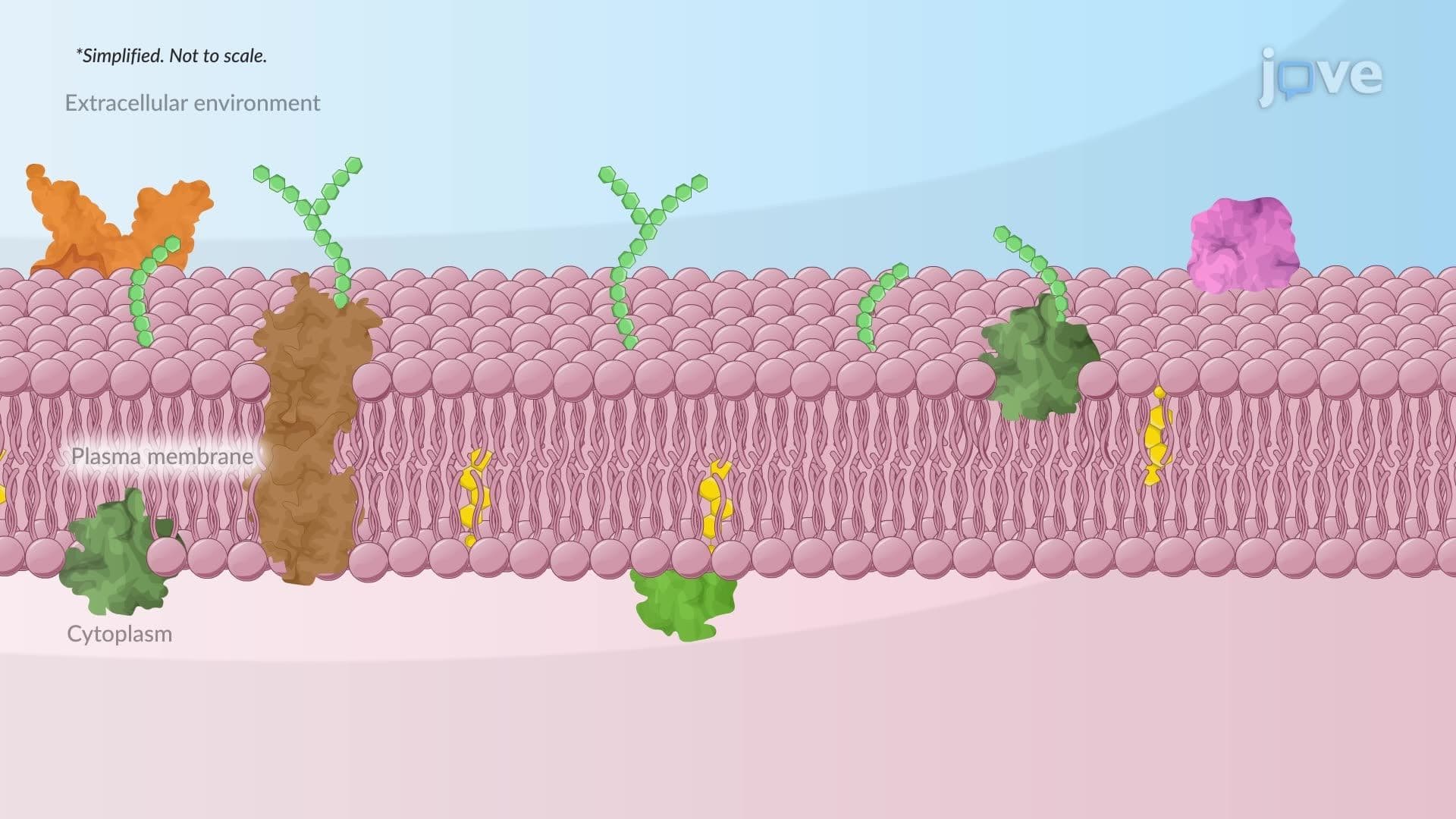
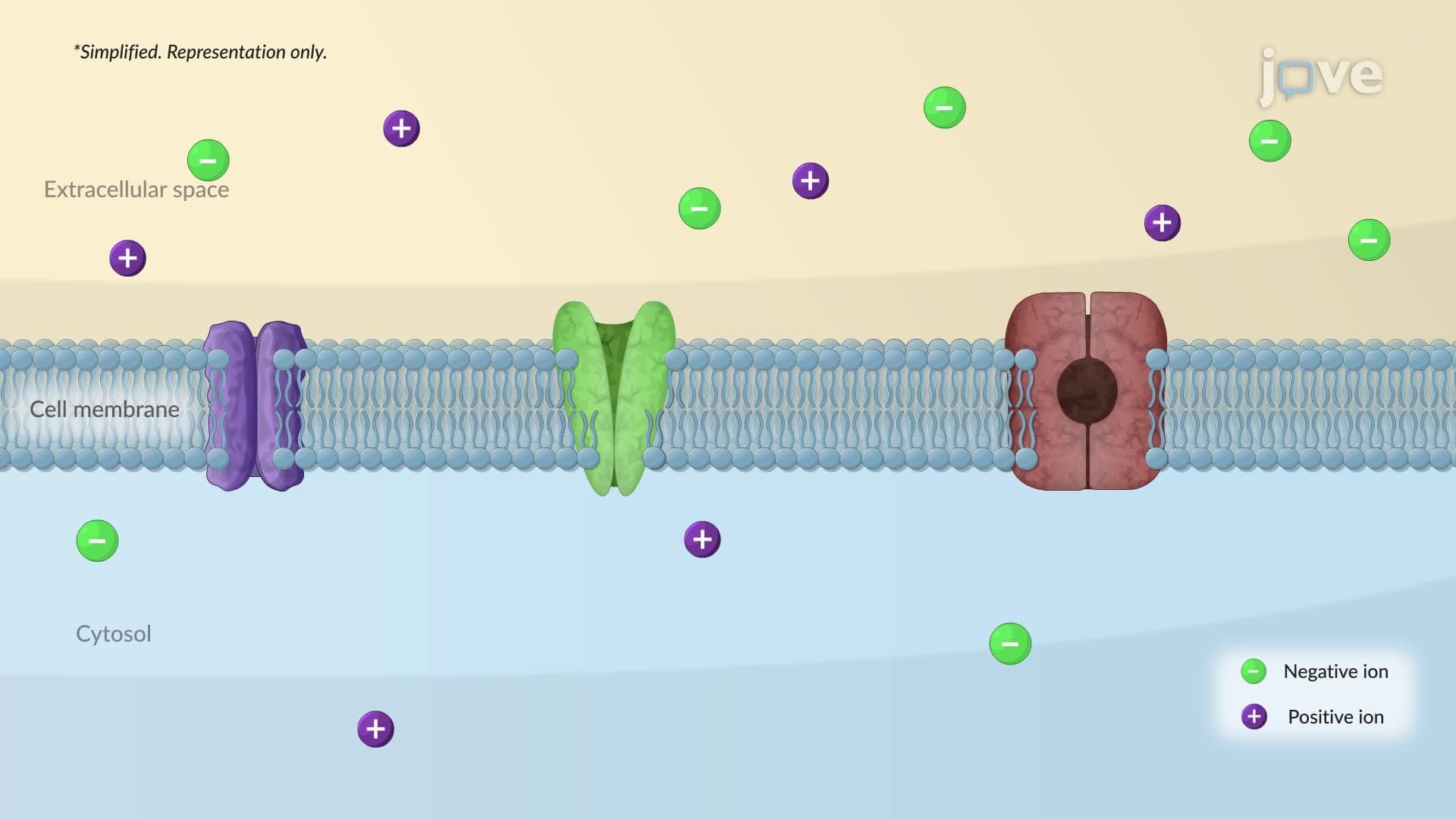
Integral proteins represent the permanent residents of cellular membranes, with their amphipathic nature allowing them to span the lipid bilayer effectively. Their hydrophilic regions face the watery environments inside and outside the cell, while their hydrophobic domains nestle comfortably within the fatty acid tails of phospholipids.
Transmembrane proteins, a subset of integral proteins, completely span the membrane and often serve as cellular gatekeepers. The cystic fibrosis transmembrane conductance regulator (CFTR) protein exemplifies this role-when functioning properly, it allows chloride ions to cross cell membranes in the lungs and other organs. Mutations in this protein cause cystic fibrosis, affecting approximately 30,000 Americans.
Structural Diversity and Functional Specialization
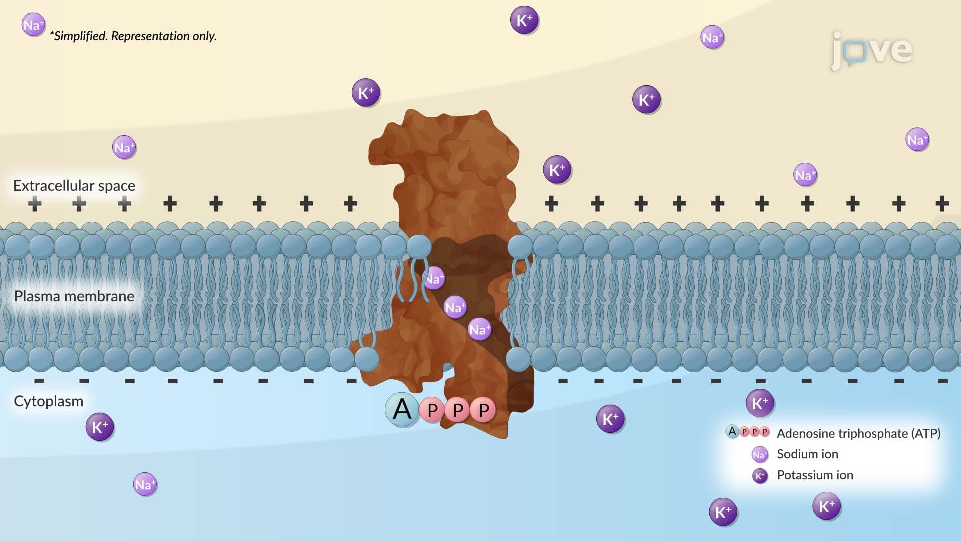
The membrane-spanning regions of transmembrane proteins display remarkable structural diversity. Single alpha helices work well for simple ion channels, while multiple alpha helices create more complex transport systems like the sodium-potassium pump crucial for nerve function. Beta barrels, with their central pores, form sophisticated channels-bacterial porins use this structure to control molecular traffic.
For AP Biology students and college undergraduates, understanding these structural patterns proves essential for exam success. The MCAT frequently tests knowledge of how protein structure relates to function, particularly in membrane transport and cell signaling contexts.
Frequently Asked Questions
Introduction to Membrane Proteins: Structure and Function explores how proteins associate with cellular membranes and perform vital cellular tasks. This foundational biology concept covers peripheral proteins that temporarily associate with membranes and integral proteins permanently embedded within lipid bilayers. Understanding these protein types is essential for grasping cellular transport, signaling, and structural organization.
Peripheral proteins associate with membrane surfaces through weak interactions and can easily detach, while integral proteins embed permanently within the lipid bilayer. Peripheral proteins often participate in signaling or provide structural support, whereas integral proteins typically function as transporters, enzymes, or receptors. This distinction is crucial for understanding cellular function and commonly appears on AP Biology and college biology exams.
The MCAT tests membrane protein concepts through passages on transport mechanisms, enzyme function, and disease pathways. AP Biology frequently includes questions about protein structure-function relationships and cellular transport. Students should focus on understanding how amphipathic properties allow proteins to span membranes and how structural defects cause diseases like cystic fibrosis.
Introductory biology, cell biology, biochemistry, and molecular biology courses all cover membrane proteins extensively. Advanced courses like membrane biophysics and structural biology explore these concepts in greater depth. Many pre-med students encounter membrane proteins again in physiology and pathology courses, making this a foundational concept for healthcare careers.
Diabetes management relies heavily on membrane proteins-glucose transporters (GLUT4) allow muscle cells to uptake glucose in response to insulin, while glucose sensors in continuous glucose monitors detect blood sugar levels. When these proteins malfunction, patients develop diabetes complications. The insulin receptor itself is also a transmembrane protein, highlighting how protein structure directly impacts human health.
While membrane proteins involve complex molecular interactions, the basic concepts build logically from simple chemistry principles. Understanding soap molecules helps explain amphipathic behavior, and familiarity with basic protein structure provides sufficient background. Most high school students successfully master these concepts with proper visual aids and step-by-step explanations.
Focus on drawing and labeling membrane protein diagrams, emphasizing the relationship between structure and function. Practice identifying protein types from descriptions and predicting functional consequences of structural changes. Create concept maps connecting membrane proteins to transport, signaling, and disease processes. Active recall through practice questions proves more effective than passive reading.
Consider studying protein folding mechanisms, X-ray crystallography of membrane proteins, and drug targeting of membrane receptors. Advanced topics include lipid-protein interactions, membrane protein evolution, and biotechnology applications like engineered ion channels. These areas connect to cutting-edge research in drug discovery and synthetic biology.
Related Micro-courses