Loading subject page…

- Biology
- Membranes and Cellular Transport
- What are Membranes?
Micro-course: Membranes and Cellular Transport
2,101,760 views
Learn key concepts and earn your certificate.



What are Membranes?


00:00
01:54
Video Summary: What are Membranes Structure Composition and Function
Did you know that every single cell in your body-from neurons firing in your brain to muscle cells contracting in your heart-depends on incredibly thin barriers just 7 nanometers thick? Understanding cell membrane structure function is fundamental to grasping how life operates at the molecular level. Consider how insulin must cross cell membranes to regulate blood sugar in diabetic patients-this process relies entirely on the sophisticated architecture of What Are Membranes? Structure, Composition, and Function. Watch the full video on JoVE Coach to master this concept with expert-led visuals and step-by-step explanations.
- Understand the fundamental components of What Are Membranes? Structure, Composition, and Function, including phospholipids, proteins, and carbohydrates
- Identify the arrangement of phospholipids in the bilayer structure and explain why hydrophilic heads face outward while hydrophobic tails cluster inward
- Analyze how membrane proteins function as receptors, transporters, and structural connectors within cellular systems
- Compare the roles of glycolipids and glycoproteins in cellular recognition and signaling processes
- Evaluate the selective permeability properties that allow membranes to control molecular transport
- Apply knowledge of membrane dynamics to predict how different molecules will interact with cellular boundaries
- Recognize the relationship between membrane composition and specific cellular functions
- Explore how membrane structure enables compartmentalization within eukaryotic cells
Understanding Cell Membrane Structure Function
The cell membrane structure function represents one of biology's most elegant solutions to cellular organization. These dynamic barriers solve a fundamental problem: how can cells maintain their internal environment while still communicating with the outside world? The answer lies in the sophisticated architecture of biological membrane explained through the fluid mosaic model.
The Phospholipid Bilayer Foundation
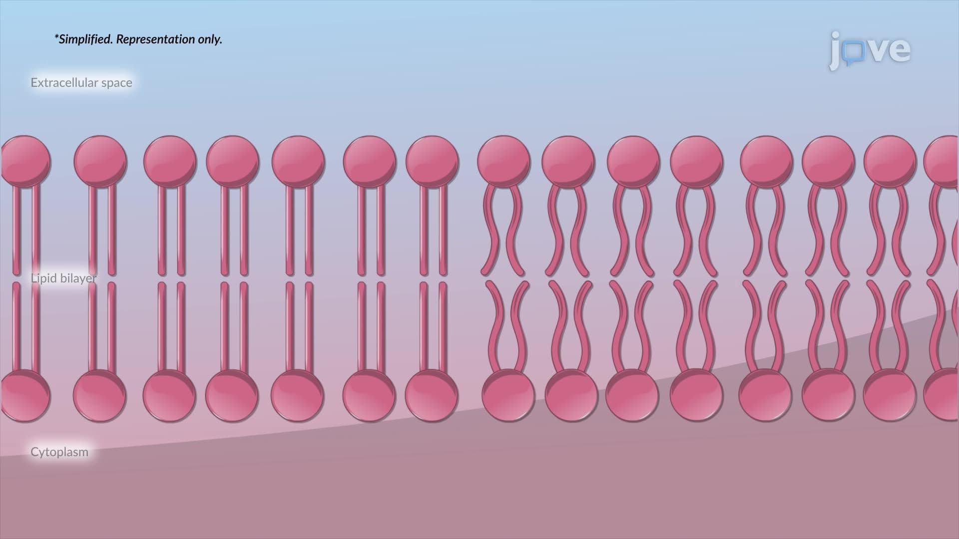
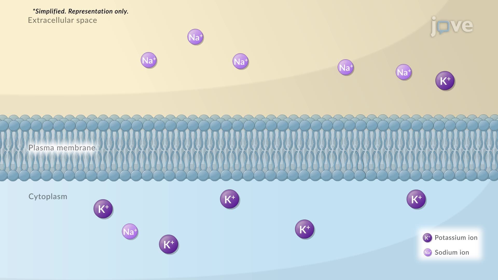
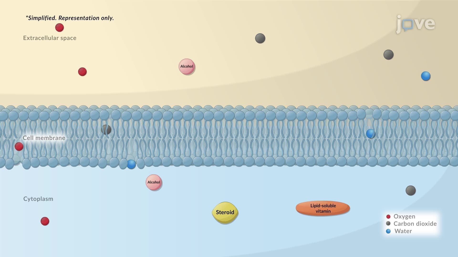
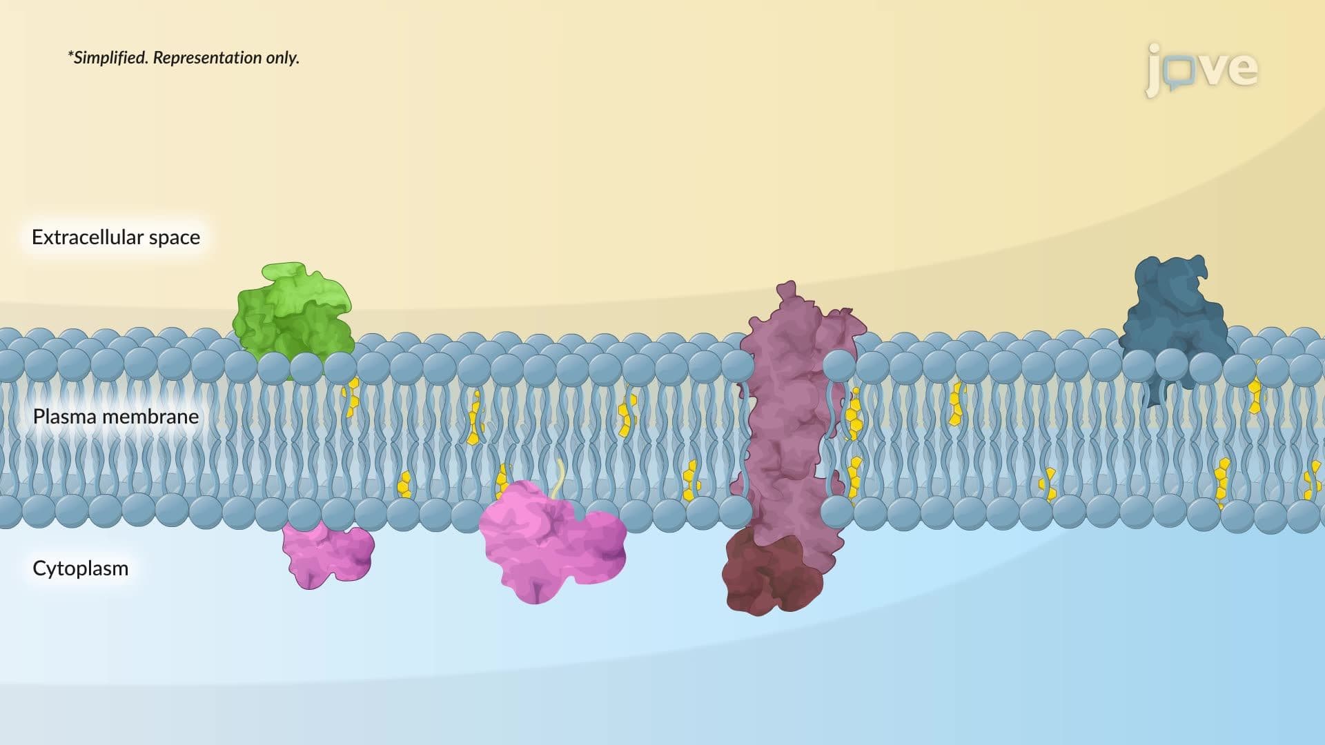
At the heart of every membrane composition biology lies the phospholipid bilayer-a self-assembling structure that forms spontaneously in aqueous environments. Each phospholipid molecule contains a hydrophilic (water-loving) phosphate head and two hydrophobic (water-fearing) fatty acid tails. This amphipathic nature drives the formation of the bilayer, with heads facing the watery environments inside and outside the cell, while tails hide in the membrane's interior.
This phospholipid bilayer membrane serves as more than just a barrier-it's a dynamic platform. The fatty acid composition determines membrane fluidity, which varies based on cellular needs. For instance, neurons require highly fluid membranes for rapid signal transmission, while plant cell membranes need stability to withstand environmental stress.
Membrane Proteins: The Functional Workhorses
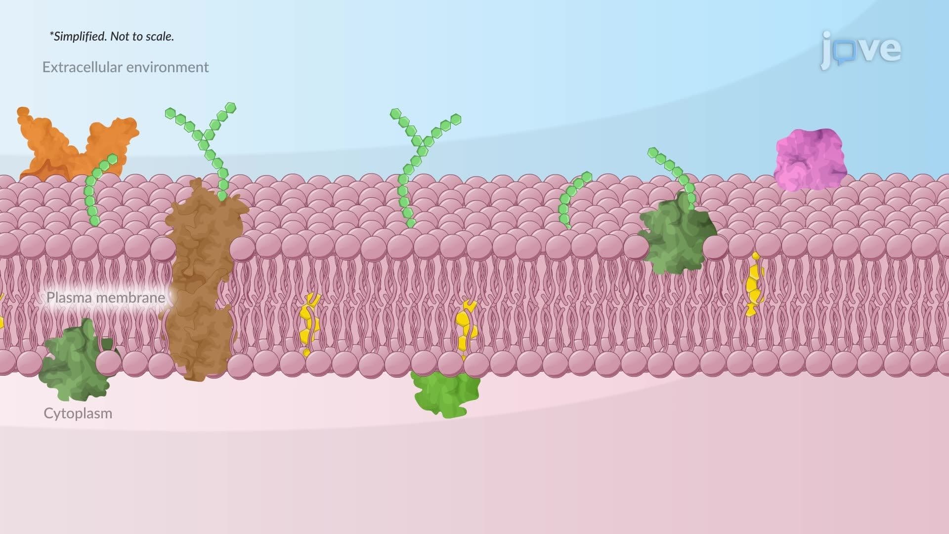
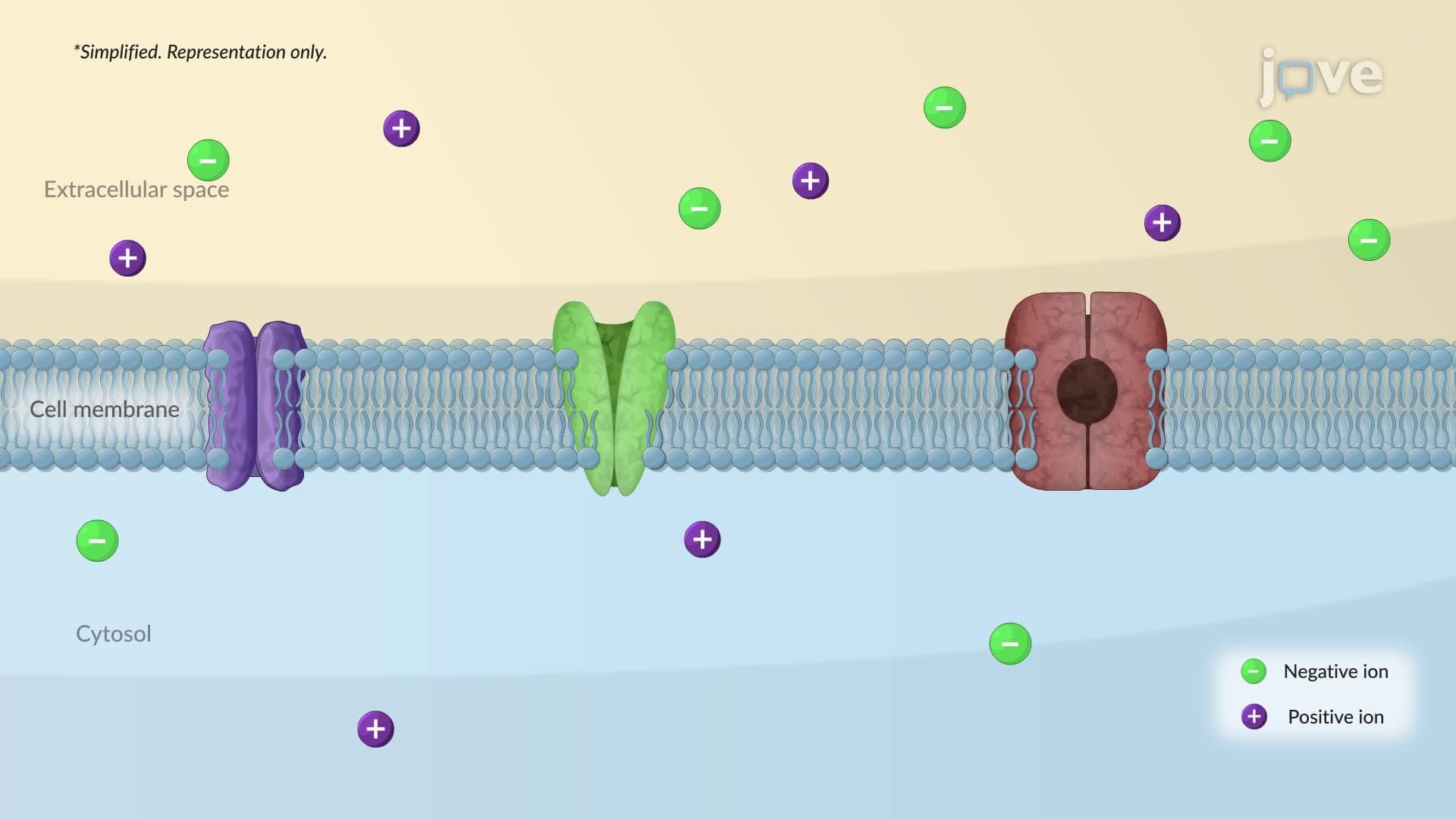
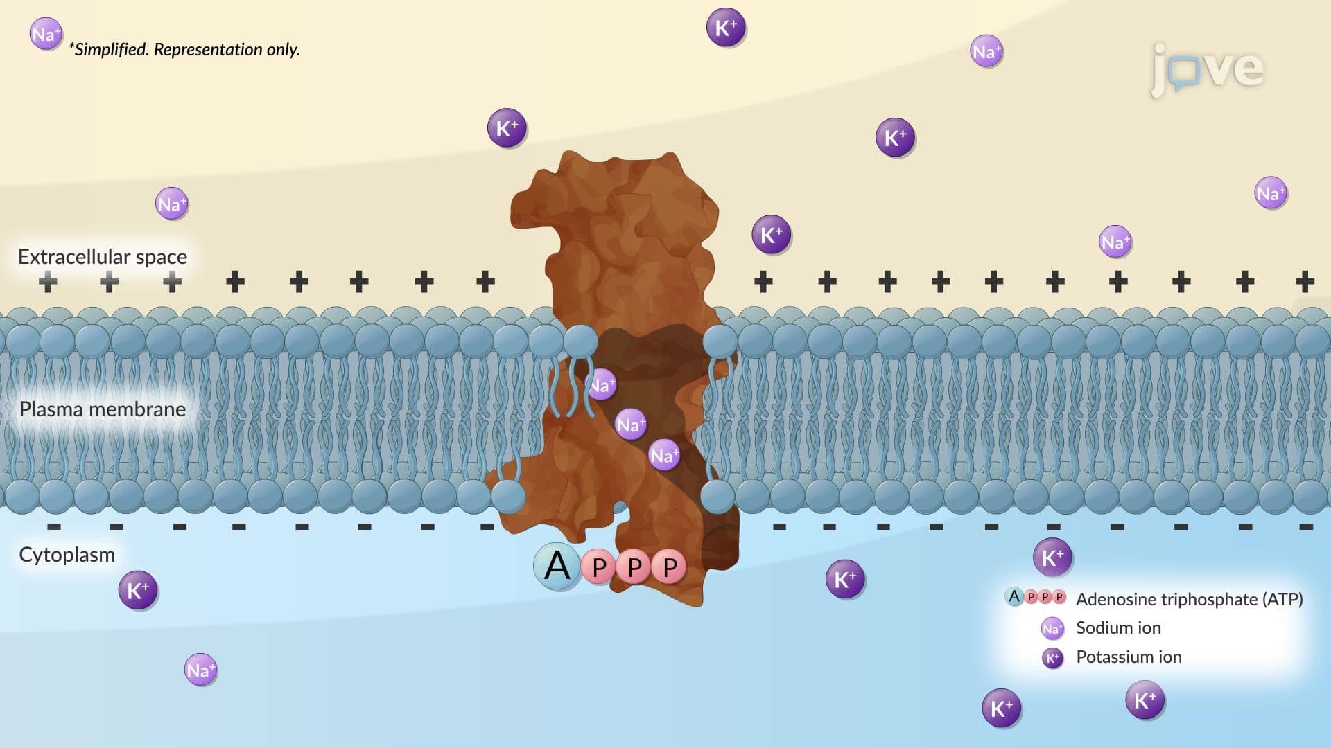
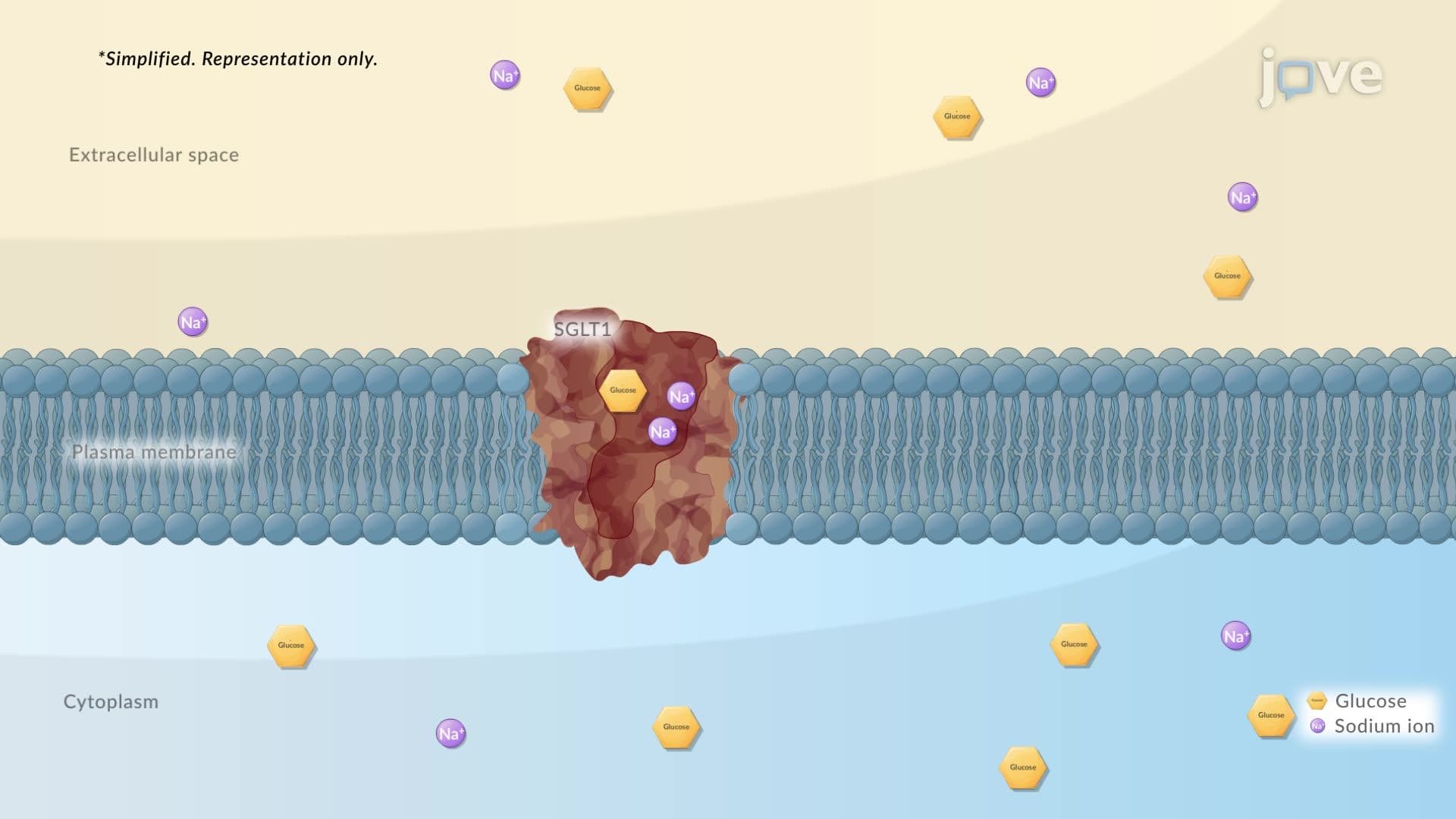
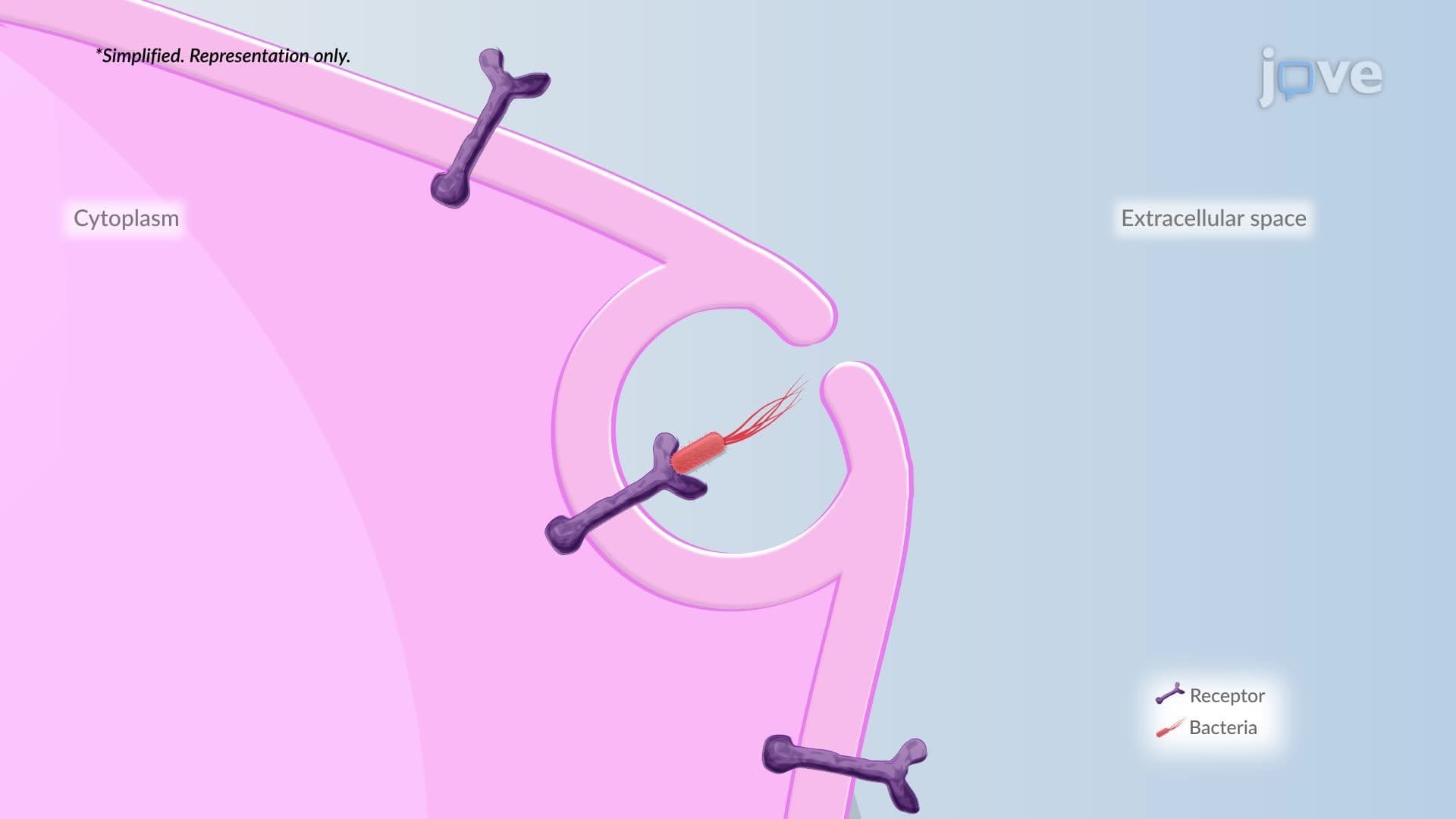
While phospholipids provide structure, membrane protein function drives cellular activity. These proteins fall into several categories that students encounter on the MCAT and AP Biology exams. Receptor proteins, like insulin receptors in muscle cells, bind specific signaling molecules and trigger intracellular cascades. Transport proteins create selective channels-think of the sodium-potassium pump that maintains nerve cell electrical potential.
Integral proteins span the entire membrane thickness, while peripheral proteins associate with membrane surfaces. This distinction becomes crucial when studying diseases like cystic fibrosis, where defective chloride channels (integral proteins) cause serious health complications.
Carbohydrate Components and Cellular Recognition
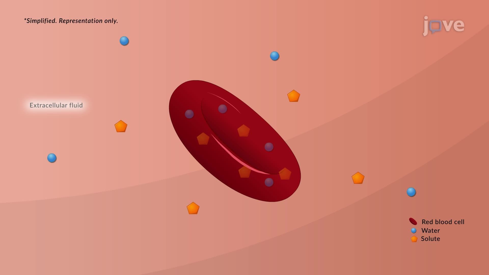
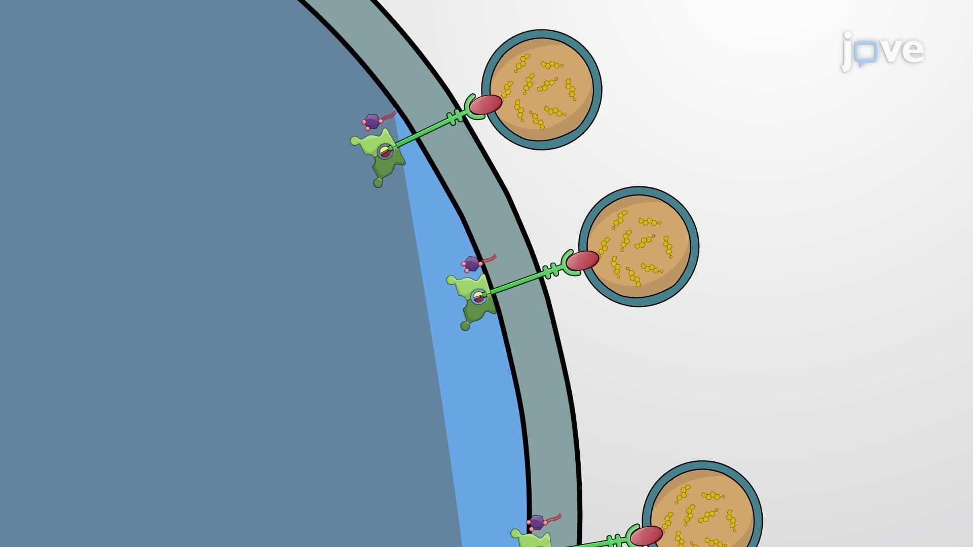
The fluid mosaic model membrane incorporates carbohydrates as glycolipids and glycoproteins, creating the cell's "identity card." These sugar-containing molecules enable cellular recognition-essential for immune system function and tissue formation. Blood type antigens exemplify this concept: A, B, and O blood types differ based on specific carbohydrate structures on red blood cell membranes.
Medical students studying for the USMLE encounter this concept when learning about organ transplant compatibility and autoimmune diseases, where cellular recognition goes awry.
Frequently Asked Questions
Membranes are thin, flexible boundaries made of phospholipids, proteins, and carbohydrates that surround cells and internal compartments. They act like selective gatekeepers, controlling what enters and exits while maintaining cellular organization. The phospholipid bilayer provides the basic structure, while proteins handle transport and signaling functions. Carbohydrates help cells recognize each other and bind important molecules like hormones.
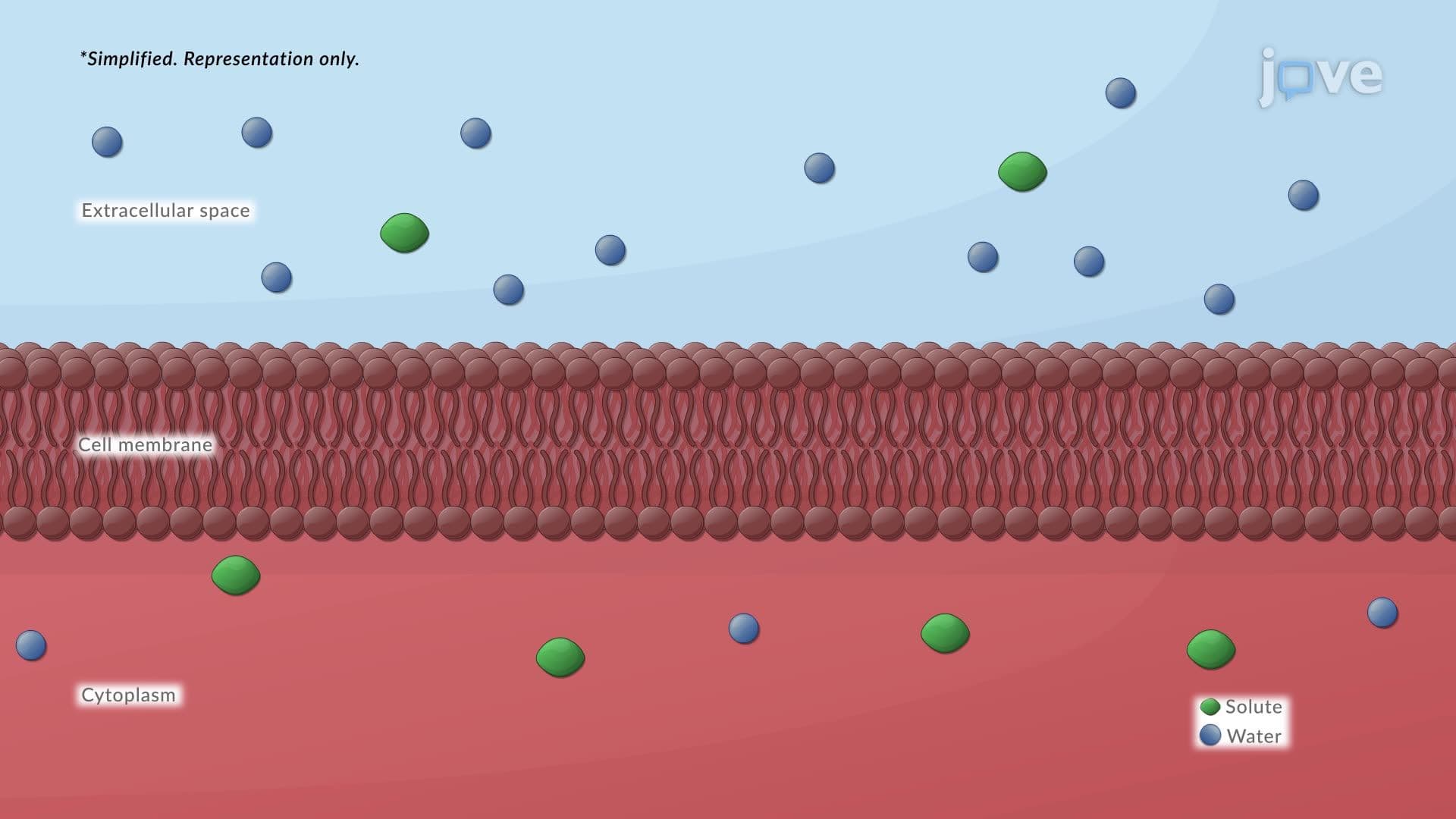
The selective permeability concept is crucial-membranes allow some substances through while blocking others based on size, charge, and polarity. Understanding how this relates to the fluid mosaic model, where proteins float in a phospholipid sea, helps explain transport mechanisms. Focus on active vs. passive transport, protein channels, and how membrane composition affects function. This concept appears frequently in AP Biology FRQs and multiple-choice questions.
MCAT Biological and Biochemical Foundations sections heavily emphasize membrane transport, signal transduction, and membrane potential. Expect questions linking membrane structure to function, especially regarding nerve conduction, hormone signaling, and cellular energetics. Understanding how membrane proteins facilitate facilitated diffusion versus active transport is essential. The exam often connects membrane function to disease states and pharmacology.
Drug delivery systems rely on understanding how medications cross cell membranes-from oral medications absorbed in the intestine to chemotherapy drugs targeting cancer cells. Diabetes management involves insulin binding to membrane receptors, while cholesterol-lowering statins affect membrane composition. Anesthetics work by altering nerve membrane properties, and many antibiotics target bacterial cell wall synthesis or membrane integrity.
Not at all-membrane concepts build logically from basic chemistry principles you already know. If you understand that oil and water don't mix, you can grasp why phospholipid tails cluster together. Think of membranes like a crowd of people holding hands (phospholipids) with some individuals (proteins) moving through the group. Start with the big picture, then add details gradually as your confidence grows.
Create visual diagrams showing the bilayer with different protein types, then practice drawing them from memory. Use active recall by explaining how insulin signaling works or why cystic fibrosis affects chloride transport. Connect structure to function constantly-don't just memorize components. Practice with released AP Biology questions or MCAT passages to see how concepts integrate with other topics like cellular respiration and photosynthesis.
Membrane concepts connect to virtually every biological process-cellular respiration occurs across mitochondrial membranes, photosynthesis happens in chloroplast membranes, and nerve conduction depends on membrane potential changes. Understanding membranes prepares you for advanced topics like bioenergetics, signal transduction cascades, and membrane trafficking in cell biology courses. This foundation becomes essential for understanding how drugs work in pharmacology and how diseases affect cellular function.
Explore membrane transport mechanisms in detail-facilitated diffusion, active transport, and bulk transport processes like endocytosis and exocytosis. Then investigate membrane potential and action potentials in neurons, followed by signal transduction pathways. These advanced topics build directly on membrane structure knowledge and appear prominently in upper-level biology courses and medical school curricula.
Related Micro-courses