Loading subject page…

2,101,787 views

Video Summary: What Is the Fluid Mosaic Model
Ever wondered how a cell membrane can be both protective and selective, like a high-tech security system? The fluid mosaic model explains this remarkable cellular structure as a dynamic arrangement of phospholipids, proteins, and carbohydrates that work together to control what enters and exits cells. Think of how medications like insulin must cross cell membranes to treat diabetic patients across the US healthcare system. Watch the full video on JoVE Coach to master this concept with expert-led visuals and step-by-step explanations.
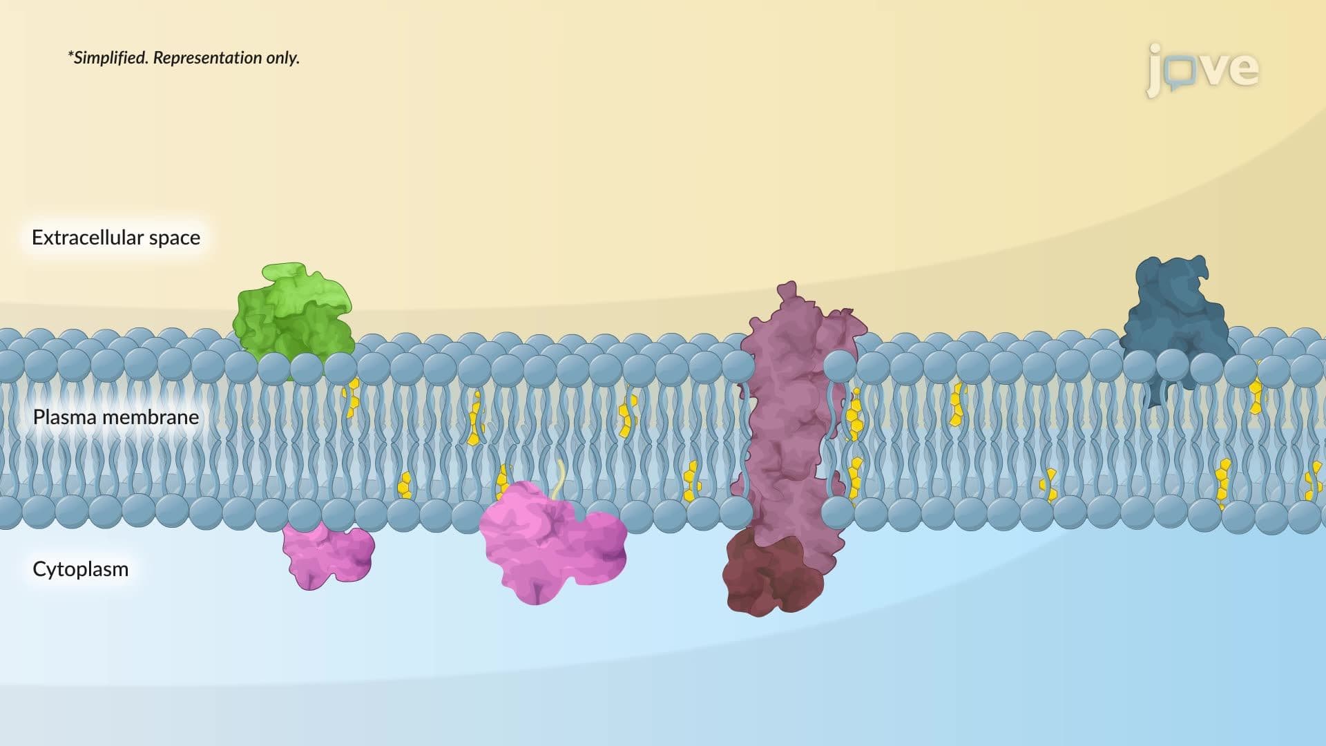
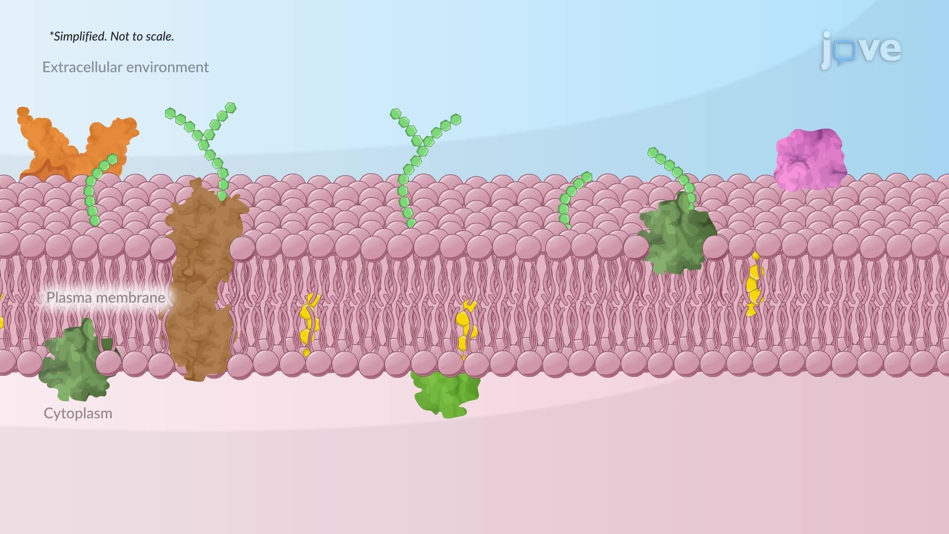
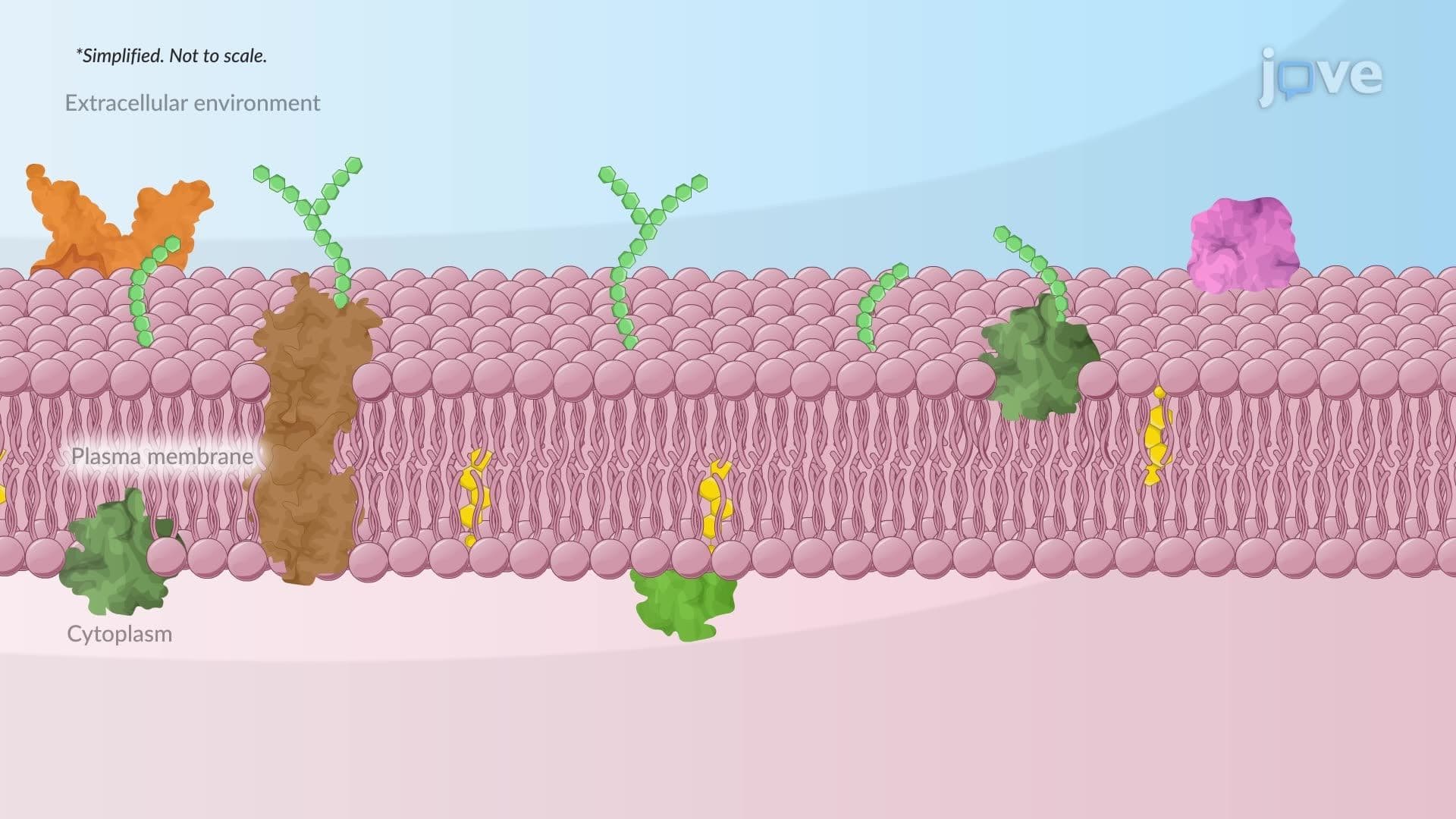
The fluid mosaic model revolutionized our understanding of cell membrane architecture when proposed by Singer and Nicolson in 1972. This model describes biological membranes as dynamic, two-dimensional fluids where lipids and proteins move laterally while maintaining structural integrity. The "mosaic" aspect refers to the diverse protein components embedded within or associated with the lipid bilayer, creating a complex pattern of functional units.
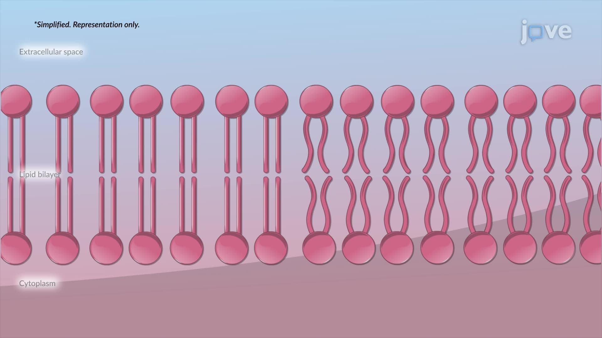
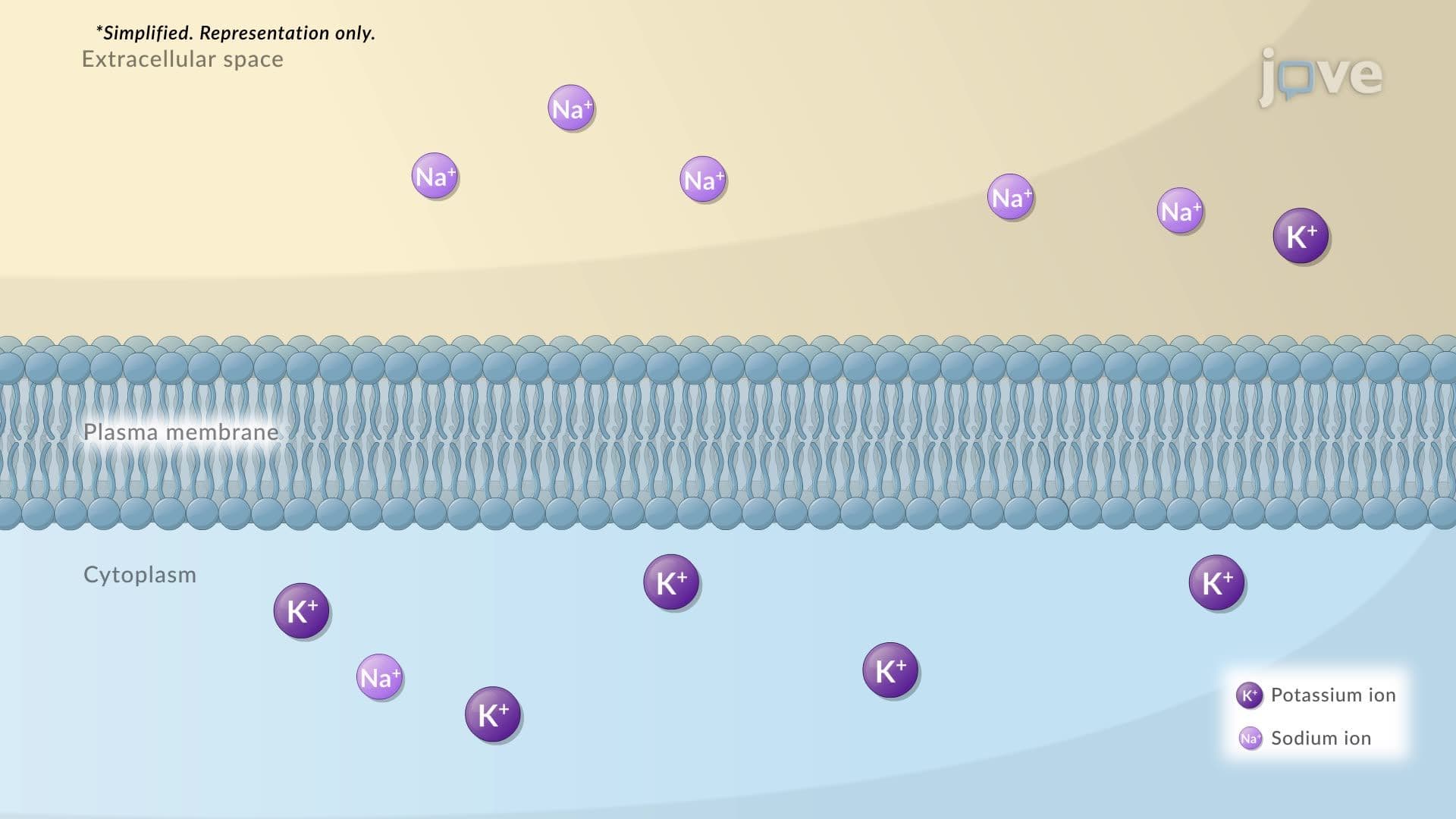
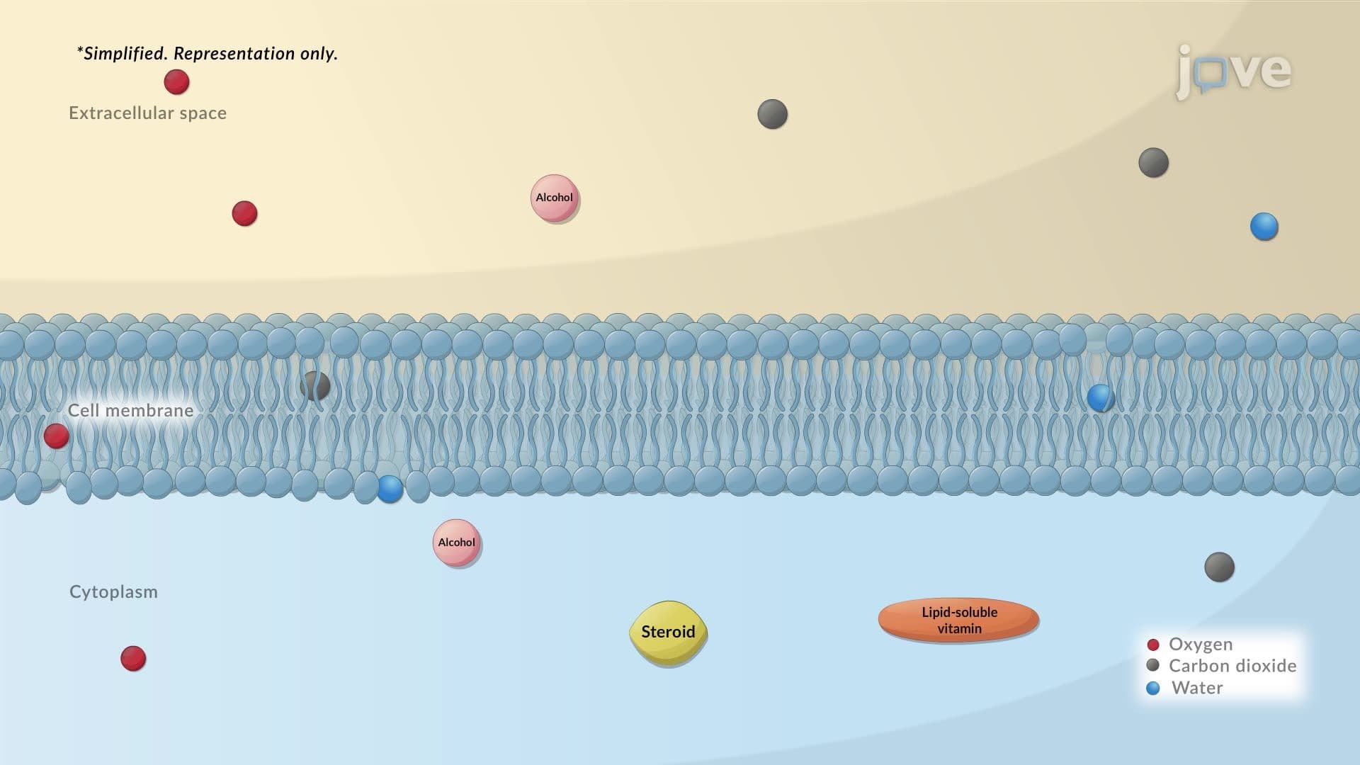
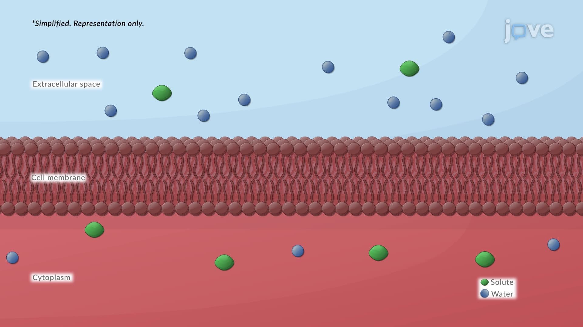
Phospholipids form the structural backbone through their unique amphipathic properties. Each phospholipid contains a hydrophilic phosphate head group and two hydrophobic fatty acid tails. In aqueous environments, these molecules spontaneously arrange into bilayers, with hydrophobic tails facing inward and hydrophilic heads facing the aqueous environments inside and outside the cell. This arrangement creates a selective barrier that prevents most water-soluble substances from freely crossing while allowing lipid-soluble molecules to pass through.
Cholesterol, another crucial lipid component, intercalates between phospholipids to modulate membrane fluidity. At body temperature (37°C), cholesterol prevents excessive fluidity while maintaining enough flexibility for proper membrane function. This is critical for processes like neurotransmitter release at synapses in the human nervous system.
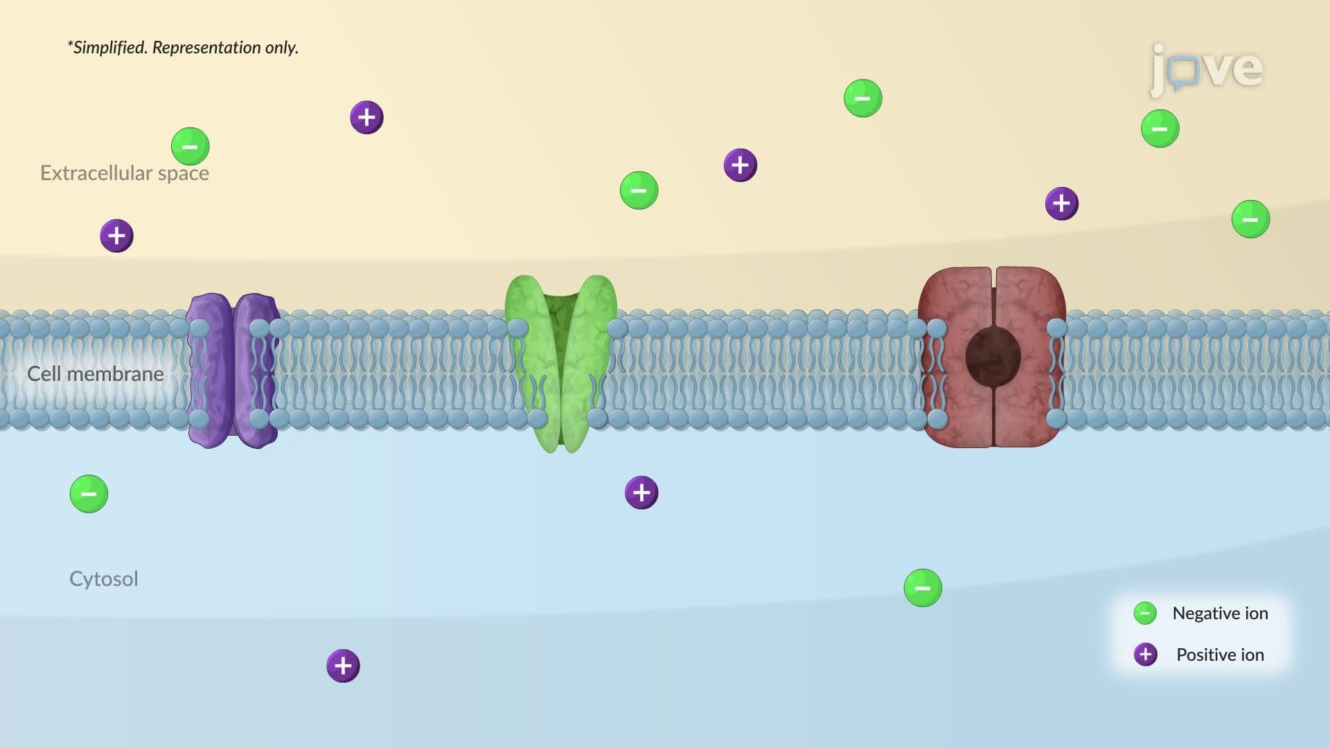
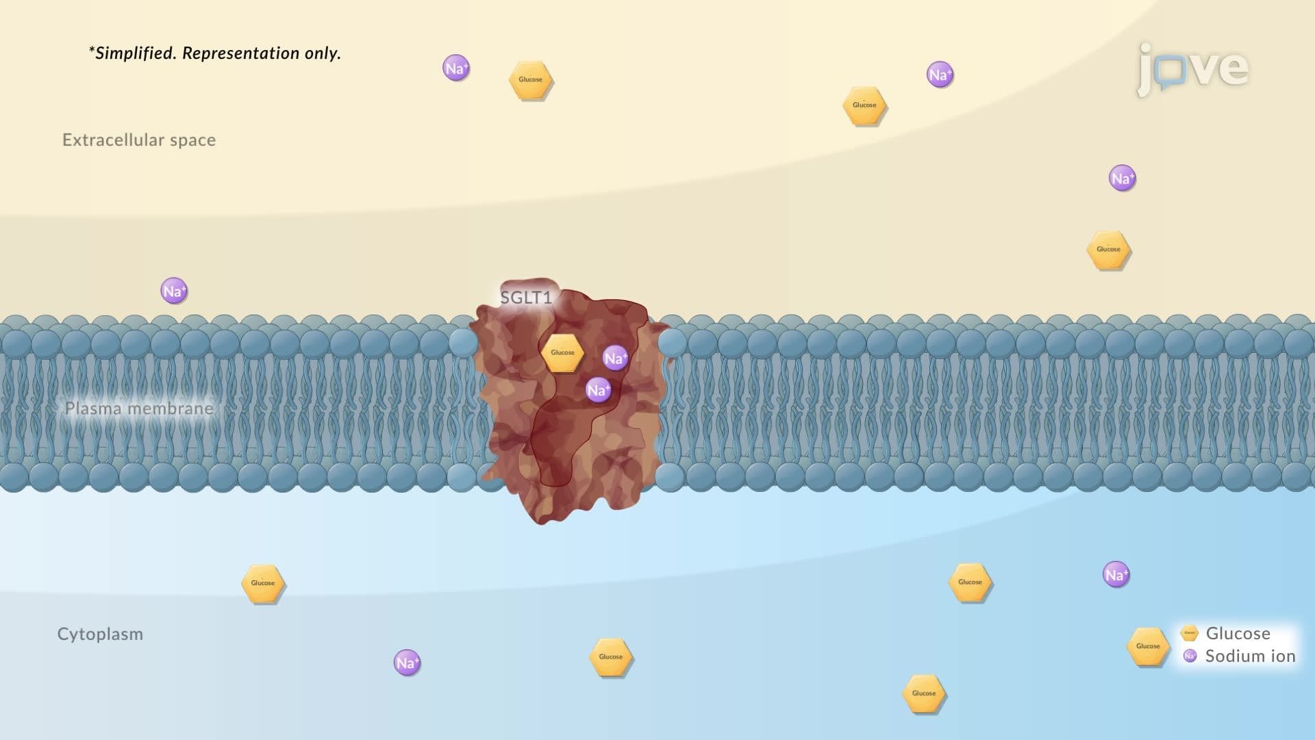
Membrane proteins serve diverse functions from transport to cell recognition. Integral membrane proteins, such as channel proteins and carrier proteins, span the entire bilayer and facilitate selective transport. For example, glucose transporters (GLUT proteins) enable glucose uptake in muscle cells, crucial for energy metabolism during exercise.
Peripheral proteins associate with membrane surfaces without spanning the bilayer. These include many enzymes and structural proteins that anchor the cytoskeleton. The estrogen receptor, mentioned in transcripts, exemplifies how some receptors can be both membrane-associated and cytosolic, depending on cellular conditions.
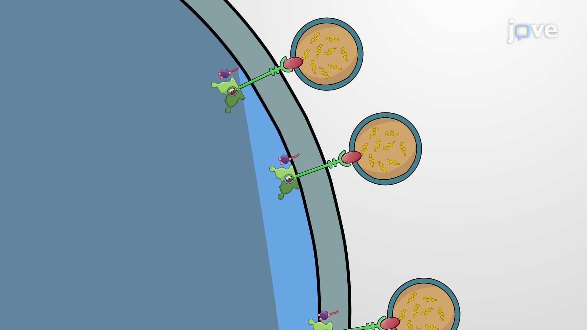
The glycocalyx represents the carbohydrate-rich outer layer formed by glycoproteins and glycolipids. This "sugar coating" plays essential roles in cell recognition, adhesion, and immune system function. In medical settings, ABO blood typing depends on glycoprotein differences in red blood cell membranes, determining transfusion compatibility for patients in US hospitals.
Students preparing for AP Biology exams should understand how membrane structure relates to function, particularly in contexts like nerve signal transmission, hormone action, and drug delivery mechanisms. The MCAT frequently tests membrane protein classification and transport mechanisms, making this foundational knowledge essential for pre-med students.
Related Micro-courses