Loading subject page…

- Biology
- Reproduction and Development
- Cleavage and Blastulation
Micro-course: Reproduction and Development
442,001 views
Learn key concepts and earn your certificate.
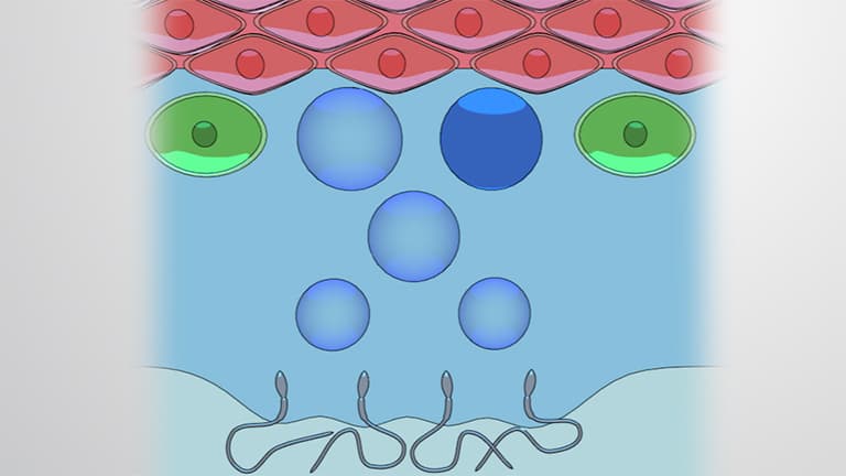

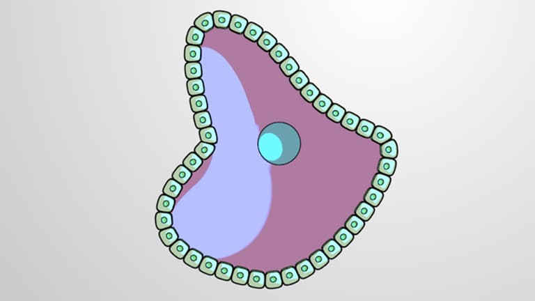
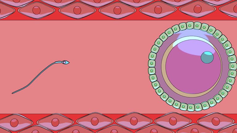

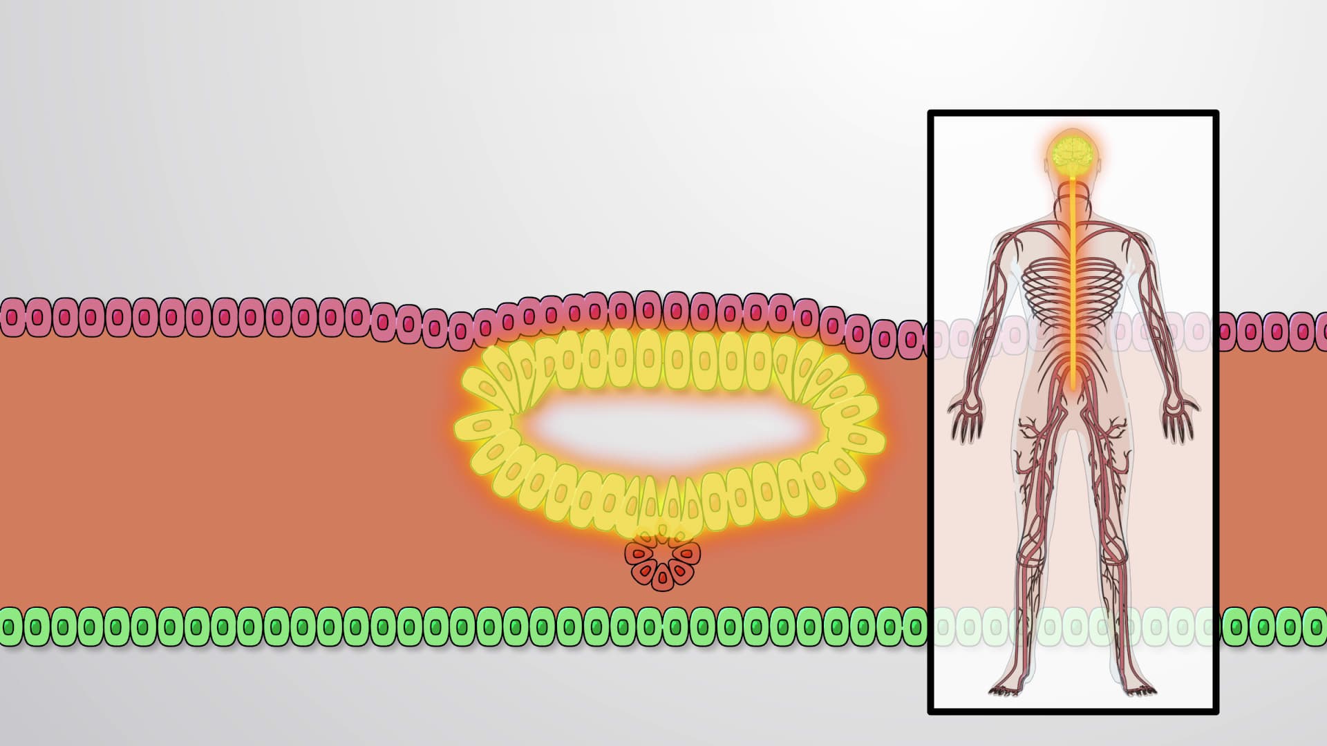
Cleavage and Blastulation

00:00
01:33
Video Summary: What Is Cleavage and Blastulation
Did you know that within just one week of fertilization, a single human cell transforms into a hollow ball containing over 100 specialized cells? Cleavage blastulation represents the remarkable first steps of human development, where rapid cell division without growth creates the foundation for all future organ systems. At fertility clinics like those at Johns Hopkins and Mayo Clinic, embryologists carefully monitor these precise developmental stages to optimize IVF success rates. Understanding What is Cleavage And Blastulation reveals how life's blueprint unfolds through carefully orchestrated cellular events. Watch the full video on JoVE Coach to master this concept with expert-led visuals and step-by-step explanations.
- Understand What is Cleavage And Blastulation and distinguish between these two sequential developmental processes in human embryogenesis
- Identify the key cellular structures formed during cleavage, including blastomeres, morula, and blastocyst stages
- Analyze how compaction at the eight-cell stage establishes the first cell-to-cell communications through tight junctions
- Compare the roles of trophoblast and inner cell mass in establishing placental and embryonic tissues respectively
- Evaluate the significance of zona pellucida dissolution in enabling successful uterine implantation
- Apply knowledge of blastulation timing to understand fertility treatment protocols and early pregnancy detection
- Recognize the transition from cleavage to blastulation as the first major differentiation event in human development
- Explore how cleavage patterns differ between mammalian species and their evolutionary significance
Understanding Cleavage And Blastulation Fundamentals
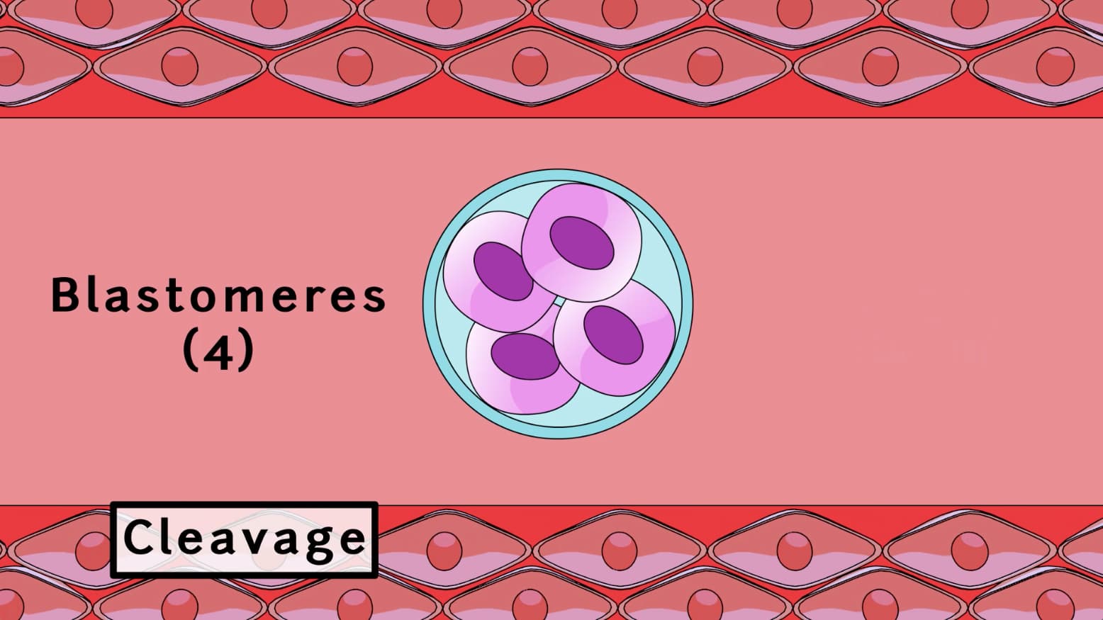
What is Cleavage And Blastulation encompasses two interconnected phases that transform a fertilized egg into an implantation-ready embryo. Cleavage represents a unique form of cell division where rapid mitotic divisions occur without intervening growth phases, effectively partitioning the zygote's cytoplasm among increasingly smaller daughter cells called blastomeres. This process defies the typical cell cycle pattern where cells grow between divisions, making it a fascinating exception in developmental biology.
The precision of early cleavage divisions has profound clinical significance. At reproductive medicine centers like those at Stanford University Medical Center, embryologists use time-lapse imaging to monitor cleavage timing, as abnormal division patterns often indicate chromosomal abnormalities that reduce IVF success rates.
Molecular Mechanisms of Compaction
Beginning at the eight-cell stage, blastomeres undergo compaction-a process where cells maximize their contact surface area and establish the first intercellular junctions. This event marks the embryo's initial polarization, creating distinct cellular domains that will guide all subsequent development. The formation of tight junctions during compaction represents the embryo's first instance of cellular cooperation, establishing the foundation for tissue formation.
Students preparing for the MCAT or AP Biology exams should understand that compaction involves specific molecular players, including E-cadherin adhesion molecules and Par proteins that establish cell polarity. These same mechanisms operate throughout embryonic development and adult tissue maintenance.
Blastulation and Cellular Differentiation
Blastulation transforms the solid morula into a hollow blastocyst, featuring the first cellular differentiation in human development. The outer trophoblast cells develop specialized transport functions necessary for implantation and placenta formation, while the inner cell mass contains pluripotent embryonic stem cells. This segregation establishes the fundamental distinction between extraembryonic support tissues and the actual embryo proper.
Research at institutions like the University of California San Francisco has revealed that blastulation timing serves as a predictor of embryo viability. Blastocysts forming too early or late often carry genetic abnormalities, making temporal assessment crucial for assisted reproductive technologies.
Clinical Applications and Exam Relevance
Understanding cleavage and blastulation proves essential for healthcare professions. USMLE and NCLEX examinations frequently test knowledge of early development, particularly regarding assisted reproductive technology and pregnancy complications. For instance, questions may address how maternal age affects cleavage division accuracy or why blastocyst transfer yields higher IVF success rates than earlier-stage embryo transfer.
Students should also recognize connections to regenerative medicine, as the principles governing blastulation inform current research into induced pluripotent stem cells and tissue engineering applications being developed at institutions like the Harvard Stem Cell Institute.
Frequently Asked Questions
Cleavage and blastulation are the first two stages of human embryonic development after fertilization. Cleavage involves rapid cell divisions that create smaller cells without overall growth, while blastulation forms a hollow ball structure with the first specialized cell types. Together, these processes prepare the embryo for implantation in the uterus within about one week of fertilization.
The MCAT Biological and Biochemical Foundations section frequently tests early development concepts, particularly cell division mechanisms and differentiation pathways. Questions often focus on comparing cleavage to normal mitosis, understanding compaction's role in establishing cell polarity, and recognizing how blastulation creates the first stem cell population. Mastering these concepts strengthens your foundation for more complex developmental biology topics.
AP Biology Unit 7 (Natural Selection) and Unit 8 (Ecology) both reference developmental patterns, while the cell division and signaling units provide essential background. Free response questions have asked students to explain how cell cycle regulation differs during cleavage versus normal development. Understanding these processes also supports Big Idea 3 about how biological systems store and transmit information across generations.
IVF laboratories at major medical centers like Cleveland Clinic monitor embryo development through these stages using specialized incubators and time-lapse imaging. Embryologists assess cleavage timing, blastomere symmetry, and blastulation success to select the healthiest embryos for transfer. Abnormal patterns often indicate genetic problems, helping doctors counsel patients and optimize treatment protocols for better pregnancy outcomes.
While these concepts involve cellular details, they follow logical progressions that build on basic mitosis knowledge from introductory biology courses. Students who understand cell division, DNA replication, and basic embryology terminology can successfully master these topics. The key is connecting the molecular events to their larger developmental significance rather than memorizing isolated facts.
Create timeline diagrams showing the progression from zygote through blastocyst, labeling key structures and molecular events at each stage. Practice drawing the transition from compacted morula to blastocyst, emphasizing how the first cell type differences emerge. Connect these developmental events to their clinical applications in reproductive medicine and stem cell research for deeper understanding.
Both processes involve rapid cell division and the establishment of cellular hierarchies-concepts central to understanding how cancers develop and spread. Research institutions like Memorial Sloan Kettering study how oncogenes can disrupt the normal controls that govern cleavage division rates. Additionally, the pluripotent properties of inner cell mass cells provide models for understanding how cancer stem cells maintain tumors and resist conventional therapies.
Natural next topics include gastrulation and the formation of the three primary germ layers, followed by neurulation and organ system development. Students should also explore comparative embryology to understand how these processes vary across species. Advanced topics include the molecular genetics of development, epigenetic regulation of cell fate decisions, and current applications in regenerative medicine and tissue engineering.
Related Micro-courses