Loading subject page…


37,923 views


Video Summary: What Is Separation of Sister Chromatids
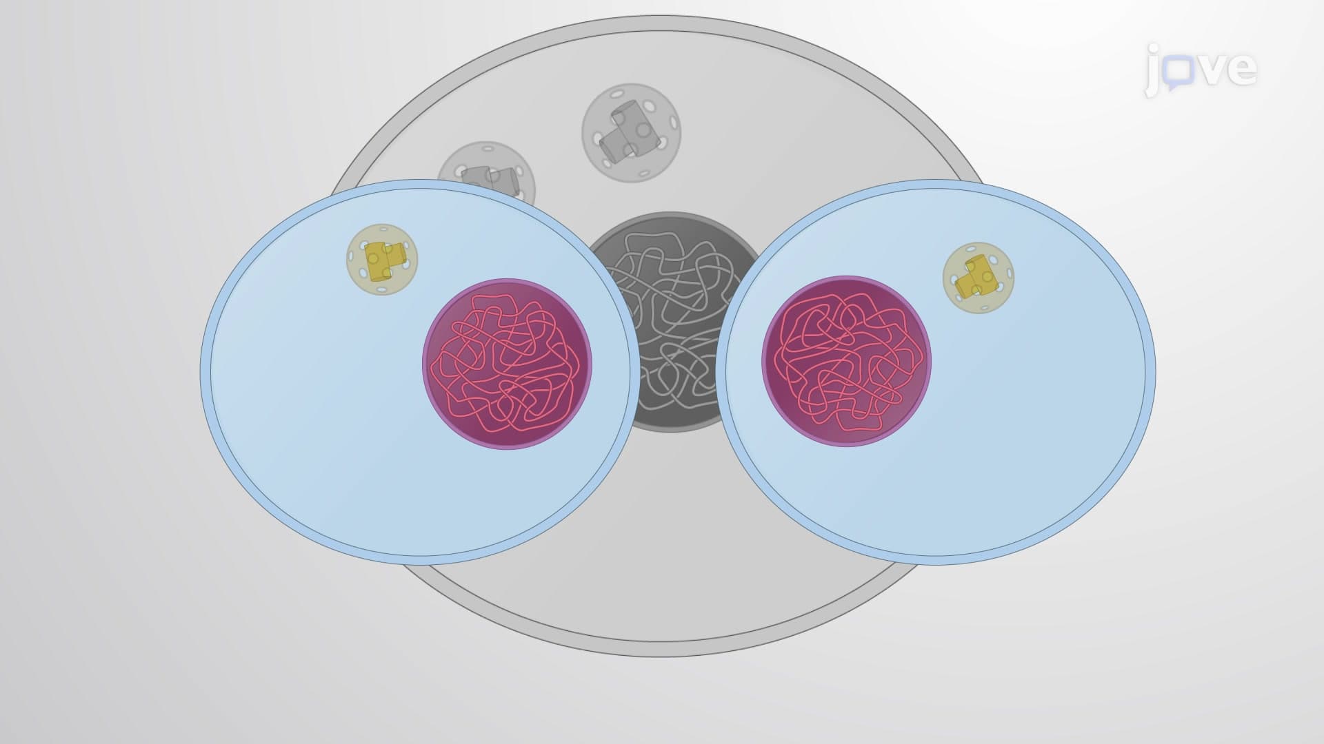
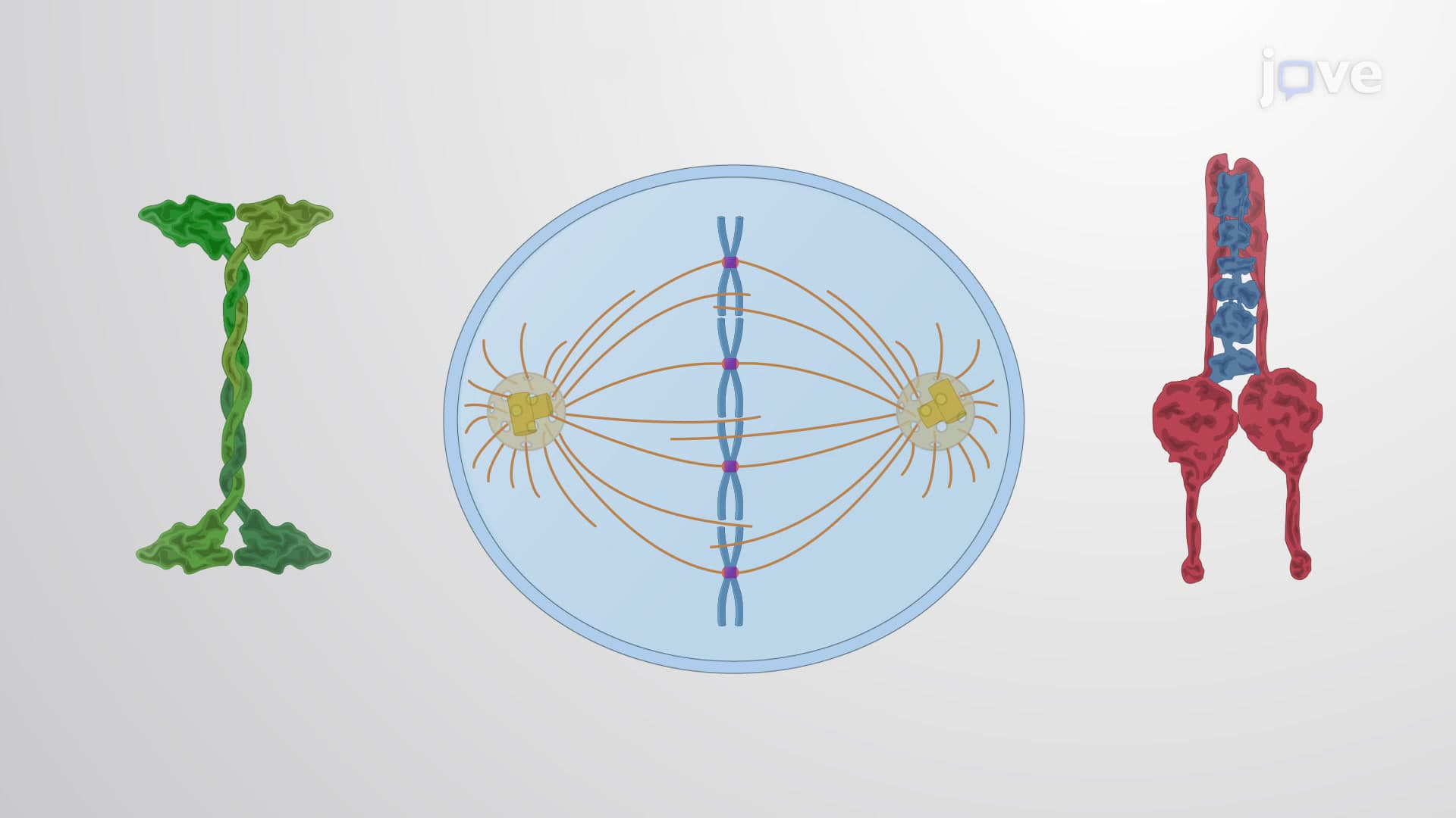
Did you know that every second, millions of cells in your body must flawlessly divide their genetic material to prevent cancer? The separation of sister chromatids is the critical moment during cell division when identical DNA copies are pulled apart to opposite ends of the cell, ensuring each daughter cell receives exactly the right amount of genetic information. When this process fails in human cells, it can lead to conditions like Down syndrome or contribute to tumor formation. Understanding what is separation of sister chromatids reveals how our bodies maintain genetic stability through billions of cell divisions throughout our lifetime. Watch the full video on JoVE Coach to master this concept with expert-led visuals and step-by-step explanations.
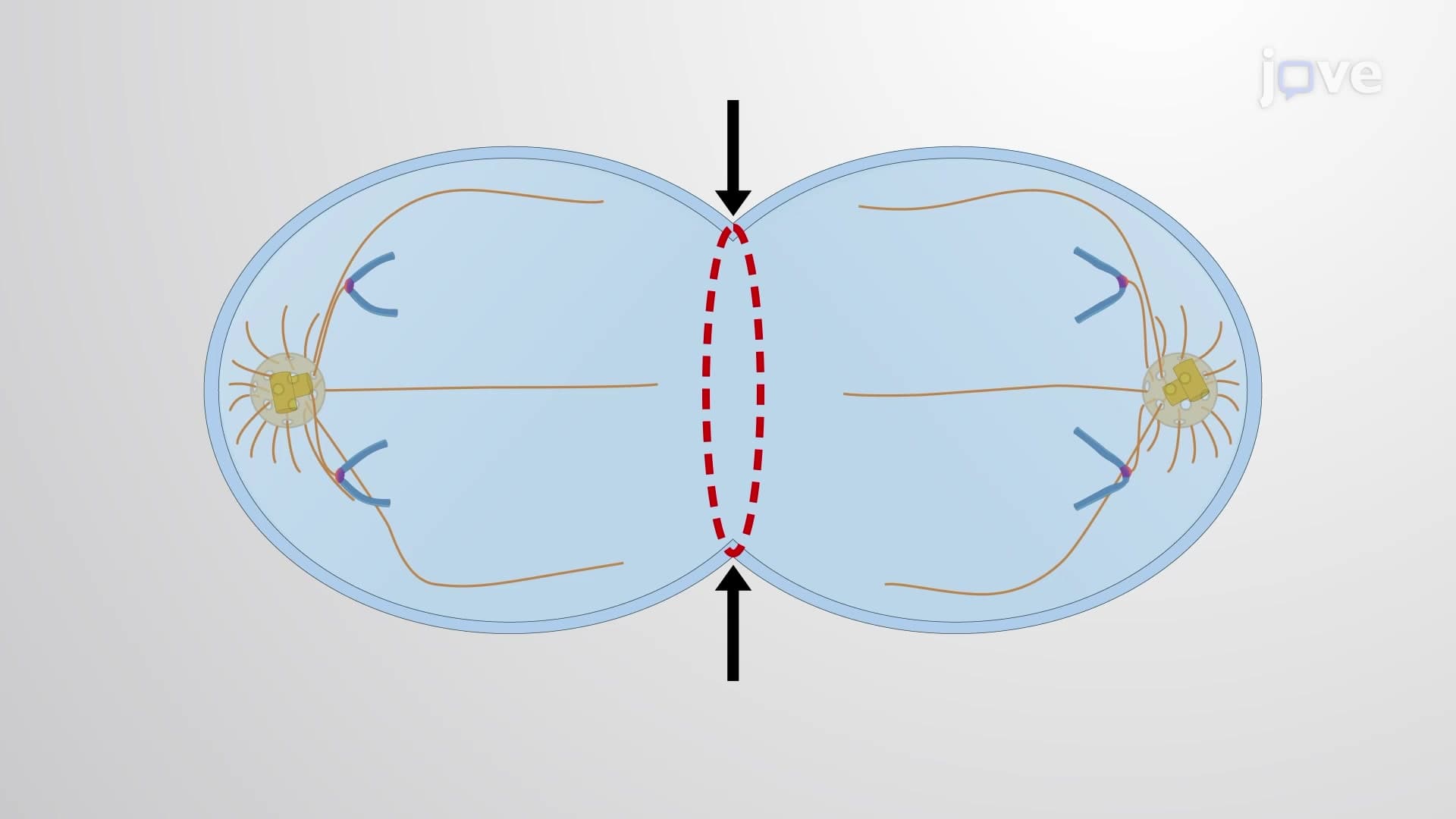
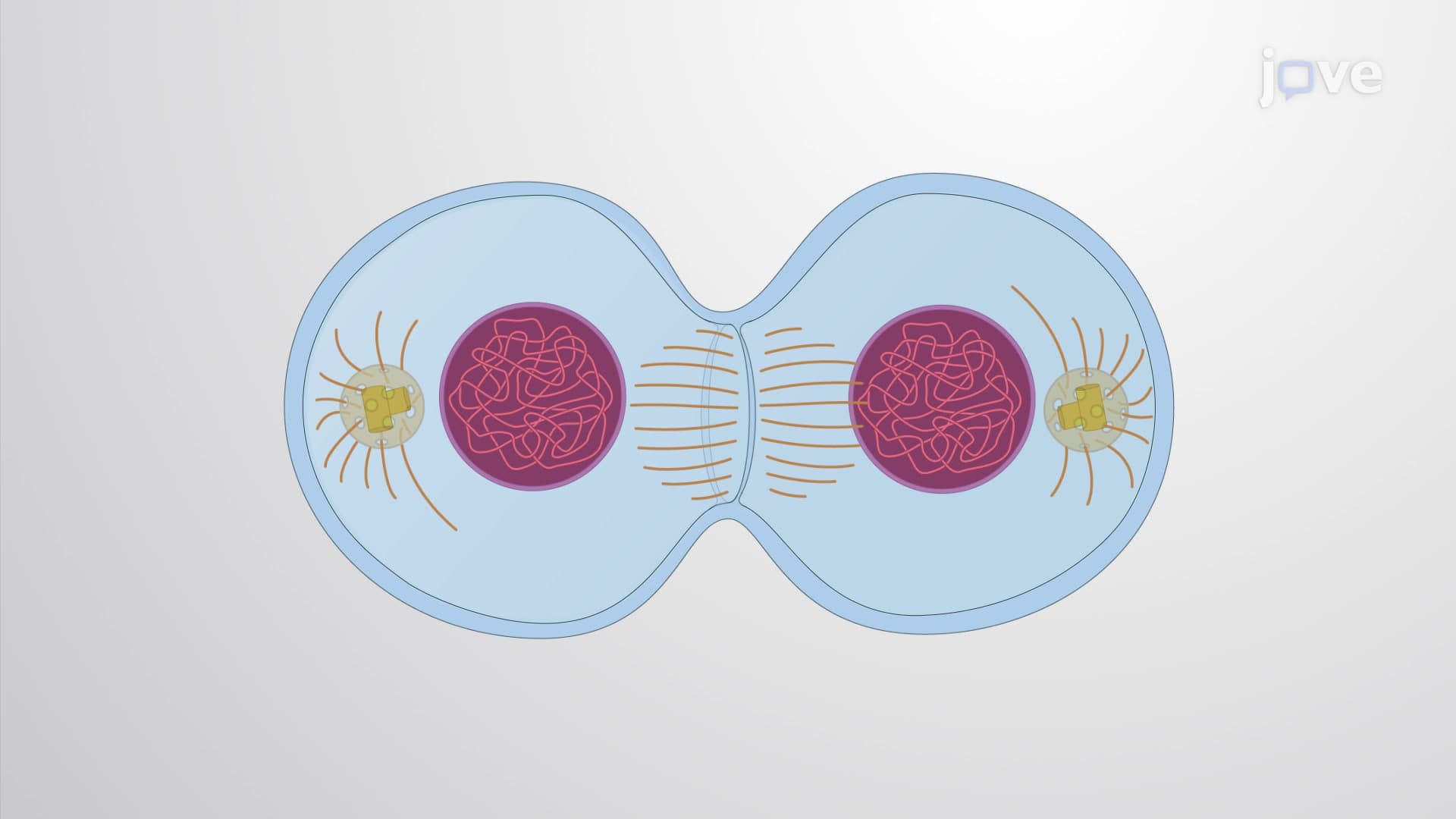
The separation of sister chromatids represents one of the most precisely controlled events in cellular biology, occurring during anaphase of mitosis when identical DNA copies are physically pulled apart to ensure accurate genetic inheritance. This process is fundamental to how multicellular organisms like humans maintain genetic stability across trillions of cell divisions throughout their lifetime.
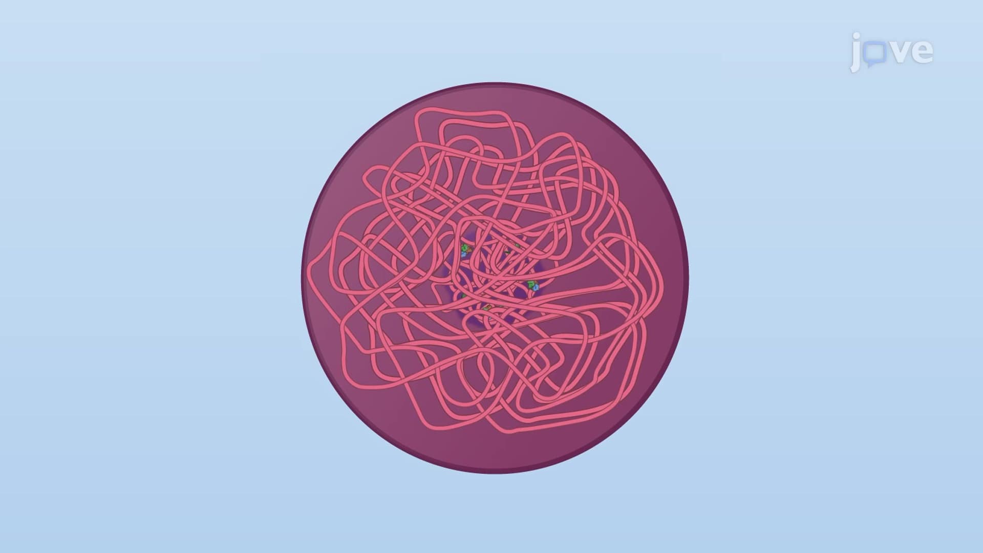
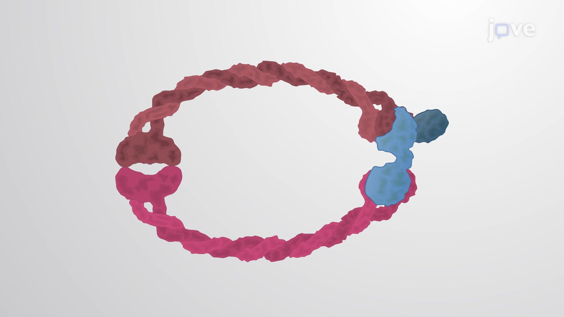
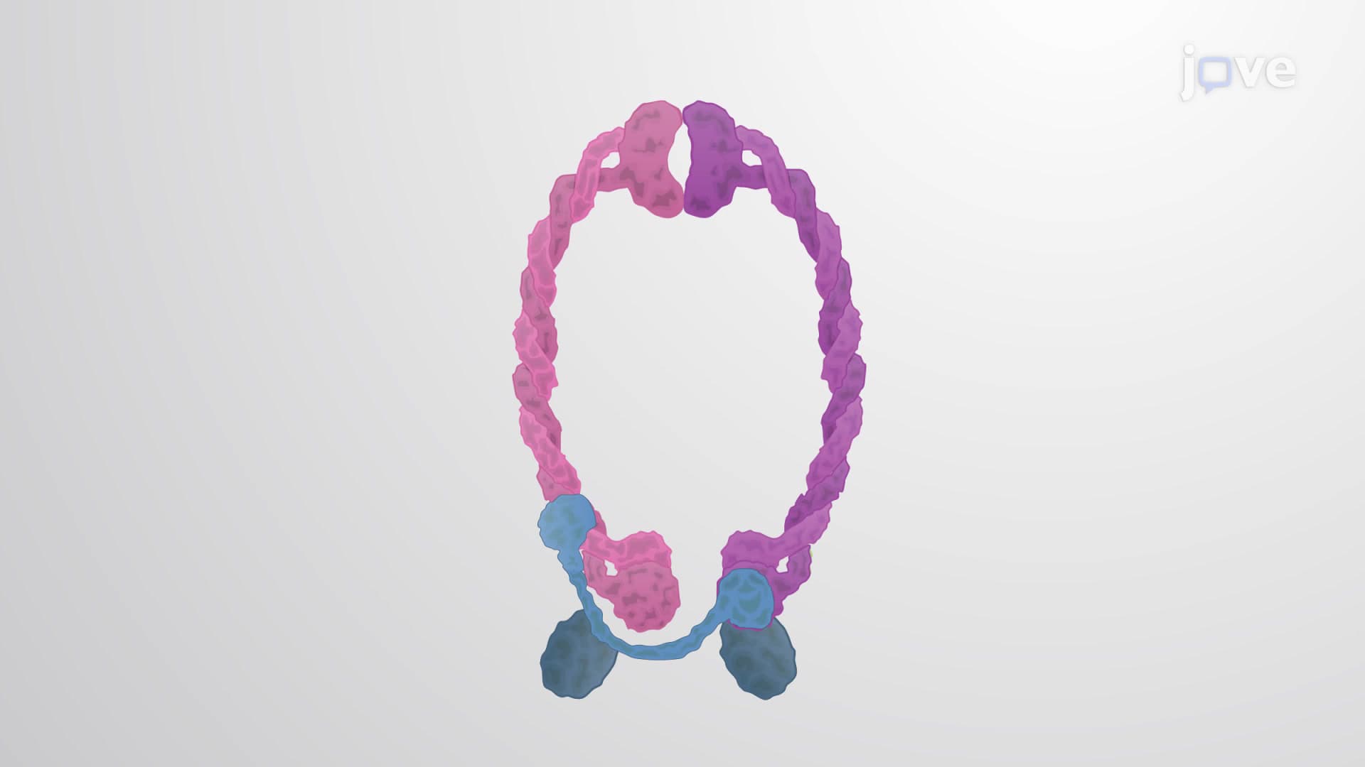
The separation process relies on sophisticated molecular machinery that has been conserved across species. Sister chromatids remain attached through protein complexes called cohesins, which form ring-like structures around the DNA. The key to separation lies in the enzyme separase, a highly regulated protease that cleaves cohesin rings precisely when conditions are optimal for division.
The timing of this cleavage is controlled by the anaphase-promoting complex/cyclosome (APC/C), which activates separase by destroying its inhibitor protein, securin. This regulatory cascade ensures that separation only occurs after all chromosomes have been properly attached to spindle microtubules from opposite poles of the cell - a safety mechanism that prevents catastrophic genetic errors.
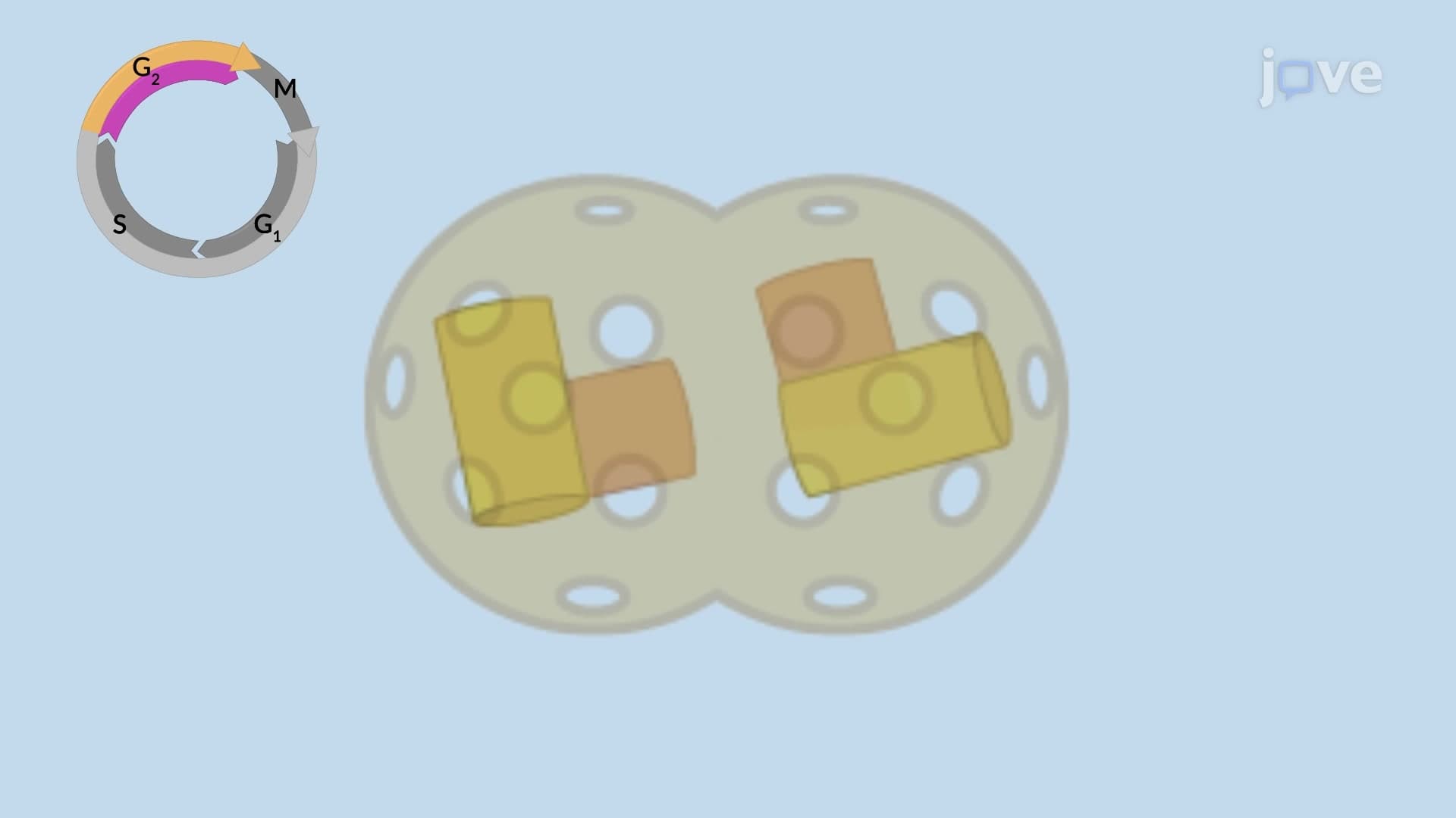
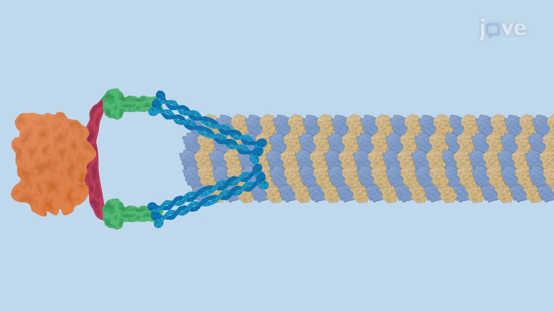
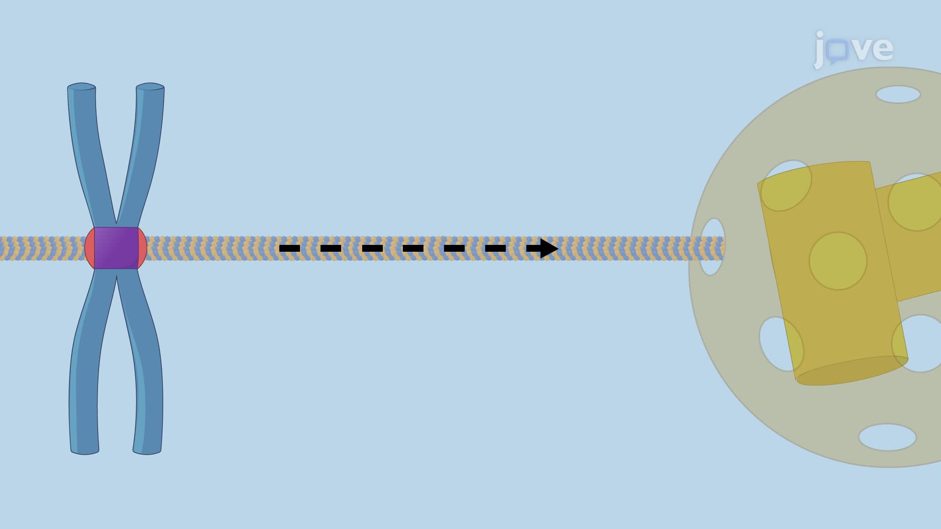
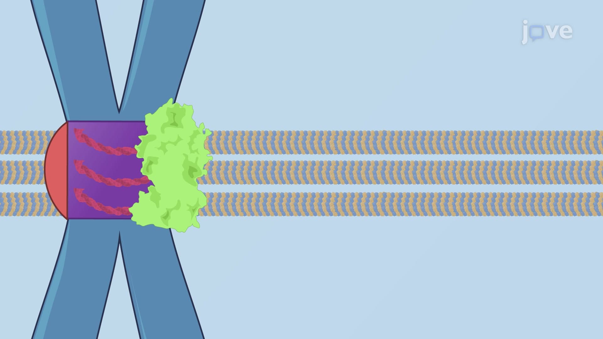
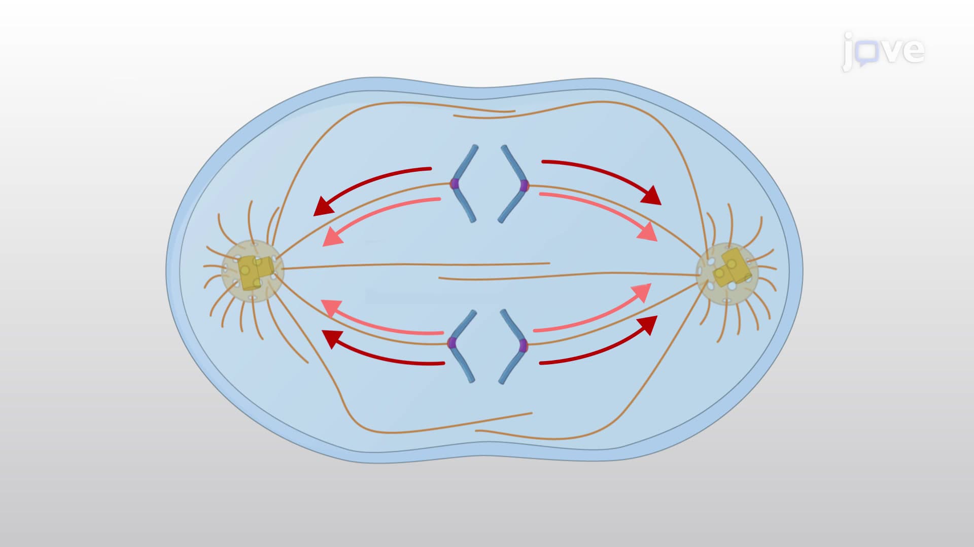
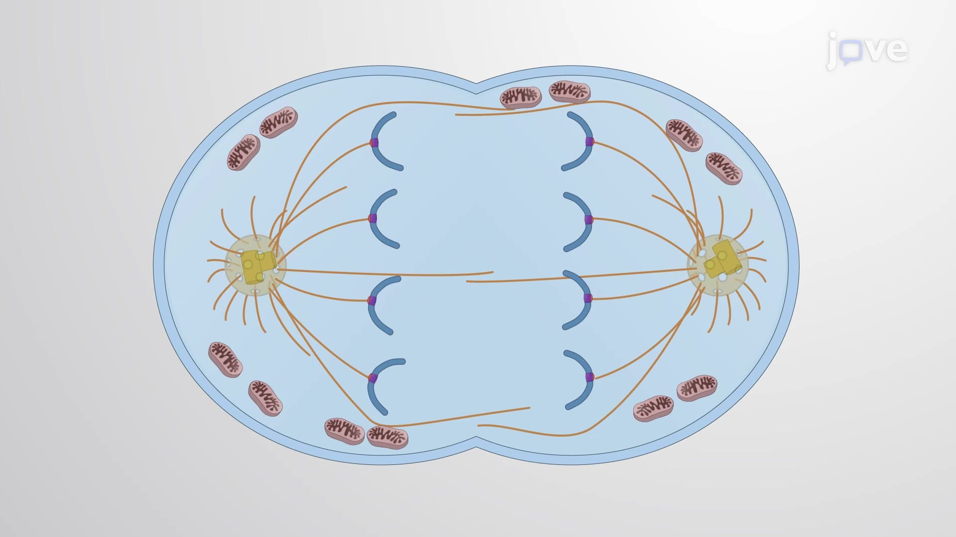
Once cohesins are cleaved, powerful mechanical forces take over to physically separate the chromatids. Kinetochore microtubules, attached to specialized protein structures at chromosome centromeres, generate pulling forces through the action of motor proteins like dynein. Simultaneously, polar microtubules elongate to push the spindle poles apart, creating a coordinated movement that efficiently distributes genetic material.
This process occurs with remarkable precision - in human cells, 46 chromosomes (92 chromatids) must be accurately separated in every division. The forces involved are substantial enough to move chromosomes measuring several micrometers across the cell in just minutes.
Understanding sister chromatid separation has profound implications for human health and medical research. Errors in this process, called nondisjunction, can result in cells with abnormal chromosome numbers (aneuploidy). In reproductive cells, such errors can lead to genetic conditions like Down syndrome, which affects approximately 1 in 700 births in the United States.
In cancer research, defects in separation machinery are increasingly recognized as drivers of tumor development. Many cancer cells show chromosomal instability linked to faulty separation mechanisms, making this pathway a target for therapeutic intervention. Students preparing for the MCAT or AP Biology exams frequently encounter questions about how separation errors contribute to genetic disorders and cancer progression.
The spindle assembly checkpoint, which monitors proper chromosome attachment before allowing separation, represents a critical quality control mechanism that prevents these errors under normal circumstances.
Related Micro-courses