Loading subject page…

- Cell Biology
- Cell Division
- The Mitotic Spindle
Micro-course: Cell Division
37,915 views
Learn key concepts and earn your certificate.
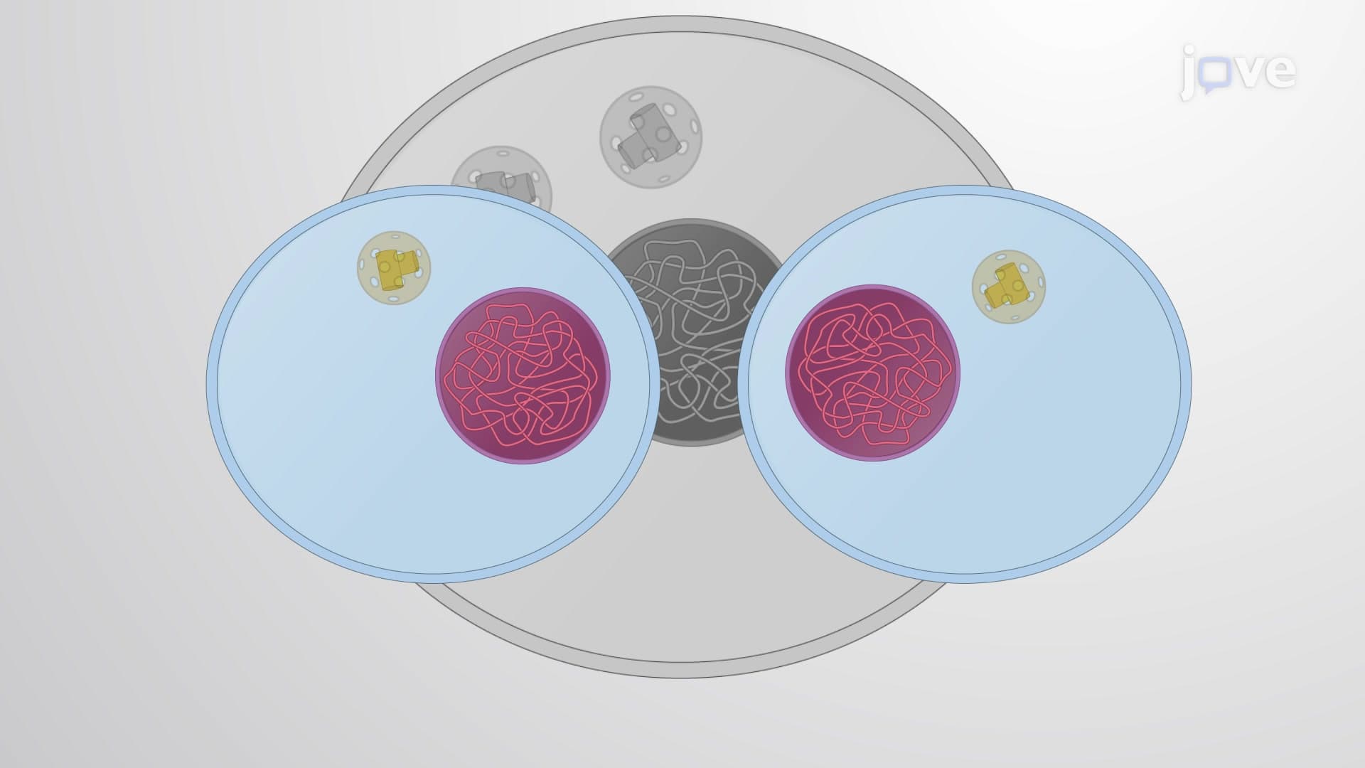

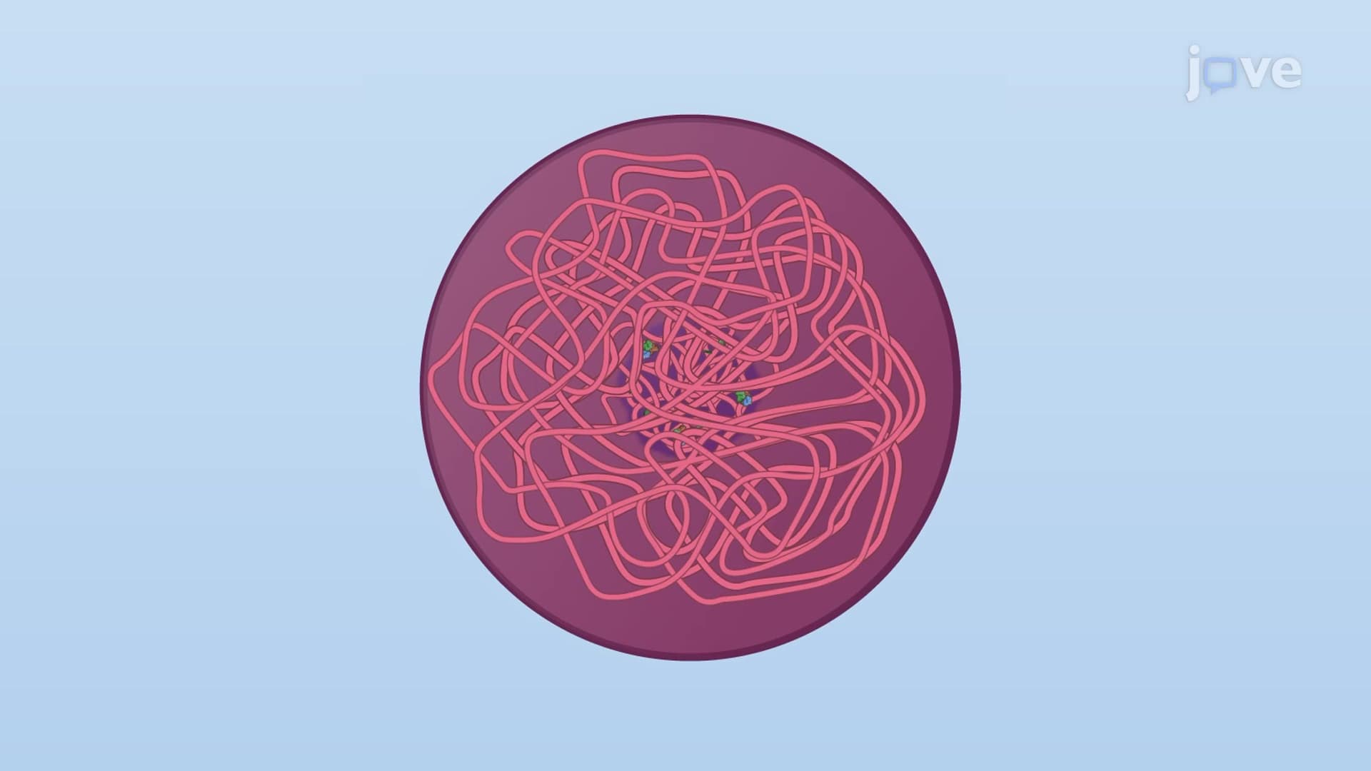
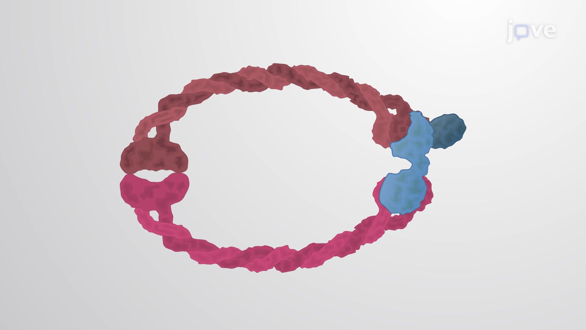
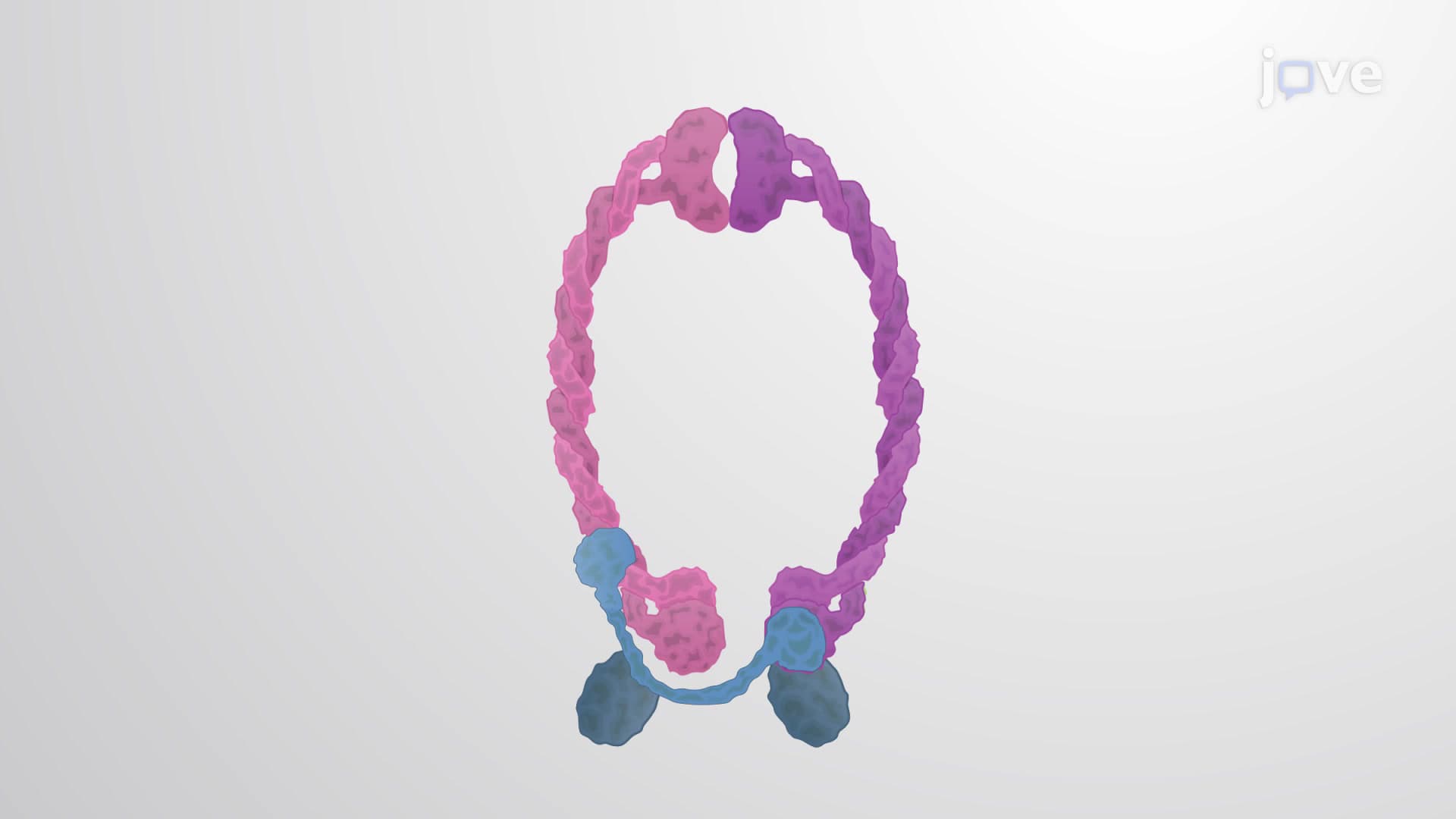



The Mitotic Spindle

00:00
02:27
Video Summary: What Is the Mitotic Spindle
Cancer treatments like Taxol work by targeting the mitotic spindle, the cellular machinery that divides chromosomes during cell division. The mitotic spindle is a dynamic structure made of microtubules and motor proteins that ensures each daughter cell receives an exact copy of genetic material. This process is so critical that errors can lead to cancer or cell death. Watch the full video on JoVE Coach to master this concept with expert-led visuals and step-by-step explanations.
- Understand what is the mitotic spindle and its essential role in chromosome segregation during cell division
- Identify the three main types of microtubules that compose the spindle apparatus and their specific functions
- Analyze how motor proteins like dynein and kinesin coordinate chromosome movement throughout mitosis
- Recognize the spindle checkpoint mechanism that prevents errors in chromosome distribution
- Evaluate how chemotherapy drugs target spindle function to treat cancer patients
- Apply knowledge of spindle dynamics to predict outcomes when cellular components malfunction
- Explore the connection between spindle defects and genetic disorders or cancer development
- Compare normal spindle function with pathological conditions affecting cell division
What is The Mitotic Spindle: Core Structure and Function
The mitotic spindle represents one of biology's most sophisticated molecular machines, orchestrating the precise distribution of genetic material during cell division. This temporary structure emerges during mitosis, transforming the cell into a highly organized system capable of separating duplicated chromosomes with remarkable accuracy. Understanding this process is fundamental for students preparing for AP Biology exams, college cell biology courses, and pre-med requirements like the MCAT.
Architecture of the Spindle Apparatus
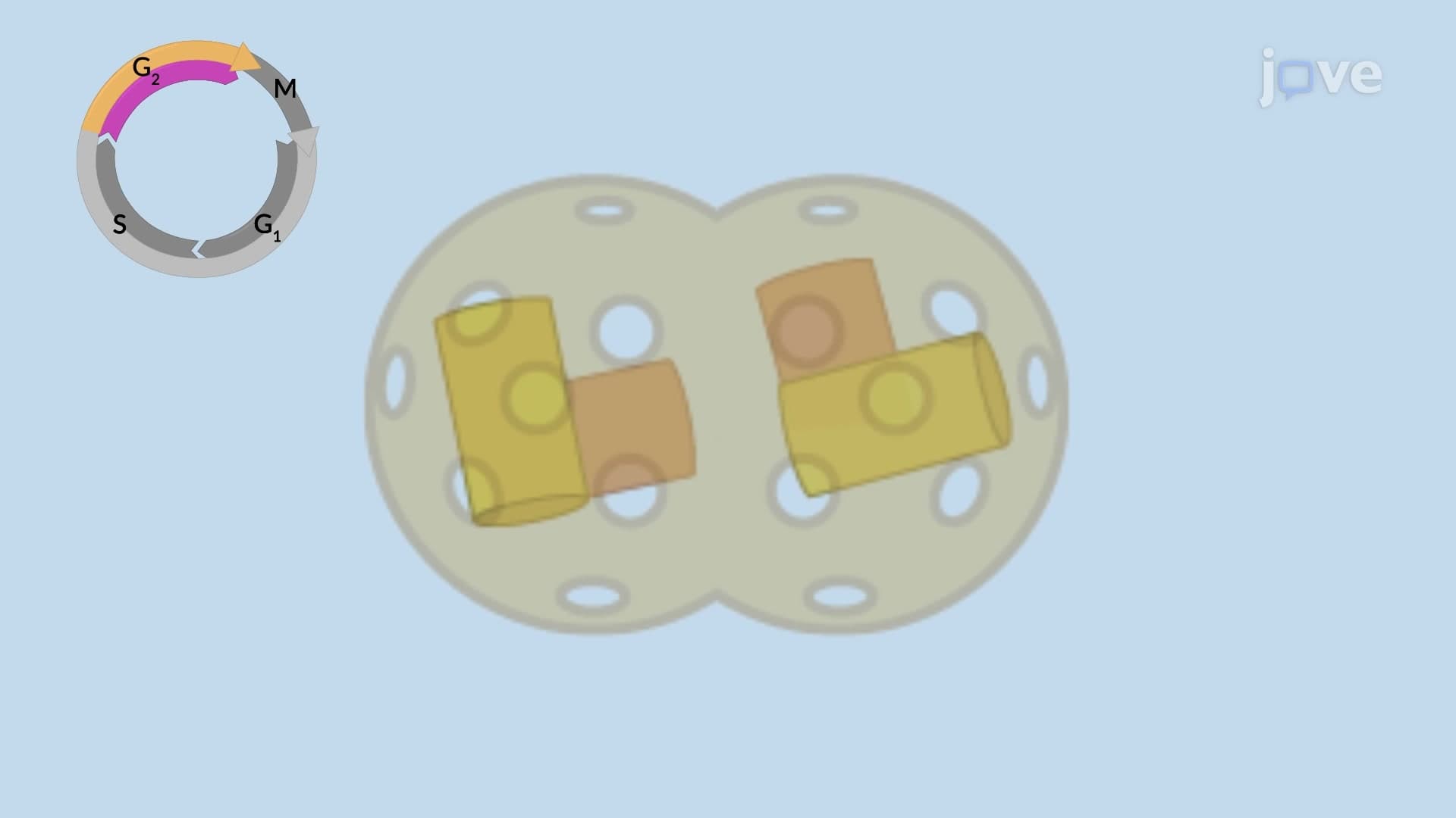
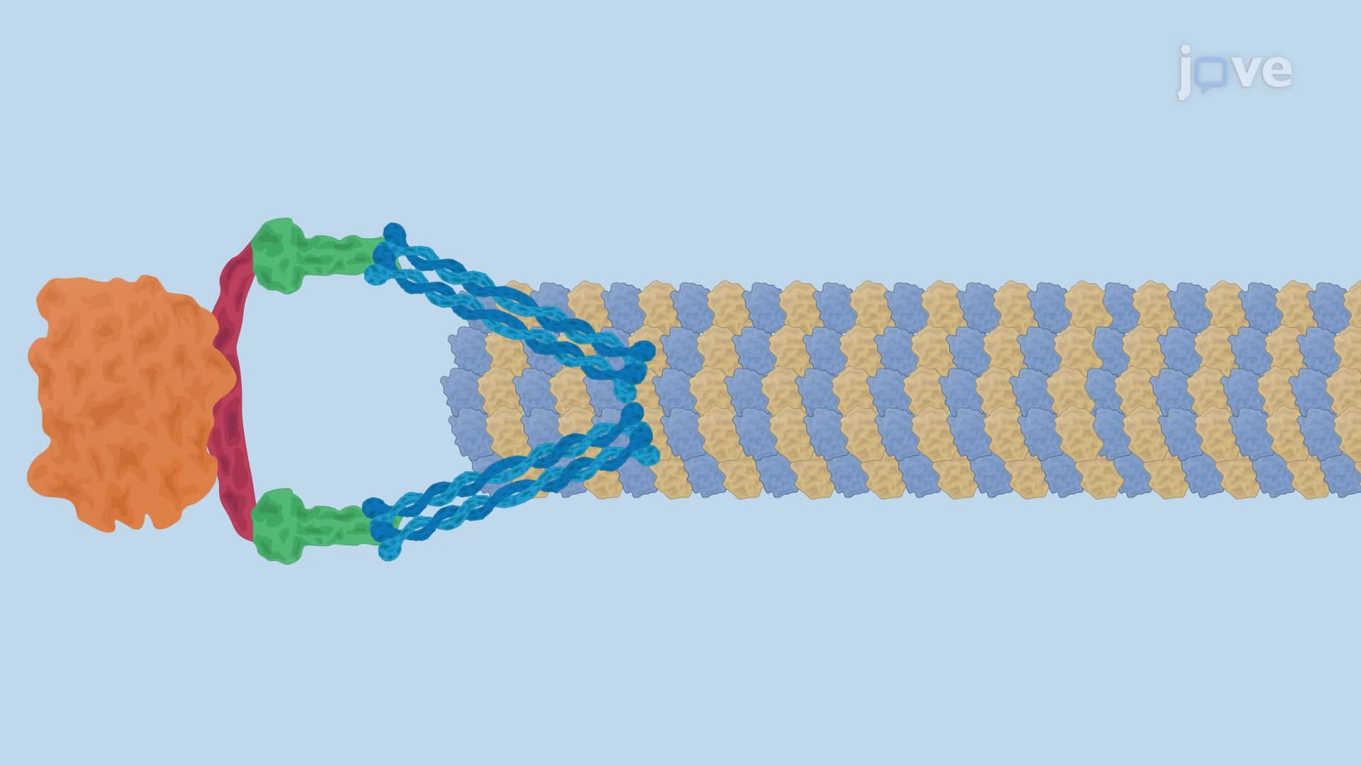
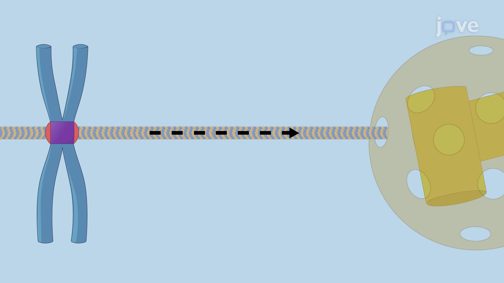
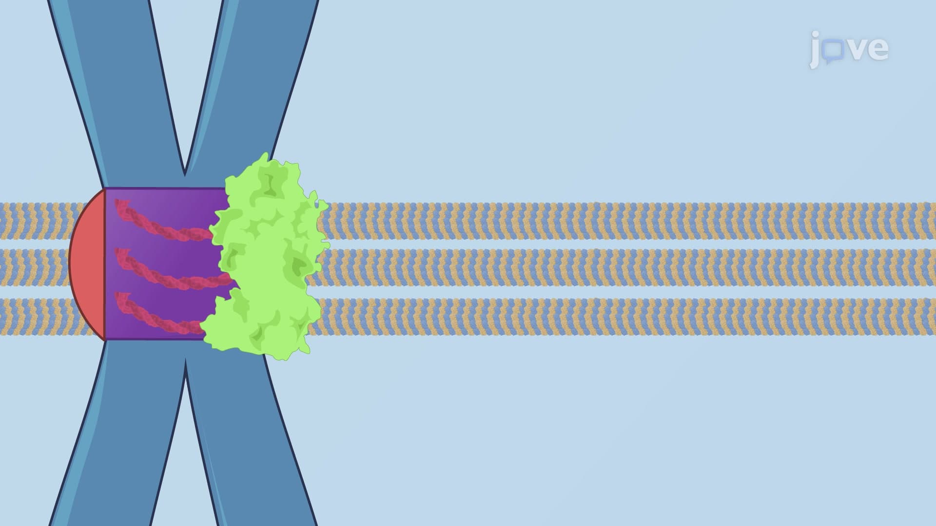
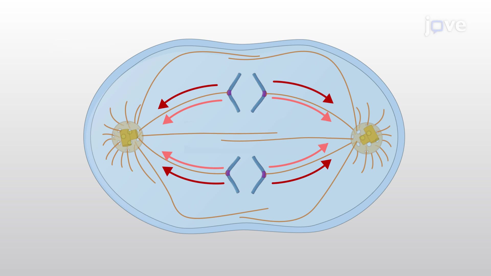
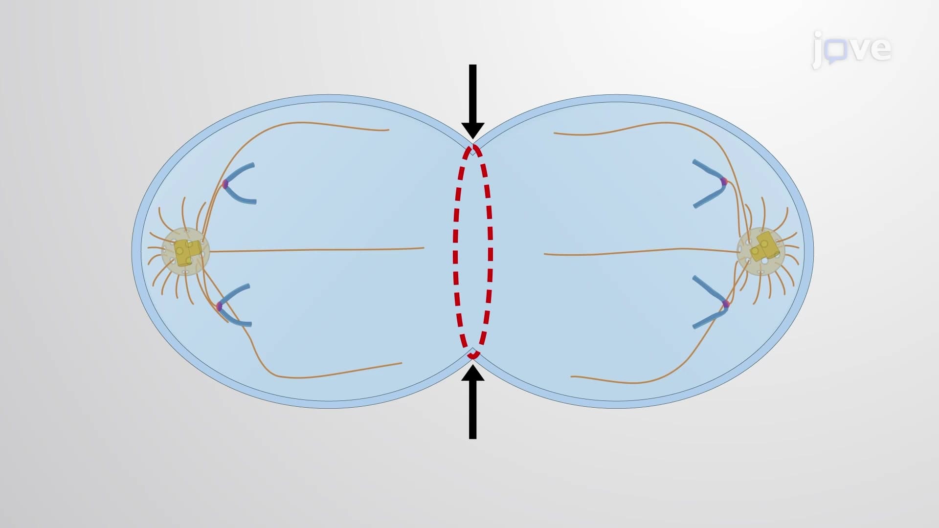
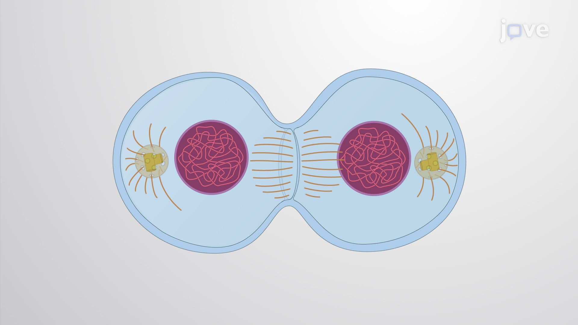
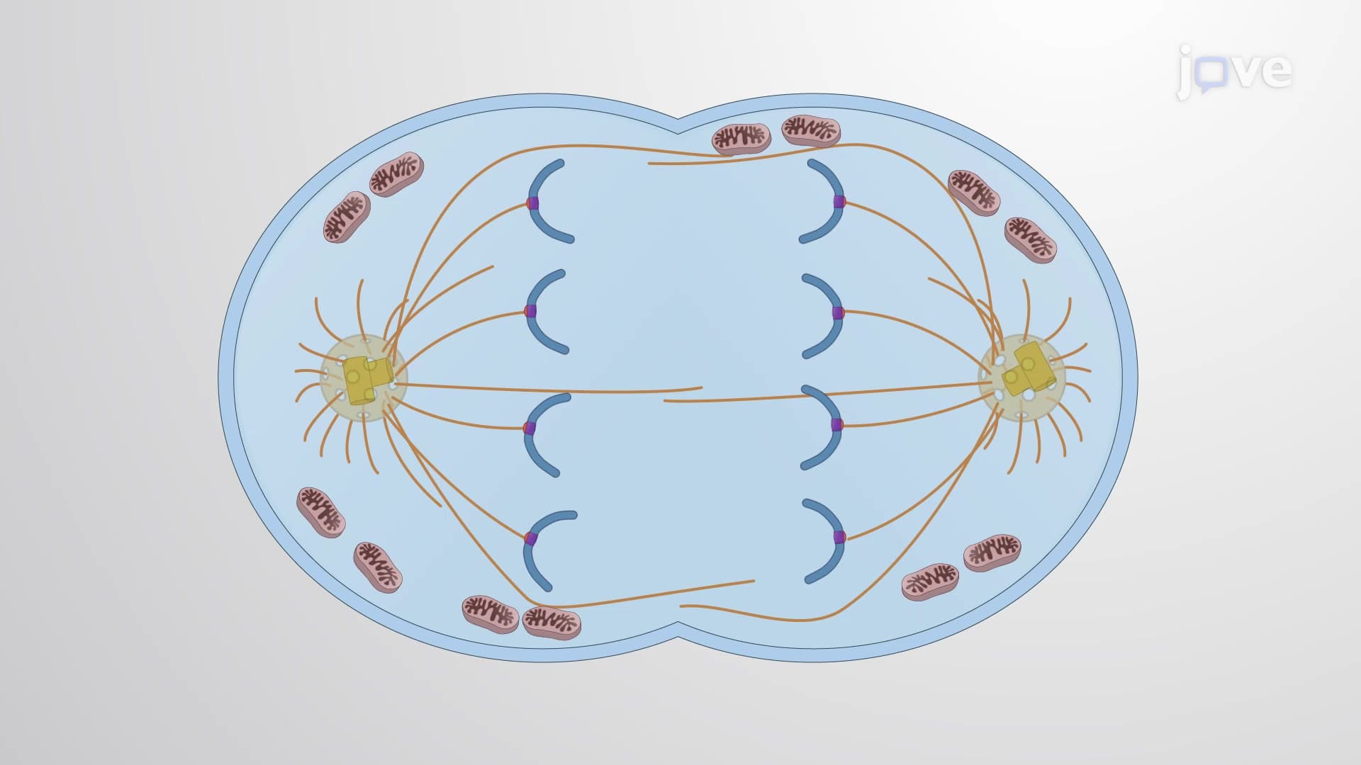
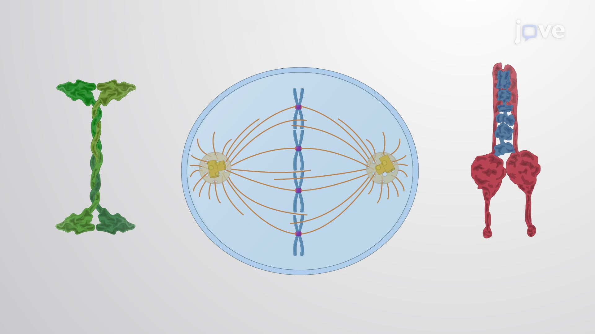
The spindle consists of three distinct microtubule networks, each serving specific functions. Kinetochore microtubules directly attach to chromosomes at specialized protein complexes called kinetochores, acting like molecular ropes that pull sister chromatids apart. Interpolar microtubules extend from opposite spindle poles, overlapping at the cell's center to provide structural stability and help elongate the cell during division. Astral microtubules radiate outward from centrosomes, anchoring the entire apparatus to the cell membrane and positioning the spindle correctly within the cell.
This organization becomes particularly important when studying cancer biology. Researchers at institutions like Johns Hopkins and Memorial Sloan Kettering have shown how disrupting any of these microtubule types can halt cell division, making the spindle a prime target for chemotherapy drugs.
Molecular Motors and Dynamic Assembly
Motor proteins serve as the spindle's "engines," converting chemical energy into mechanical force. Dynein motors primarily drive chromosome movement toward spindle poles, while kinesin motors facilitate various transport functions and contribute to spindle organization. These proteins work in concert with microtubule dynamics-the constant growing and shrinking of microtubule ends-to generate the forces necessary for chromosome separation.
The spindle's assembly begins at centrosomes, cellular structures containing γ-tubulin ring complexes that nucleate new microtubules. This process, studied extensively at research universities like MIT and Stanford, demonstrates how cells can rapidly construct complex molecular machinery from relatively simple components.
Clinical and Research Applications
Beyond its fundamental biological importance, spindle research directly impacts human health. Chemotherapy drugs like paclitaxel (Taxol) stabilize microtubules, preventing their normal dynamics and effectively freezing the spindle in place. Conversely, vinca alkaloids destabilize microtubules, causing spindle collapse. Both approaches prevent cancer cells from completing division, leading to cell death.
Students studying for medical school entrance exams should note how spindle checkpoint defects contribute to chromosomal instability, a hallmark of cancer. This connection between basic cell biology and clinical medicine exemplifies why understanding the mitotic spindle remains essential for future healthcare professionals and researchers.
Frequently Asked Questions
The mitotic spindle is a temporary cellular structure made of protein fibers that separates chromosomes during cell division. Think of it as microscopic machinery that ensures each new cell gets an exact copy of genetic information. It assembles before division begins and disassembles once chromosomes are properly distributed.
The MCAT frequently tests spindle function in cancer biology contexts and drug mechanisms. AP Biology exams often include questions about spindle checkpoint failures and their consequences. Expect diagrams showing spindle organization and questions connecting spindle defects to genetic disorders or cancer development.
Understanding spindle function is fundamental to cell biology, genetics, and cancer research. Most pre-med and biology majors encounter detailed spindle studies in courses like Cell Biology, Genetics, and Molecular Biology. This knowledge directly applies to medical school topics including oncology and developmental biology.
Taxol binds to microtubules and prevents them from disassembling normally, freezing the spindle in place. Cancer cells attempting to divide get stuck in mitosis and eventually die. This targeted approach explains why chemotherapy affects rapidly dividing cells like cancer cells more than normal, slowly dividing cells.
While the spindle involves complex molecular interactions, the basic concept is straightforward-it's cellular machinery for chromosome separation. Students with basic knowledge of cell structure and DNA can grasp spindle function. Visual aids and step-by-step diagrams make the process much more accessible.
Focus on drawing and labeling spindle diagrams repeatedly until you can sketch them from memory. Create flashcards for the three microtubule types and their functions. Practice connecting spindle defects to real diseases, and always relate spindle function back to the fundamental goal of accurate chromosome segregation.
Spindle research directly impacts drug development, as many cancer treatments target this cellular machinery. Students interested in oncology, pharmaceutical research, or biotechnology will encounter spindle biology throughout their careers. Research institutions like the National Cancer Institute actively study spindle mechanisms for therapeutic applications.
Explore cell cycle checkpoints and their regulation, particularly the spindle checkpoint mechanism. Study cancer biology to understand how spindle defects contribute to tumor development. Advanced topics include motor protein biochemistry and microtubule-targeting drug development for deeper molecular understanding.
Related Micro-courses


