Loading subject page…

- Cell Biology
- Cell Proliferation
- M-Cdk Drives Transition Into Mitosis
Micro-course: Cell Proliferation
51,501 views
Learn key concepts and earn your certificate.

M-Cdk Drives Transition Into Mitosis

00:00
02:15
Video Summary: M Cdk Drives Transition Into Mitosis Explained
Cancer cells divide uncontrollably because they've lost the ability to properly regulate when mitosis begins-a process where M Cdk drives transition into mitosis through precise molecular timing. The cyclin B-CDK1 complex acts like a molecular switch, phosphorylating hundreds of proteins to trigger chromosome condensation and nuclear envelope breakdown. At the National Cancer Institute, researchers study how disrupted M-Cdk regulation leads to the rapid cell division seen in aggressive tumors like glioblastoma. Watch the full video on JoVE Coach to master this concept with expert-led visuals and step-by-step explanations.
- Understand how M Cdk drives transition into mitosis by phosphorylating key substrate proteins that initiate chromosomal and structural changes
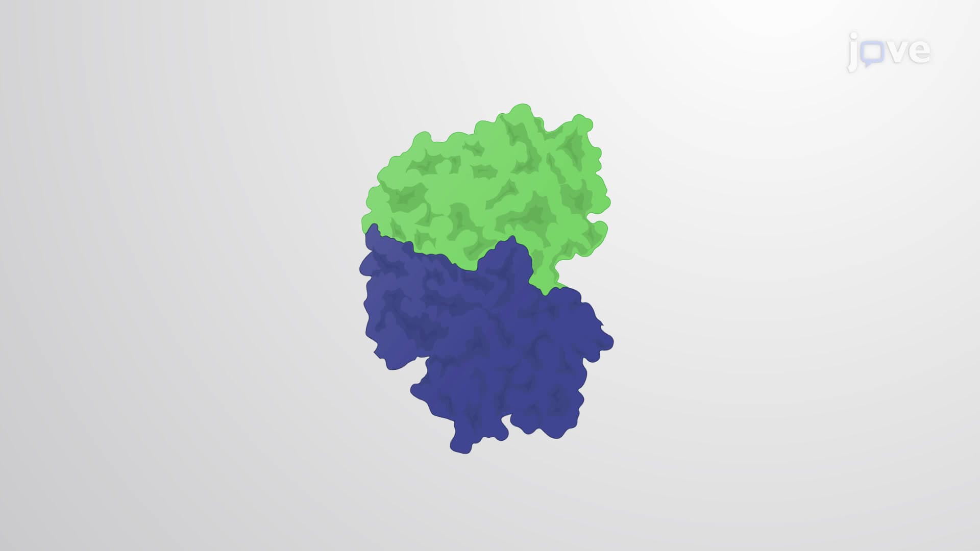
- Identify the cyclin B-CDK1 complex as the primary M-Cdk that controls mitotic entry and regulates timing of cell division
- Analyze the regulatory mechanisms including Cdc25 phosphatases and Wee1 kinases that control M-Cdk activation and prevent premature mitosis
- Evaluate how the spindle assembly checkpoint works with M-Cdk to ensure proper chromosome attachment before division proceeds
- Apply knowledge of M-Cdk dysfunction to explain how cancer cells bypass normal cell cycle controls
- Recognize the clinical significance of M-Cdk inhibitors as potential cancer therapeutics in current drug development
- Compare the molecular events of M-Cdk activation with other cell cycle checkpoints to understand integrated cell cycle control
- Explore how aberrant M-Cdk activity leads to aneuploidy and chromosomal instability in disease states
Understanding How M Cdk Drives Transition Into Mitosis
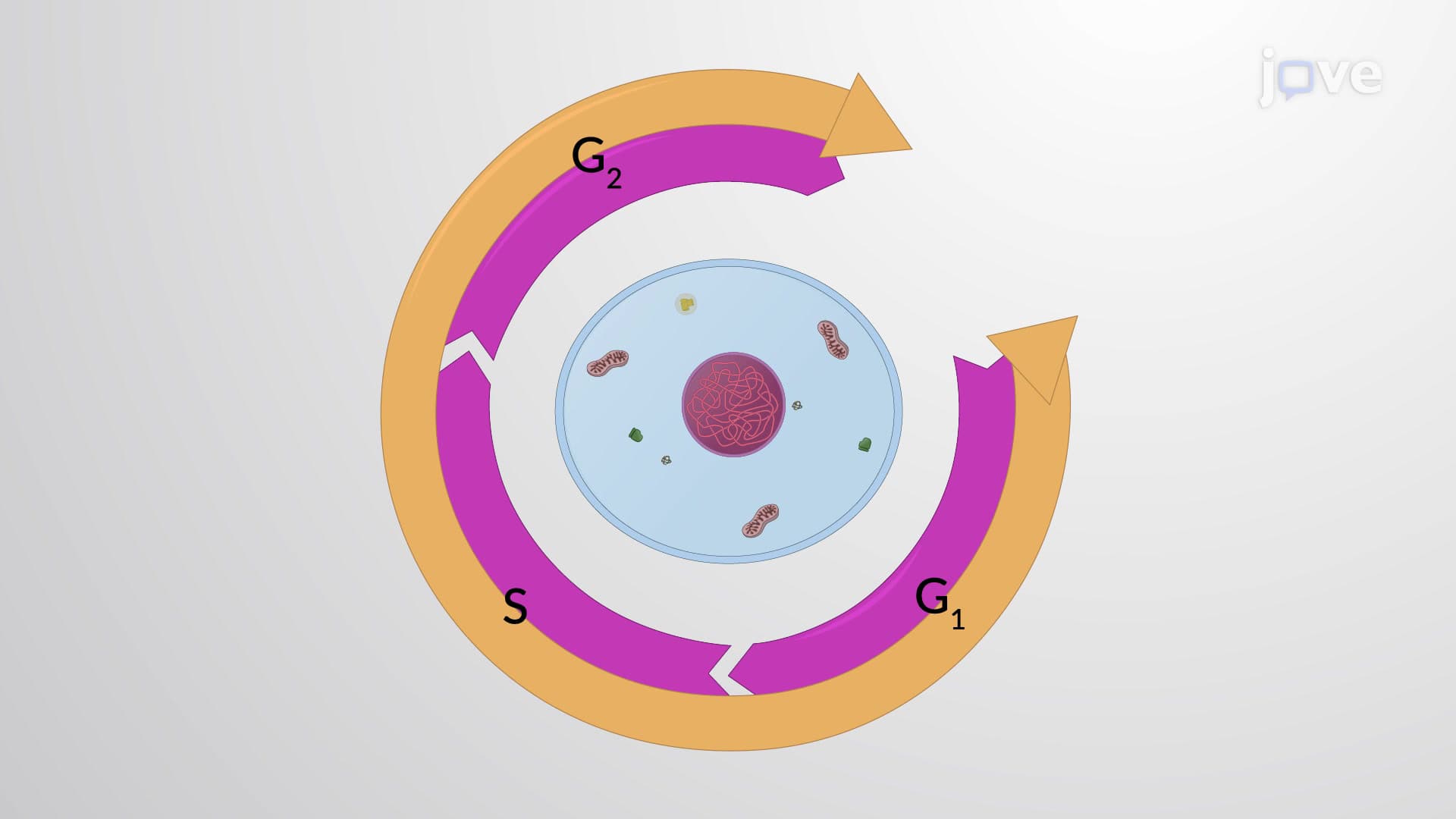
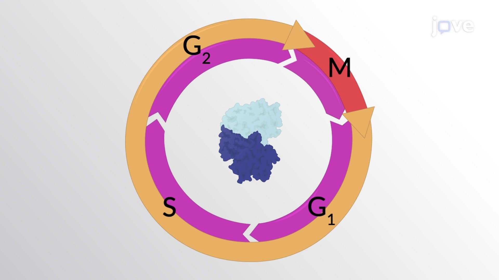
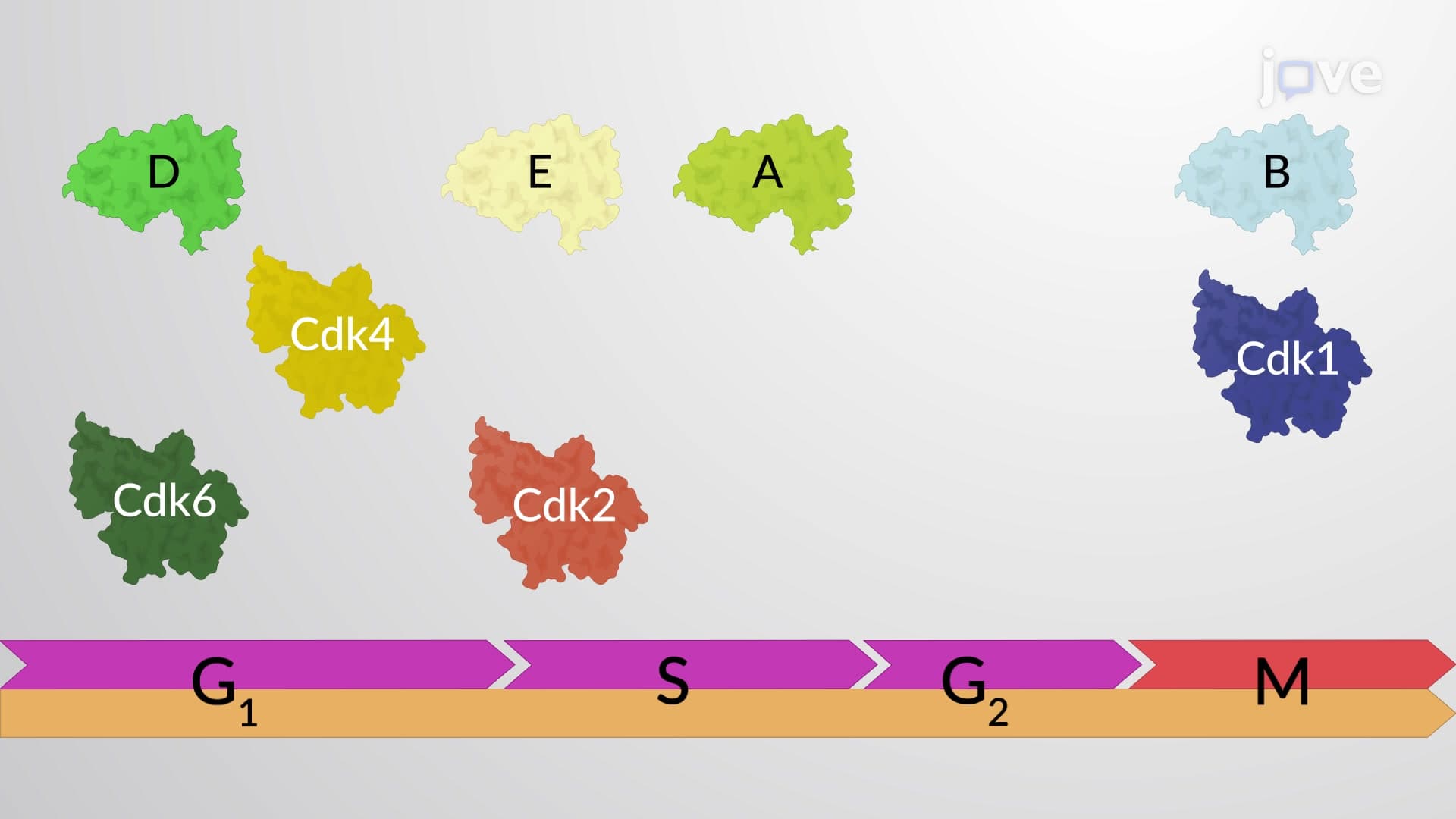
The transition from G2 phase into mitosis represents one of the most dramatic transformations in cell biology. Within minutes, a cell reorganizes its entire architecture-condensing DNA into visible chromosomes, dismantling the nuclear envelope, and assembling the mitotic spindle. This remarkable coordination depends on M-Cdk (mitotic cyclin-dependent kinase), primarily the cyclin B-CDK1 complex, which acts as the master regulator orchestrating mitotic entry.
Molecular Mechanism of M-Cdk Activation
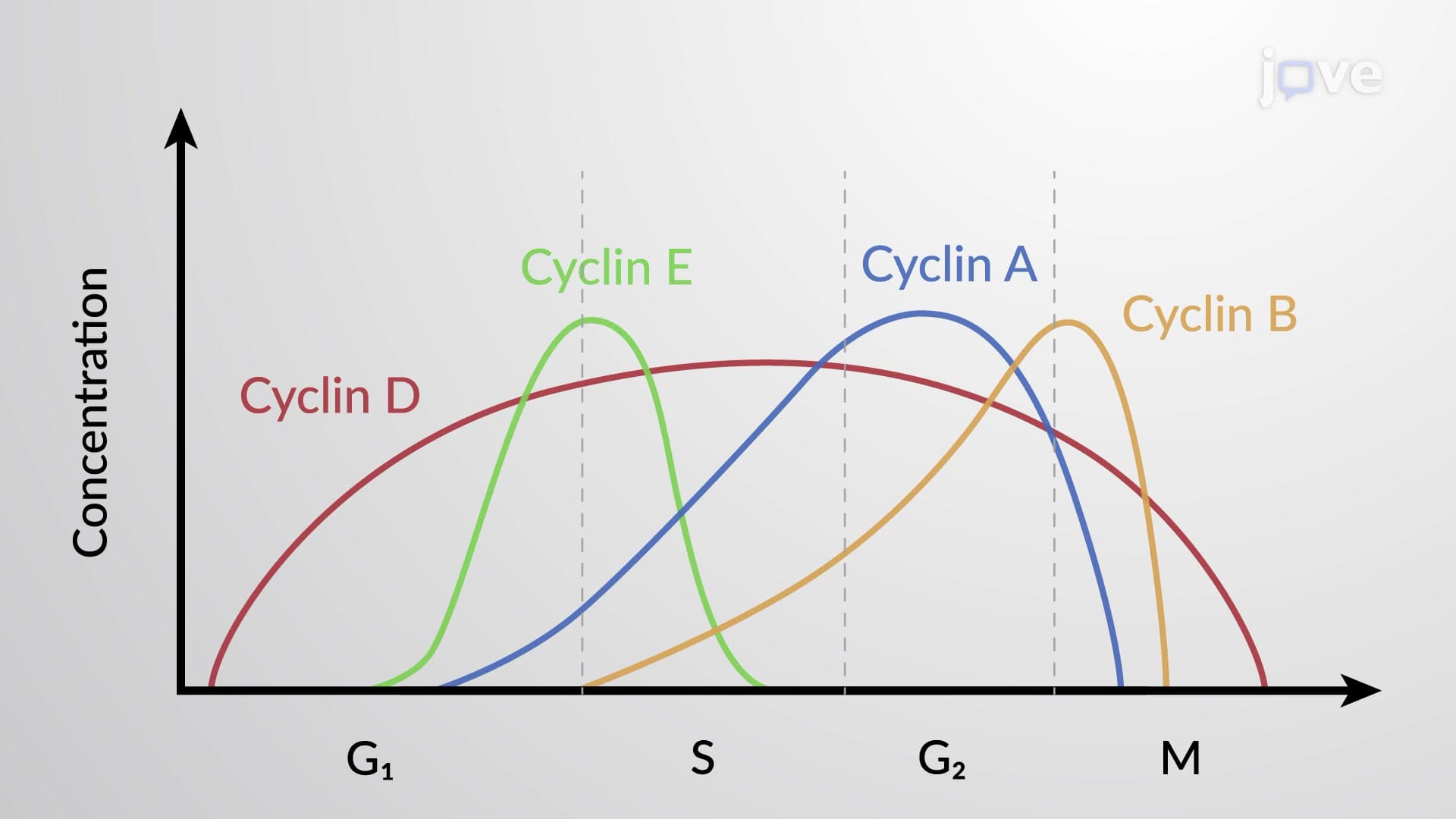
M-Cdk activation follows a precisely timed molecular cascade. During S and G2 phases, CDK1 protein accumulates but remains inactive due to inhibitory phosphorylation by Wee1 kinase at threonine 14 and tyrosine 15. Simultaneously, cyclin B protein levels rise throughout G2 phase, forming inactive cyclin B-CDK1 complexes. The critical switch occurs when Cdc25C phosphatase removes these inhibitory phosphate groups, while additional phosphorylation at threonine 161 fully activates the kinase. This creates a positive feedback loop-active M-Cdk phosphorylates and activates more Cdc25C while simultaneously phosphorylating and inactivating Wee1.
Substrate Phosphorylation and Mitotic Events
Once activated, M-Cdk phosphorylates over 1,000 substrate proteins, triggering the hallmark events of mitotic entry. Phosphorylation of histone H3 at serine 10 promotes chromatin condensation, making chromosomes visible under microscopy. Nuclear envelope breakdown results from M-Cdk phosphorylation of nuclear lamins, causing the nuclear membrane to fragment into vesicles. Centrosome maturation and spindle formation occur through phosphorylation of proteins like NUMA and TPX2, while Golgi apparatus fragmentation ensures equal organelle distribution to daughter cells.
Clinical Significance and Therapeutic Applications
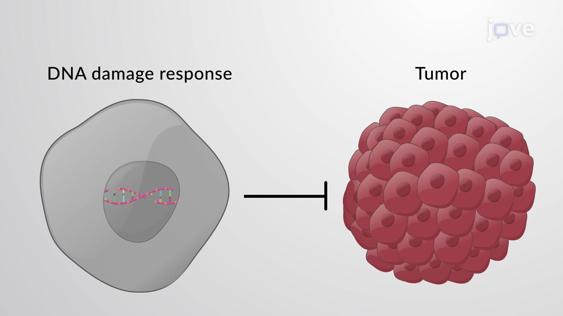
Understanding M-Cdk regulation has profound implications for cancer treatment. Many cancer cells exhibit overexpression of cyclin B or loss of checkpoint controls, leading to premature or uncontrolled mitotic entry. The MD Anderson Cancer Center has pioneered research into CDK1 inhibitors like RO-3306 and purvalanol A as potential chemotherapeutics. These drugs can arrest cancer cells in G2/M phase, preventing tumor progression. Additionally, the spindle assembly checkpoint, which monitors chromosome-spindle attachment before allowing anaphase, often becomes compromised in cancer, leading to chromosomal instability.
For students preparing for AP Biology or college-level cell biology courses, understanding M-Cdk function is essential for explaining how cells maintain genomic stability while allowing necessary cell division during development and tissue repair.
Frequently Asked Questions
M Cdk (mitotic cyclin-dependent kinase) is primarily the cyclin B-CDK1 protein complex that serves as the master switch triggering mitosis. When activated, it phosphorylates hundreds of substrate proteins simultaneously, causing chromosome condensation, nuclear envelope breakdown, and spindle formation. This coordinated phosphorylation transforms an interphase cell into a mitotic cell within minutes, ensuring proper timing of cell division.
The MCAT frequently tests M Cdk through cell cycle regulation passages, asking students to analyze graphs showing cyclin levels or predict effects of kinase inhibitors. AP Biology questions often focus on connecting M-Cdk function to cancer biology or chromosome behavior during mitosis. Students should understand both the molecular mechanism and the regulatory checkpoints that control M-Cdk activation timing.
Premature M-Cdk activation leads to mitotic catastrophe, where cells enter mitosis with incompletely replicated DNA, causing chromosome breaks and cell death. Prolonged M-Cdk activity prevents cells from exiting mitosis, blocking cytokinesis and creating multinucleated cells. Both scenarios contribute to genomic instability and can promote cancer development, which is why cells have multiple checkpoint mechanisms controlling M-Cdk timing.
At institutions like Memorial Sloan Kettering Cancer Center, researchers exploit cancer cells' dependence on rapid cell division by targeting M-Cdk pathways. CDK1 inhibitors can selectively kill cancer cells by forcing them into prolonged mitotic arrest, triggering apoptosis. Additionally, many chemotherapy drugs like taxanes work by disrupting the spindle checkpoint that normally works alongside M-Cdk, preventing proper chromosome separation.
No advanced biochemistry is required-understanding M-Cdk builds on basic concepts of protein phosphorylation and enzyme regulation covered in introductory biology. The key is recognizing that phosphorylation acts like molecular switches, turning proteins on or off. Focus on the cause-and-effect relationships: rising cyclin B levels plus Cdc25 activation leads to M-Cdk activation, which phosphorylates substrates to trigger visible mitotic changes.
Create a timeline diagram showing G2/M transition events, starting with cyclin B accumulation and ending with chromosome condensation. Practice drawing the regulatory circuit showing Wee1 (inhibitory) and Cdc25 (activating) effects on CDK1, including the positive feedback loops. Connect each molecular event to visible cellular changes, and work through practice problems involving cell cycle mutants or drug treatments affecting M-Cdk.
M-Cdk knowledge directly connects to p53 tumor suppressor pathways, DNA damage checkpoints, and apoptosis mechanisms. It also links to meiotic cell division, where similar but distinct CDK complexes control chromosome separation during gamete formation. Understanding M-Cdk provides foundation for studying stem cell biology, where cell cycle control determines when stem cells divide versus differentiate into specialized cell types.
Related Micro-courses


