Loading subject page…


167,928 views

Video Summary: What are Mechanisms of Membrane Bending
Ever wonder how your muscle cells contract or how white blood cells squeeze through tiny blood vessels? The mechanisms of membrane bending make these incredible cellular feats possible. From the formation of synaptic vesicles in neurons at Harvard Medical School research labs to the development of new drug delivery systems by pharmaceutical companies like Pfizer, understanding what are mechanisms of membrane bending is crucial for advancing medicine. These four key mechanisms-lipid-induced bending, protein scaffolding, hydrophobic insertion, and cytoskeletal forces-control how cell membranes change shape during vital processes. Watch the full video on JoVE Coach to master this concept with expert-led visuals and step-by-step explanations.
Cell membranes are dynamic structures that constantly change shape during essential biological processes. The mechanisms of membrane bending govern everything from how neurons communicate at synapses to how immune cells engulf pathogens. Understanding these mechanisms is crucial for students preparing for advanced coursework in cell biology, biochemistry, and pre-medical studies.
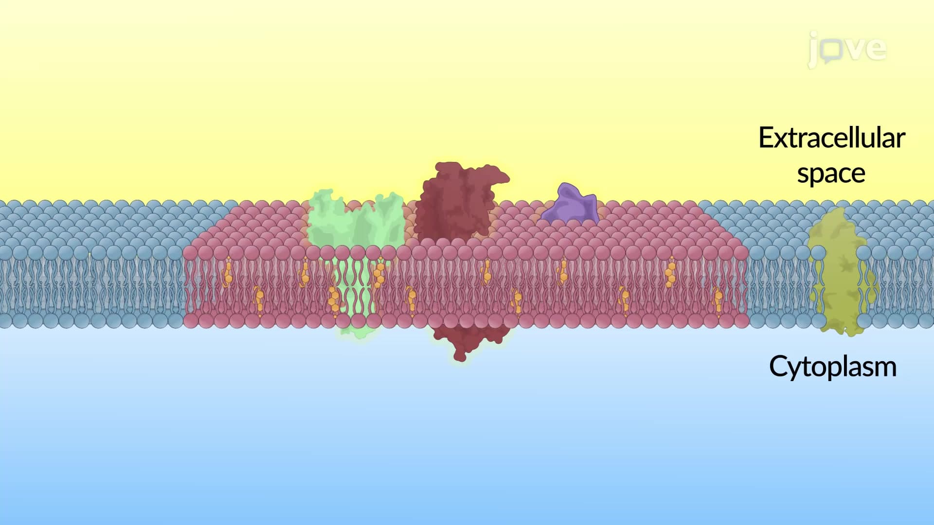
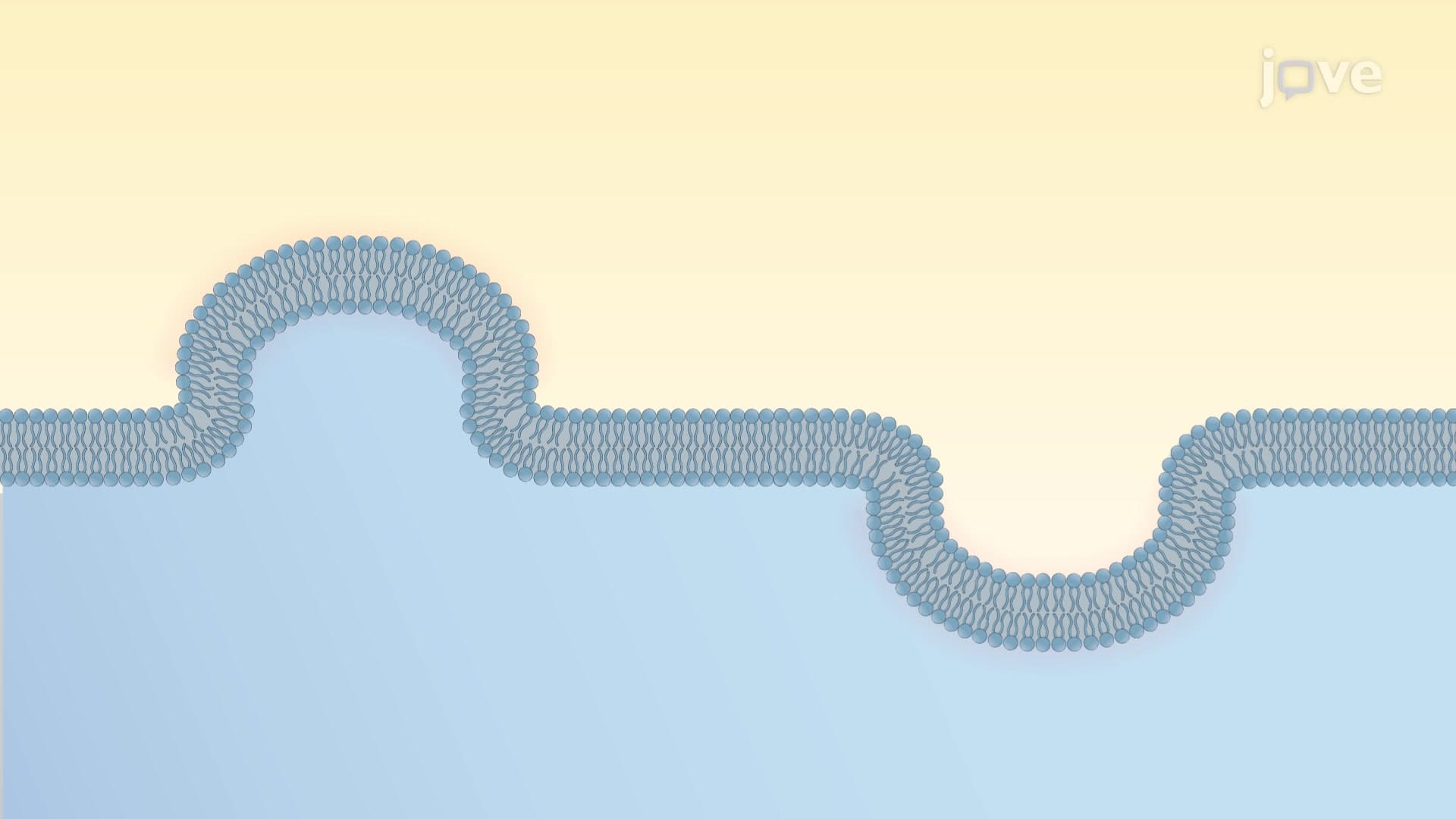
Membrane curvature direction provides the starting point for analysis. Positive curvature occurs when membranes bend toward the cytoplasmic (inner) side, while negative curvature involves bending toward the extracellular (outer) environment. This distinction becomes critical when analyzing processes like endocytosis versus exocytosis in AP Biology coursework.
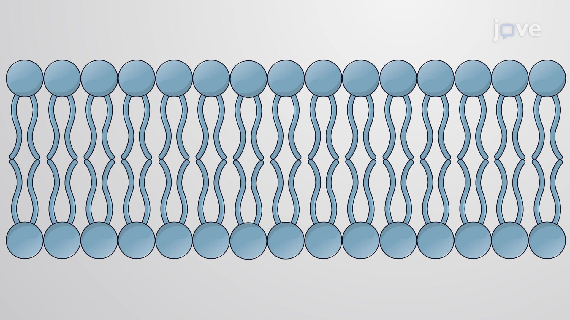
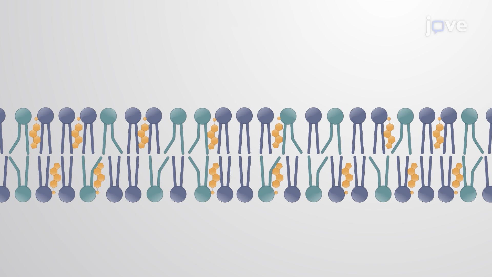
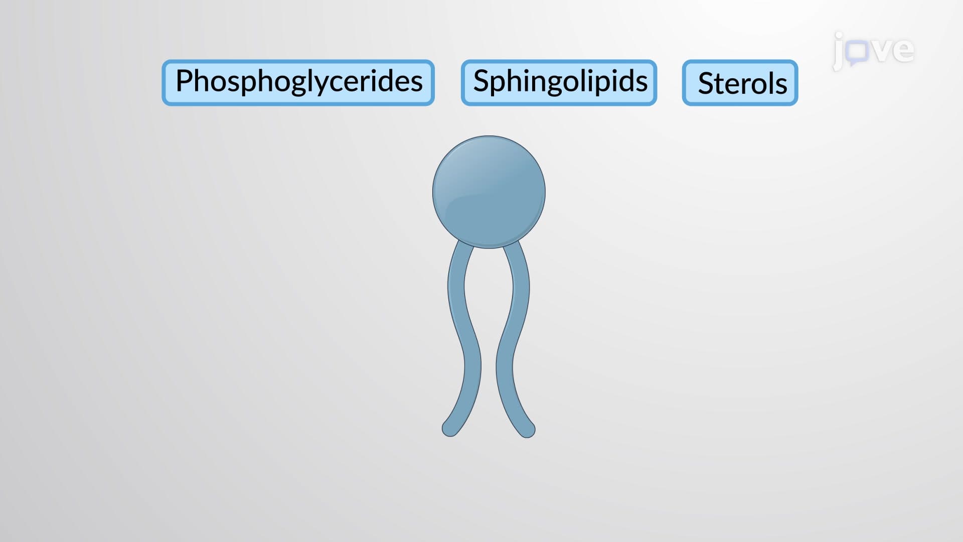
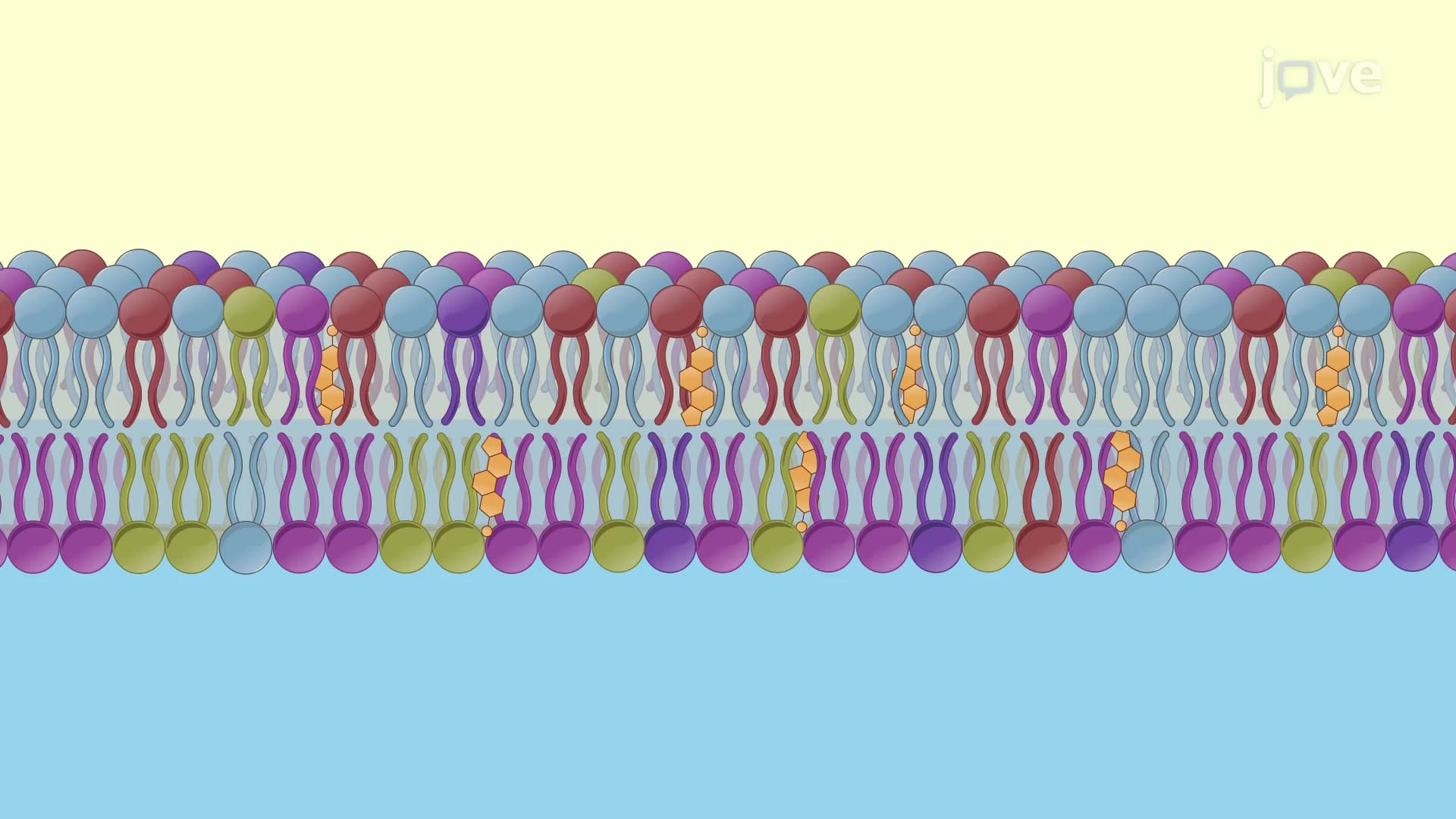
The first mechanism relies on lipid molecular geometry and asymmetric distribution. Lipids with bulky head groups, such as phosphatidylcholine, naturally create positive membrane curvature because their cone-like shape pushes the inner leaflet outward. Conversely, lipids with smaller head groups like phosphatidylethanolamine generate negative curvature through inverted cone geometry.
This principle explains how cells regulate membrane curvature during vesicle formation. Research at institutions like Johns Hopkins University has shown that cancer cells exploit lipid asymmetry to enhance metastatic potential, making this concept relevant for pre-medical students studying oncology.
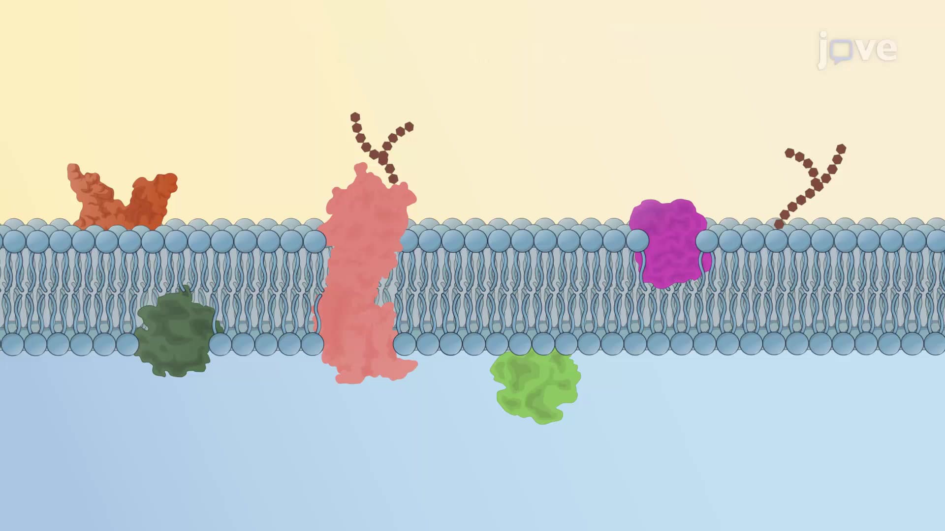
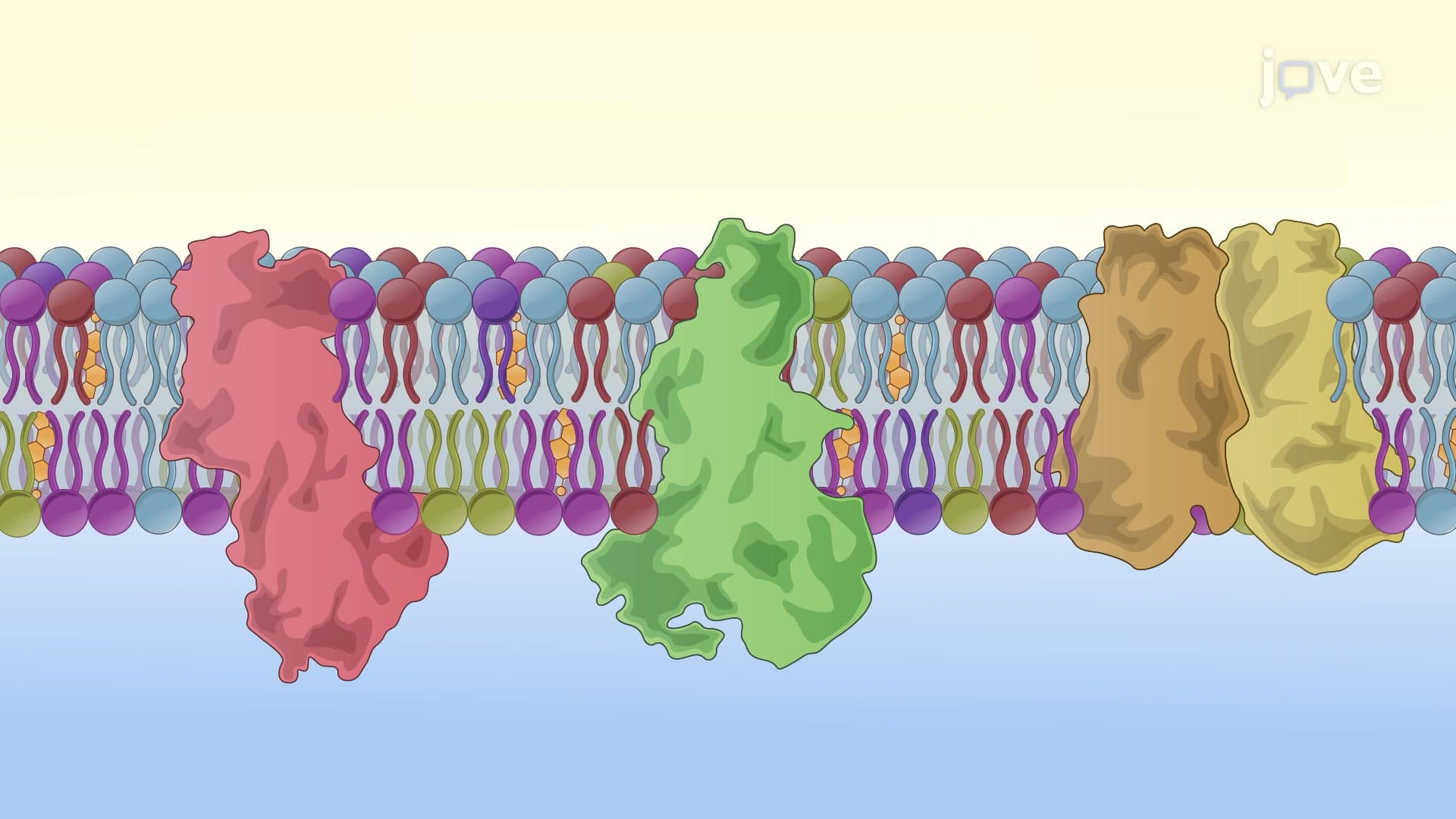
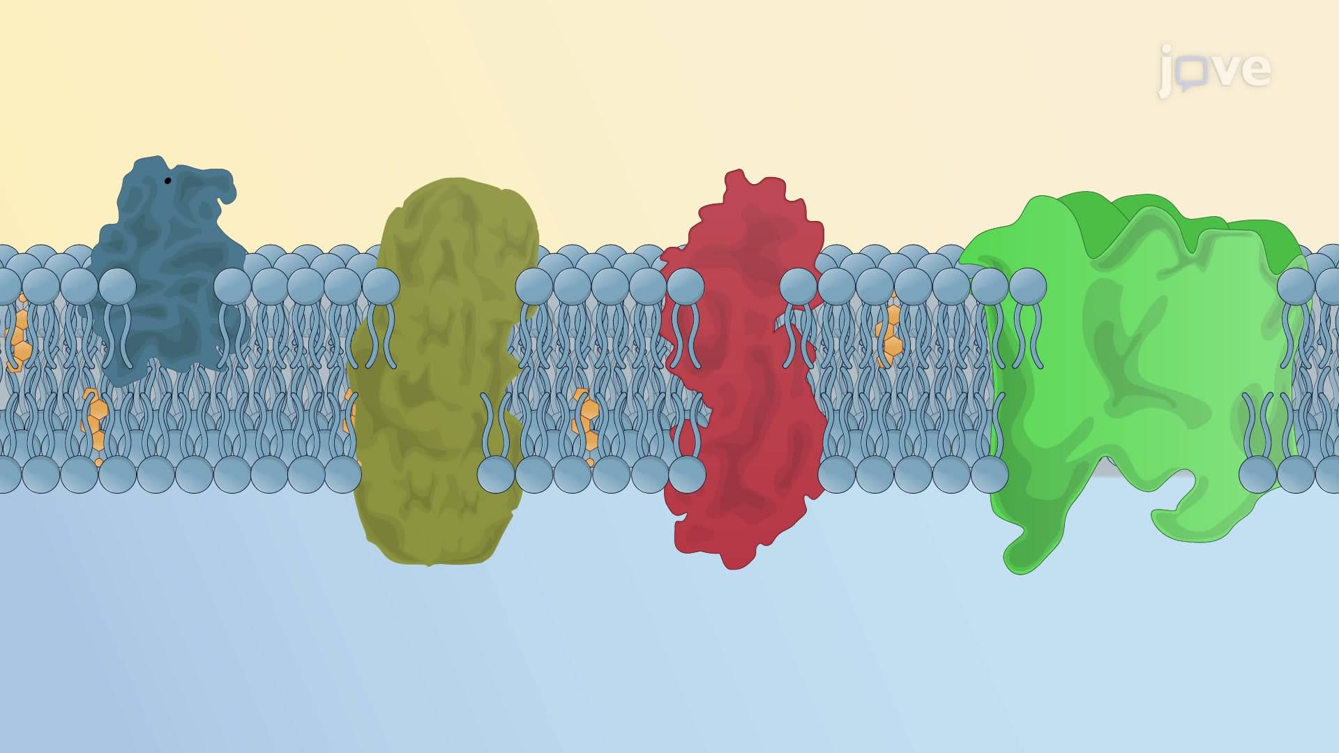
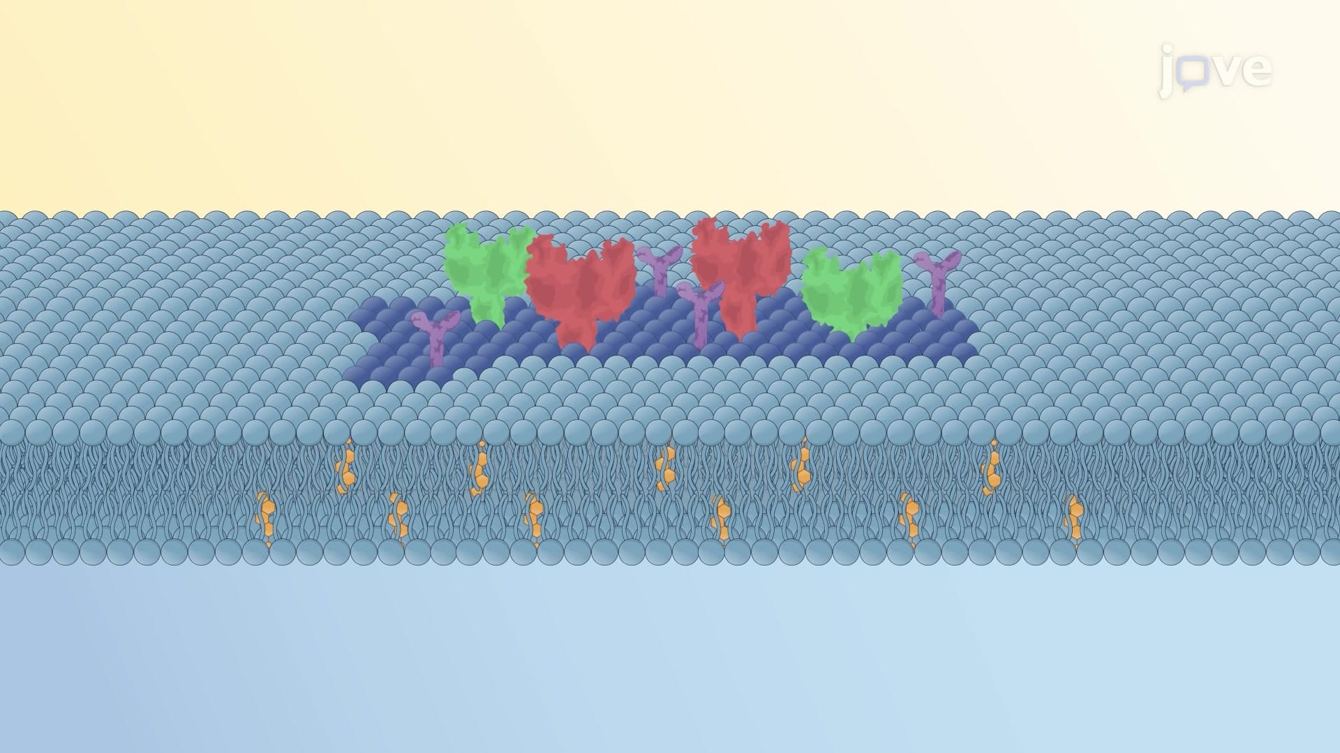
Protein scaffolding represents the second major mechanism, where specialized proteins either actively deform membranes or stabilize existing curvatures. BAR domain proteins exemplify this process-their banana-shaped structure physically molds membrane surfaces while simultaneously recruiting additional curvature-generating factors.
Clinical applications include understanding how synaptic vesicle formation occurs during neurotransmitter release. Students preparing for the MCAT will encounter this mechanism when studying neurological disorders like Alzheimer's disease, where protein scaffolding dysfunction contributes to synaptic failure.
Hydrophobic insertion involves protein domains temporarily embedding within one membrane leaflet, expanding its surface area relative to the opposite side. This asymmetric expansion forces membrane bending, as observed during viral budding processes studied at CDC laboratories.
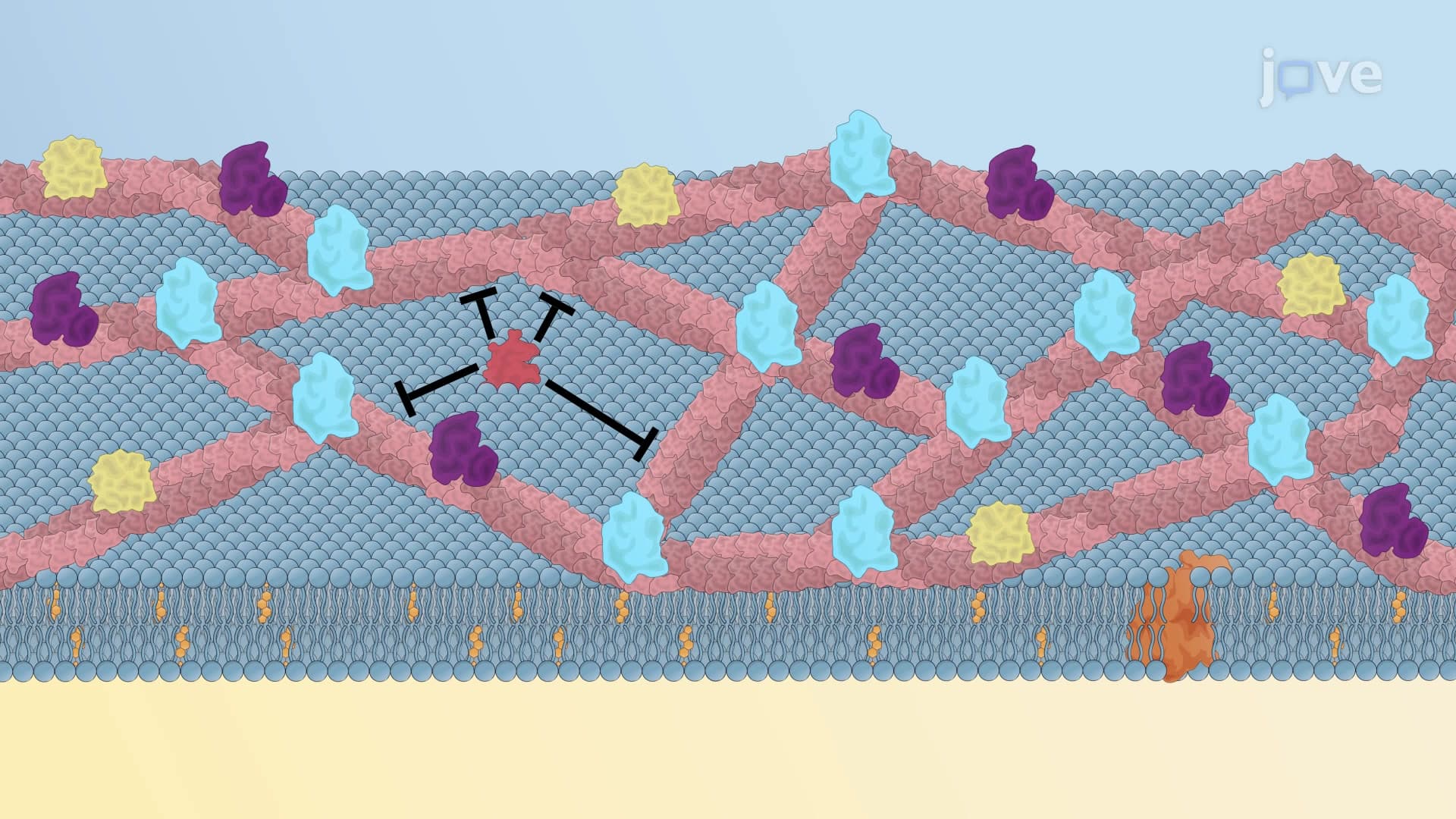
Cytoskeletal force generation provides the fourth mechanism, where actin filament assembly and myosin motor protein activity create pushing and pulling forces. These forces generate specialized membrane projections like filopodia in migrating epithelial cells, a process essential for wound healing and embryonic development.
Students can observe this mechanism in laboratory exercises using cultured cells from American Type Culture Collection (ATCC), making the concept tangible for hands-on learning experiences that reinforce theoretical knowledge for college-level examinations.
Related Micro-courses