Loading subject page…


669,501 views

Video Summary: What Is X Ray Crystallography
Did you know that the same X-rays used in hospitals helped scientists unlock the double helix structure of DNA? X ray crystallography uses X-ray diffraction patterns to reveal the atomic arrangement within crystals, from simple salt to complex proteins studied at institutions like Johns Hopkins University. This powerful technique, discovered by the Bragg father-son duo in 1913, transforms invisible atomic structures into visible spot patterns that scientists can analyze. Watch the full video on JoVE Coach to master this concept with expert-led visuals and step-by-step explanations.
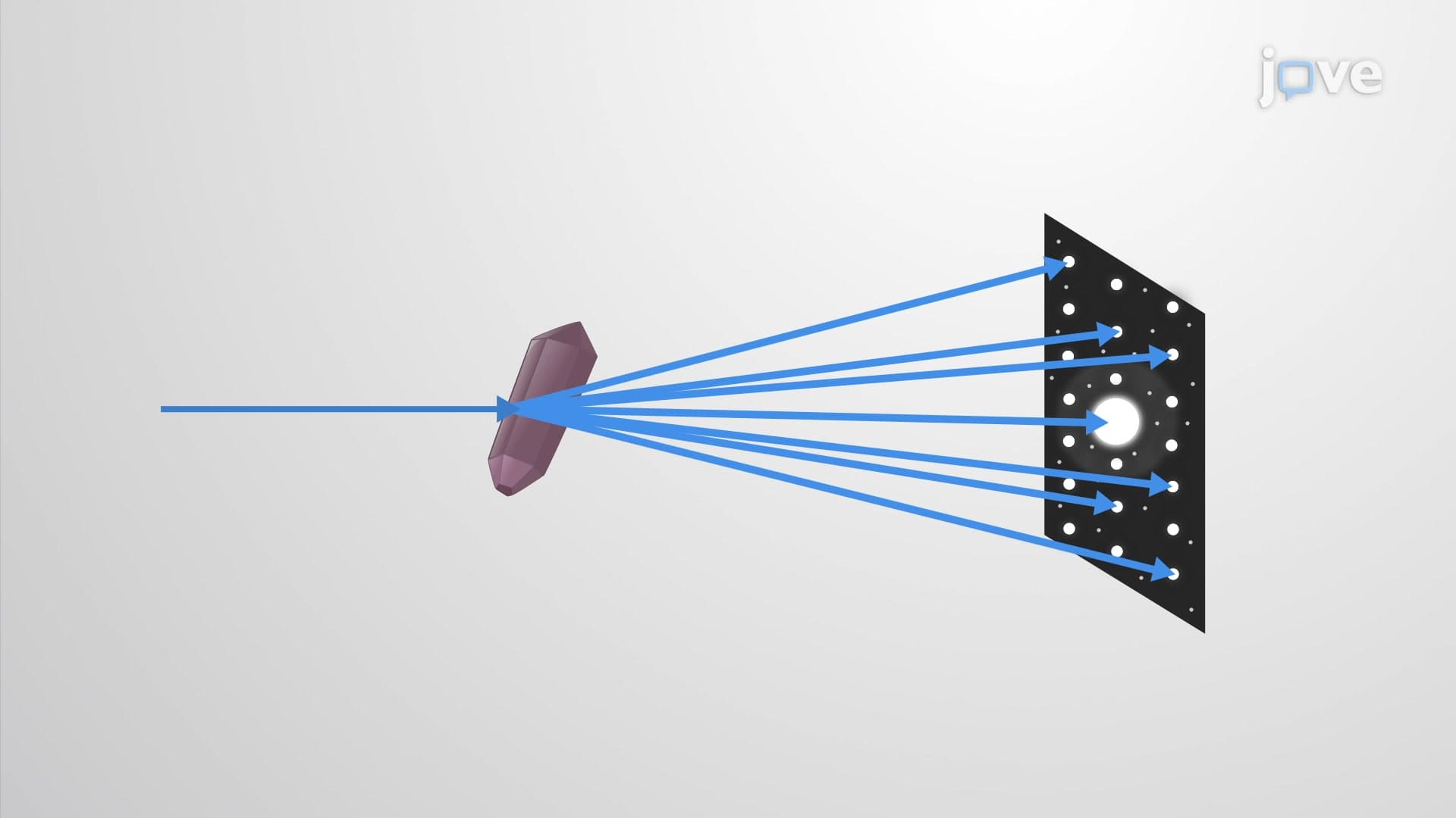
X ray crystallography represents one of the most powerful analytical techniques in modern science, allowing researchers to determine the precise three-dimensional arrangement of atoms within crystalline materials. This method exploits the wave nature of X-rays, which have wavelengths comparable to the spacing between atoms in crystals (typically 0.1 to 10 nanometers). When X-rays encounter a well-ordered crystal, they interact with the electron clouds surrounding atoms, causing the X-rays to scatter in specific directions determined by the crystal's internal structure.
The technique's foundation rests on Bragg's Law, expressed as n × λ = 2d × sin(θ), where n represents an integer, λ is the X-ray wavelength, d is the distance between parallel atomic planes, and θ is the angle of incidence. This elegant equation explains why diffracted X-rays produce constructive interference only at specific angles, creating the characteristic pattern of bright spots that scientists can analyze.
X ray crystallography plays a crucial role in numerous sectors of the US economy and research landscape. Pharmaceutical companies like Pfizer and Merck routinely use this technique during drug development to understand how potential medications interact with target proteins. For instance, researchers at the National Institutes of Health employ X-ray crystallography to study virus structures, contributing to vaccine development efforts.
In materials science, companies such as General Electric utilize crystallographic analysis to develop stronger, more efficient turbine blades for jet engines. Academic institutions across America, from MIT to Stanford University, maintain sophisticated X-ray crystallography facilities where students and researchers investigate everything from semiconductor materials to biological macromolecules.
Today's X-ray crystallography extends far beyond the simple photographic plates used by the Braggs. Modern synchrotron facilities, like the Advanced Photon Source at Argonne National Laboratory in Illinois, generate extremely intense, highly focused X-ray beams that can determine structures in minutes rather than months. These facilities enable researchers to study increasingly complex systems, including membrane proteins and large enzyme complexes.
Computer algorithms now process the thousands of diffraction spots collected from multiple crystal orientations, using mathematical techniques like Fourier transforms to reconstruct three-dimensional electron density maps. These maps reveal not only atomic positions but also provide insights into molecular dynamics and chemical bonding patterns.
Students preparing for standardized tests like the AP Chemistry exam or MCAT will encounter X-ray crystallography in contexts ranging from basic wave interference principles to protein structure analysis. College chemistry and physics courses frequently use crystallographic examples to illustrate concepts including wave-particle duality, constructive interference, and solid-state structures. Understanding this technique provides students with concrete examples of how fundamental physics principles apply to cutting-edge scientific research.
Related Micro-courses