Loading subject page…


146,122 views
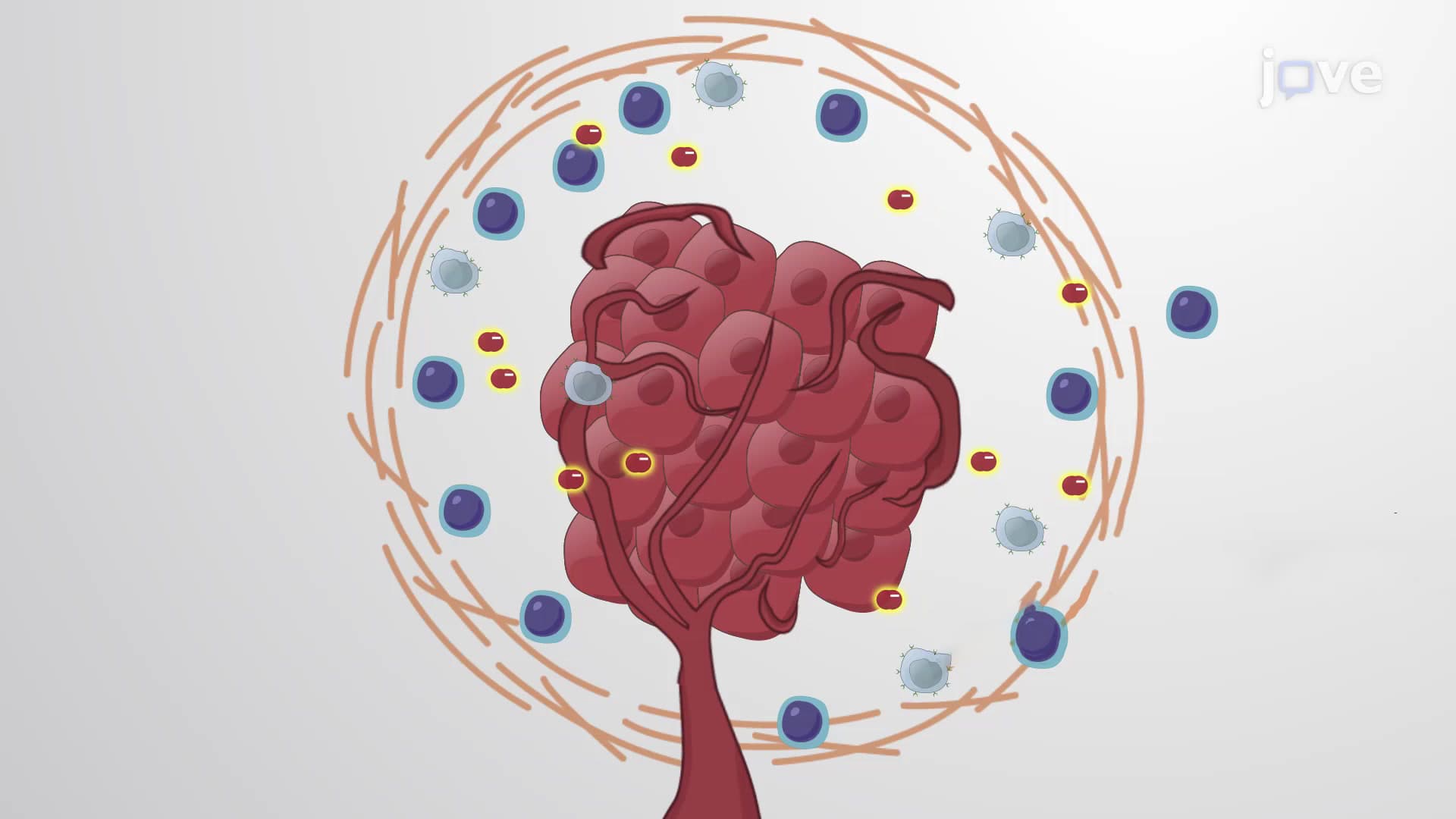

Video Summary: What Is the Tumor Microenvironment
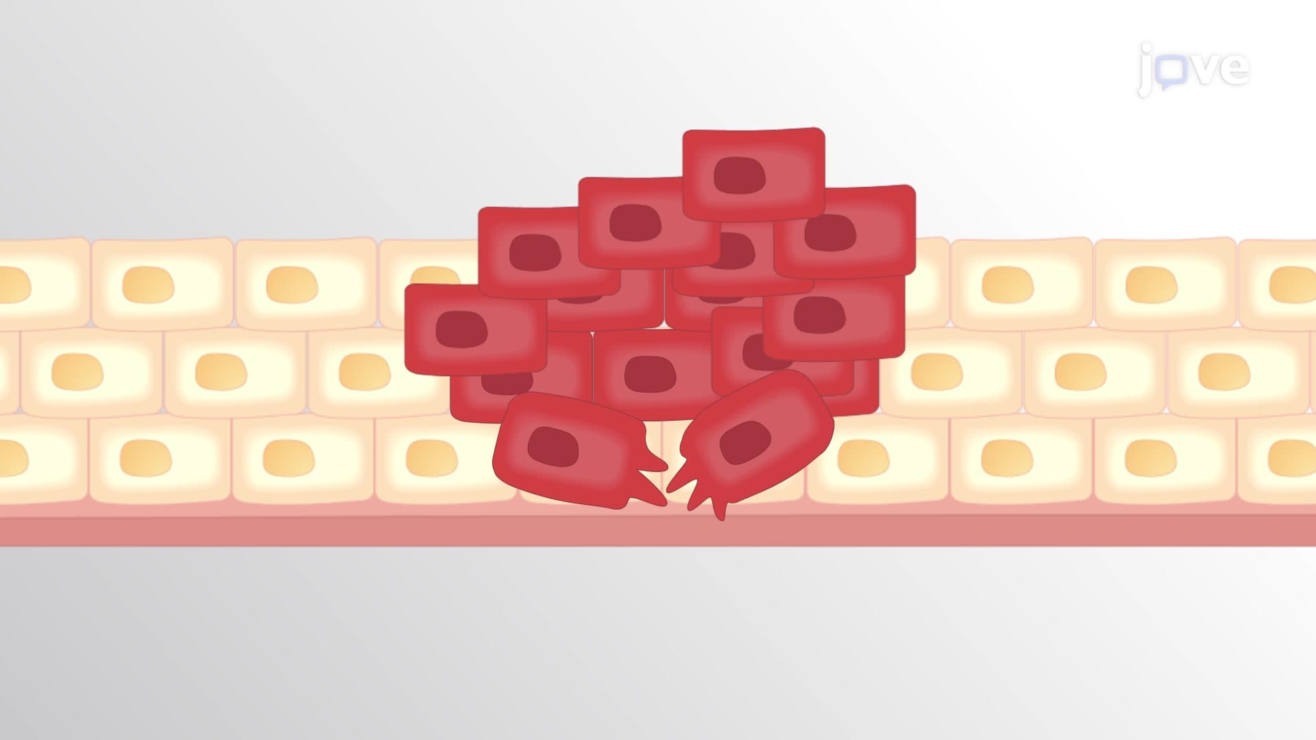
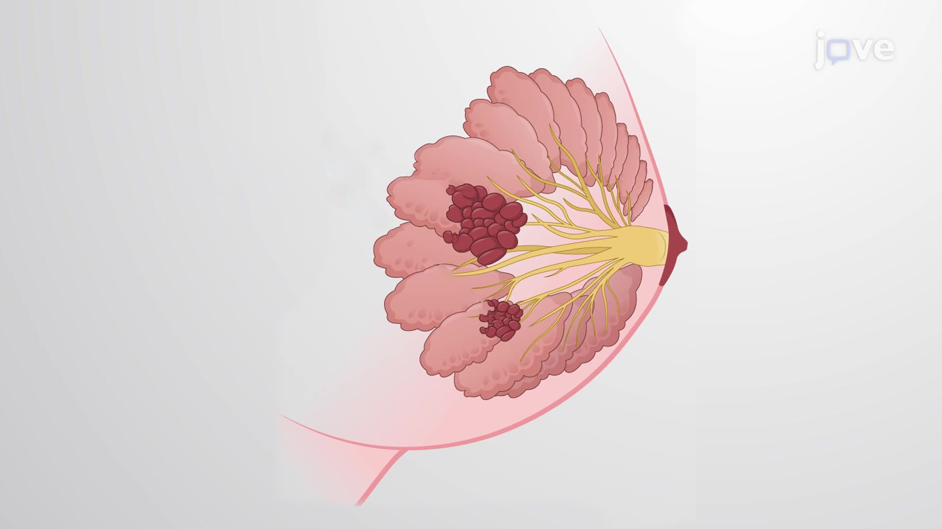
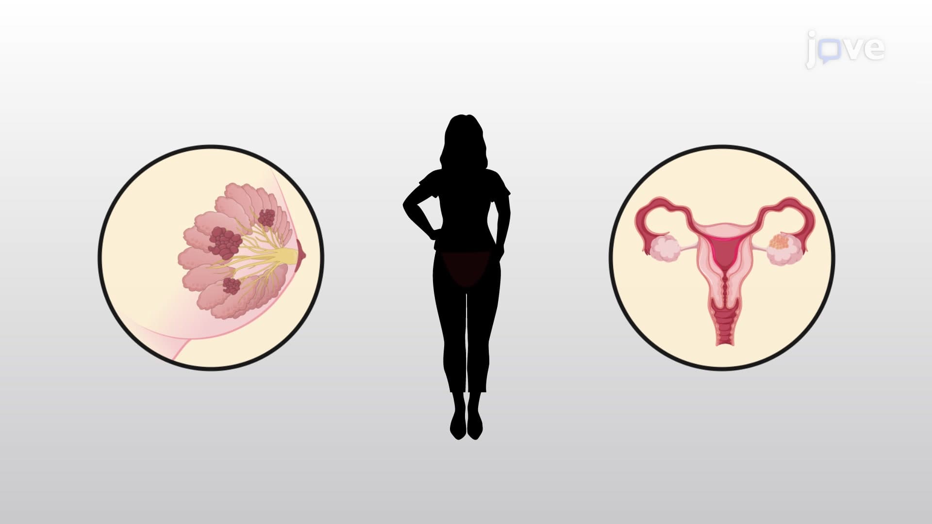
Did you know that cancer cells can't survive alone and actually recruit healthy cells to help them grow? The tumor microenvironment is the complex ecosystem surrounding cancer cells that includes supportive tissues, blood vessels, and immune cells. For instance, breast cancer tumors actively stiffen surrounding tissue to create highways for cancer cells to spread throughout the body. This cellular neighborhood plays a crucial role in cancer progression and represents a major focus in modern oncology research at institutions like MD Anderson Cancer Center. Watch the full video on JoVE Coach to master this concept with expert-led visuals and step-by-step explanations.
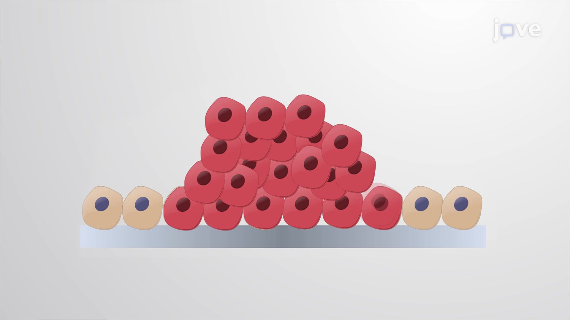
The tumor microenvironment represents one of the most fascinating and complex biological systems studied in modern cancer research. Rather than existing as isolated entities, cancer cells create and maintain intricate relationships with surrounding healthy tissues, forming what researchers call the stroma. This dynamic ecosystem fundamentally changes how we understand cancer progression and has revolutionized treatment approaches at leading US cancer centers like Memorial Sloan Kettering and the National Cancer Institute.
The extracellular matrix (ECM) undergoes dramatic structural changes in cancerous tissues. In healthy breast tissue, collagen fibers form dense, cross-linked networks that maintain tissue integrity. However, cancer cells hijack this system by increasing protease and oxidase expression, which degrades and linearizes these collagen networks. This process creates stiffer tissue pathways that function like cellular highways, allowing cancer cells to migrate more efficiently toward blood vessels and lymph nodes.
Students preparing for the MCAT or AP Biology exams should understand that this ECM remodeling represents a key mechanism of metastasis. The increased tissue stiffness doesn't just facilitate movement-it also activates cellular signaling pathways that promote aggressive cancer behavior. Research from institutions like Johns Hopkins has shown that measuring tissue stiffness can even help predict patient outcomes.
Carcinoma-associated fibroblasts (CAFs) exemplify how cancer corrupts normal cellular functions. These cells, originally designed to maintain tissue structure and repair wounds, become hijacked by nearby tumor cells. CAFs secrete numerous growth factors that directly support cancer progression, particularly when tumors face nutrient limitations.
As solid tumors grow beyond 2-3 millimeters, their innermost cells experience hypoxia due to inadequate blood supply. CAFs respond by releasing vascular endothelial growth factor (VEGF), triggering angiogenesis-the formation of new blood vessels. This process ensures continued oxygen and nutrient delivery to rapidly dividing cancer cells, enabling sustained tumor growth.
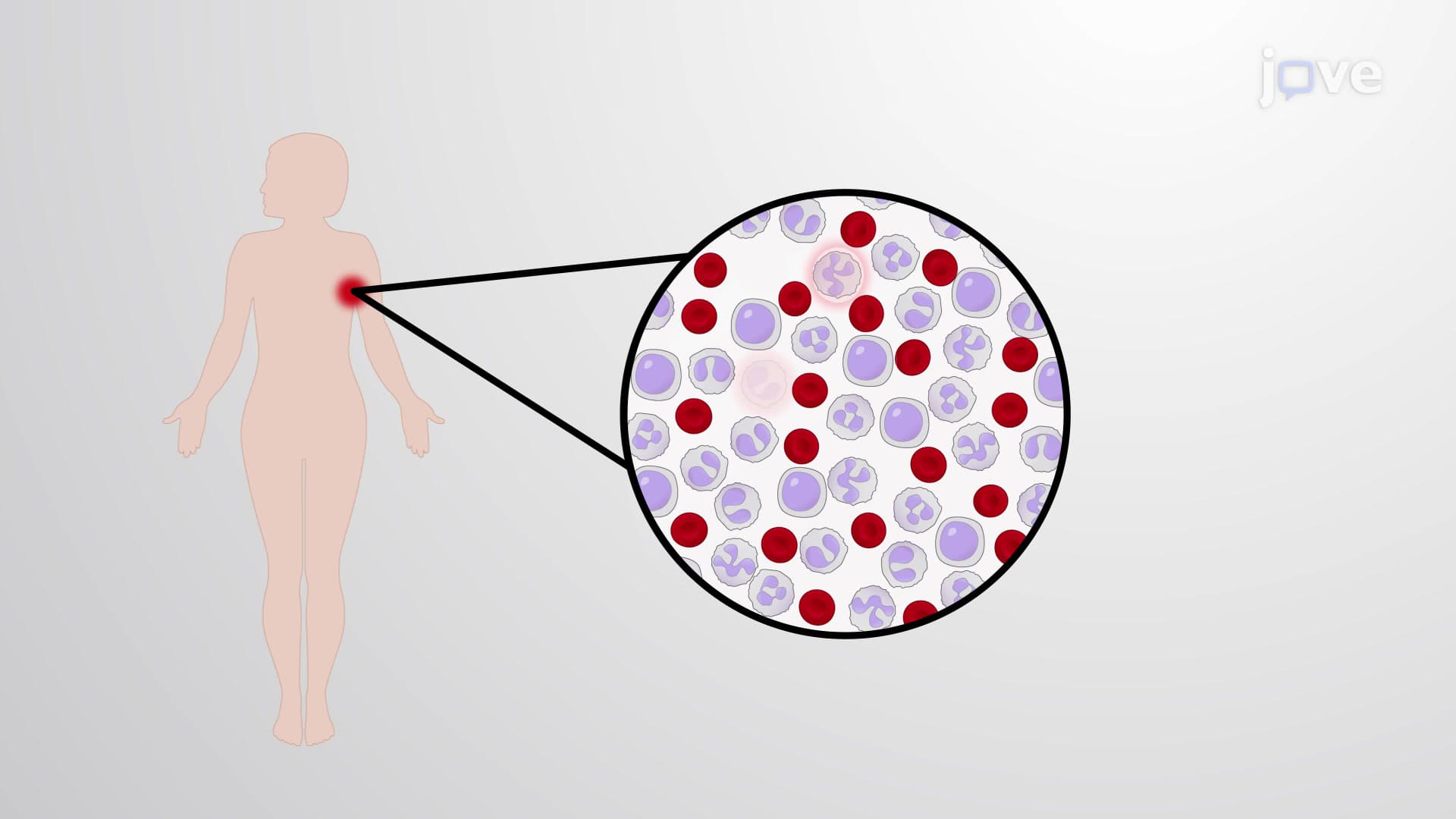
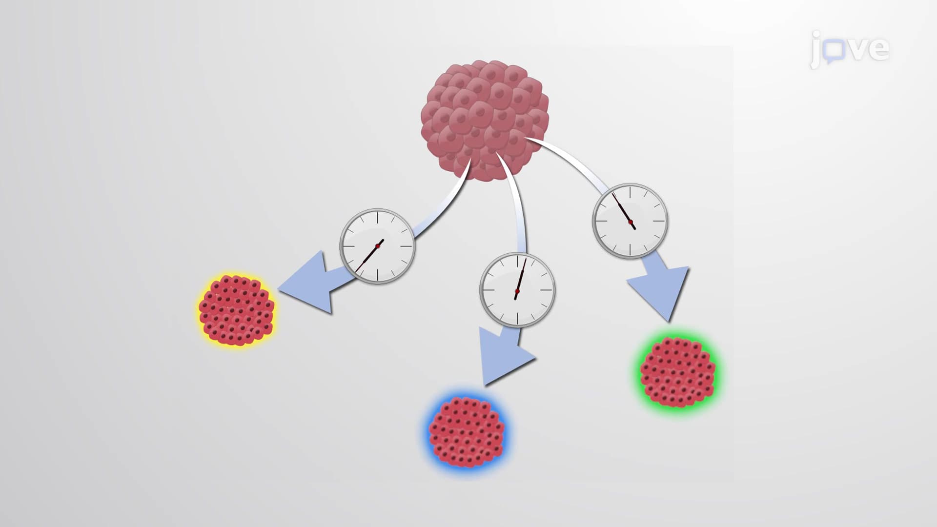
The relationship between cancer cells and immune surveillance represents a ongoing evolutionary arms race. While immune cells like macrophages, lymphocytes, and natural killer cells can initially detect and eliminate cancer cells, the genetic instability characteristic of cancer enables rapid adaptation. Tumor cells develop sophisticated immune evasion strategies, including the induction of immunosuppressive T-regulatory cells.
These corrupted immune cells produce reactive oxygen species that inhibit lymphocyte function while simultaneously increasing mutation rates in tumor cells. This creates a vicious cycle where immune suppression facilitates more aggressive cancer variants. Understanding these mechanisms proves crucial for students studying immunology or preparing for medical school entrance exams like the MCAT.
Related Micro-courses