Loading subject page…


234,001 views

Video Summary: Conservation of Protein Domains Over Explained
Did you know that despite having over 20,000 different proteins in the human body, there are fewer than 1,500 unique protein domain structures? The conservation of protein domains over evolutionary time explains why similar functional units appear across countless different proteins, from insulin receptors to immune system antibodies used in US hospitals. This remarkable efficiency allows cells to create diverse proteins by mixing and matching proven domain designs rather than reinventing molecular machinery from scratch. Watch the full video on JoVE Coach to master this concept with expert-led visuals and step-by-step explanations.
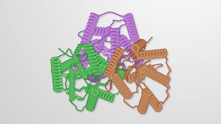
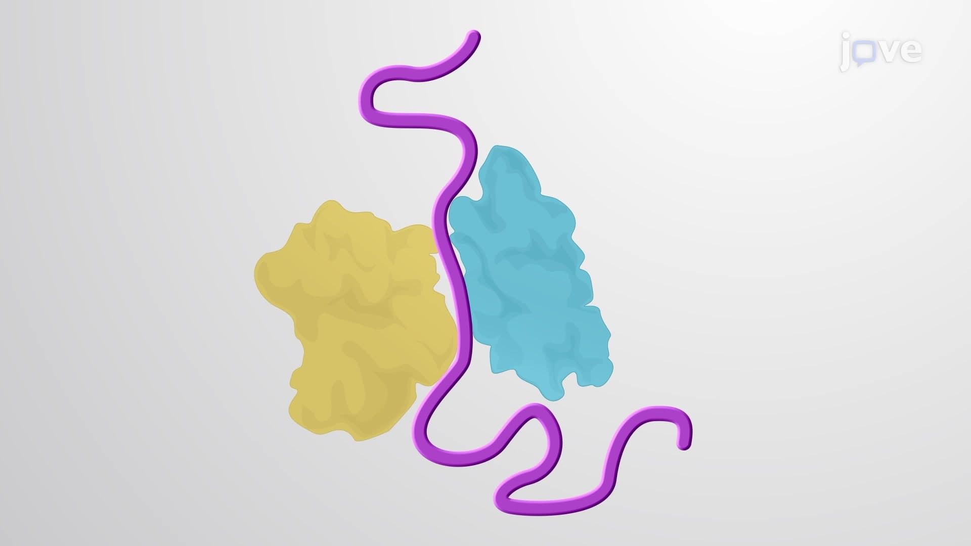
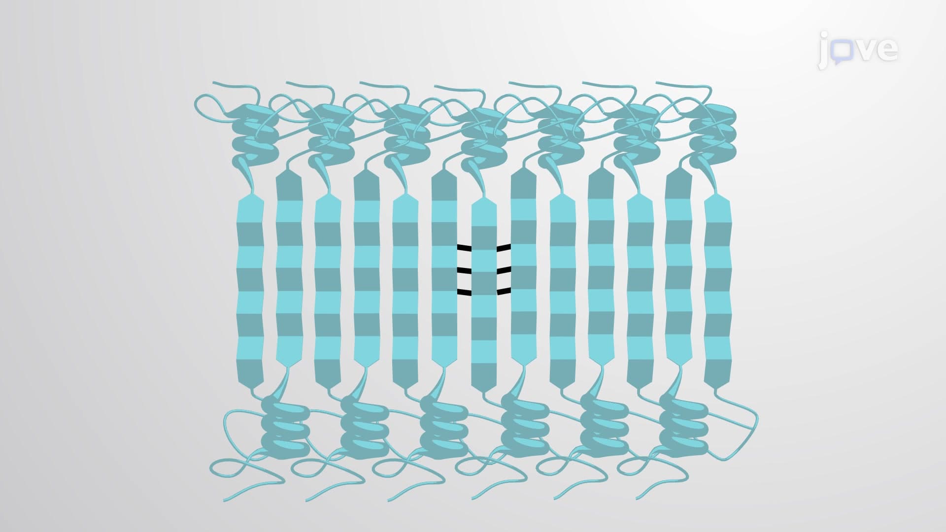
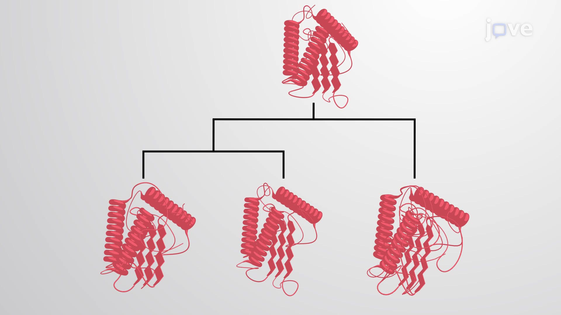
The conservation of protein domains over millions of years of evolution represents one of biology's most elegant solutions to the challenge of creating functional diversity. Rather than evolving entirely new protein structures from scratch, organisms have refined and reused successful molecular designs. This conservation occurs because certain three-dimensional arrangements of secondary structure elements-alpha helices, beta sheets, and connecting loops-provide optimal stability and function.
Protein domains face significant physical constraints that limit possible folding patterns. The laws of thermodynamics favor specific arrangements that minimize free energy while maintaining structural stability. For example, hydrophobic amino acids typically cluster in the protein core, while hydrophilic residues face the aqueous environment. These constraints mean that evolution repeatedly converges on similar solutions, explaining why we see the same domain architectures across distantly related species.
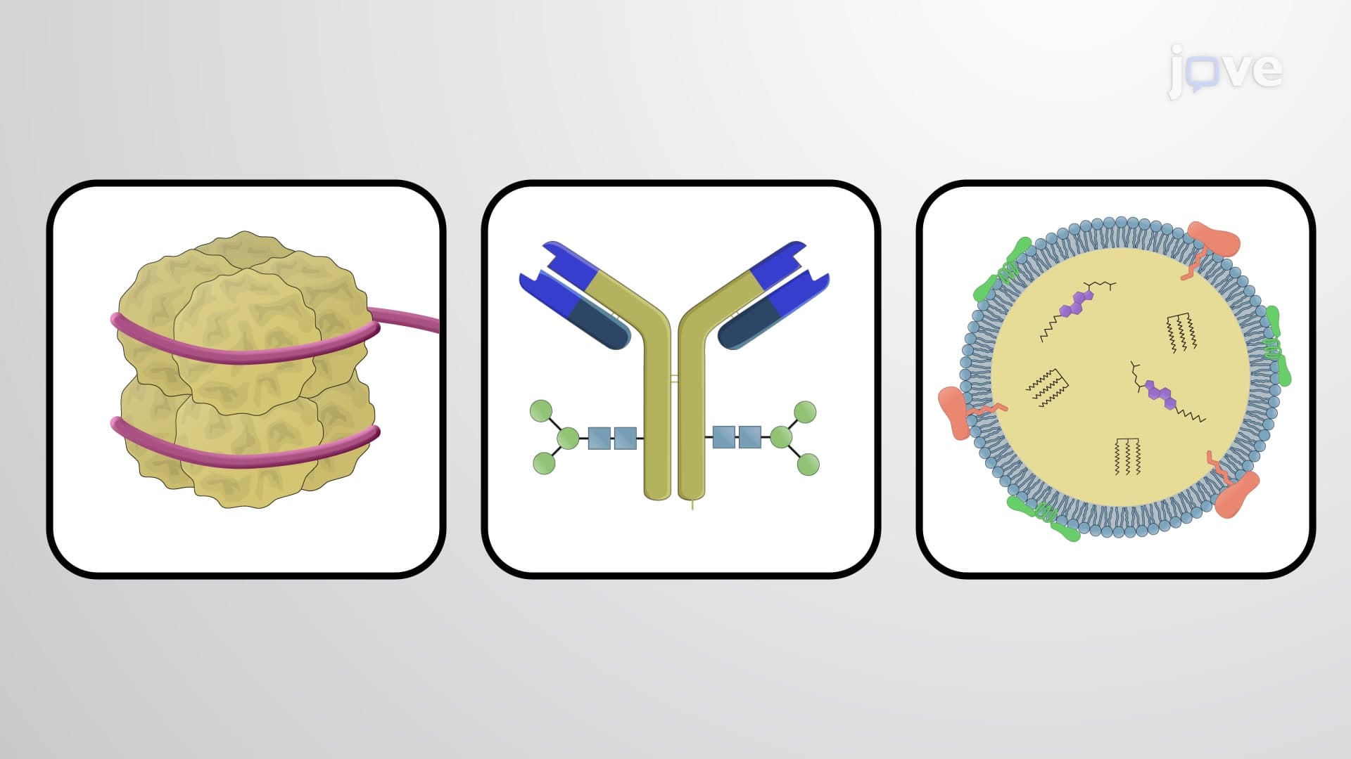
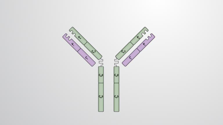
In the United States, students studying for the MCAT or AP Biology exams frequently encounter questions about domain conservation. Understanding that structural constraints limit folding possibilities helps explain why certain domain families, like the immunoglobulin fold found in antibodies, appear throughout the immune system and even in cell adhesion molecules.
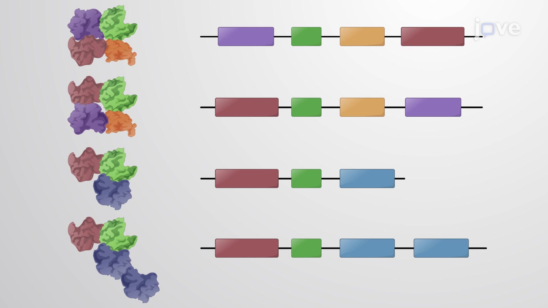
Domain shuffling represents evolution's modular approach to creating new proteins. This process involves recombining existing domains in novel arrangements, similar to how engineers might combine proven components to create new machines. The Src protein family exemplifies this principle, containing conserved SH2 and SH3 domains that appear in over 400 different human proteins.
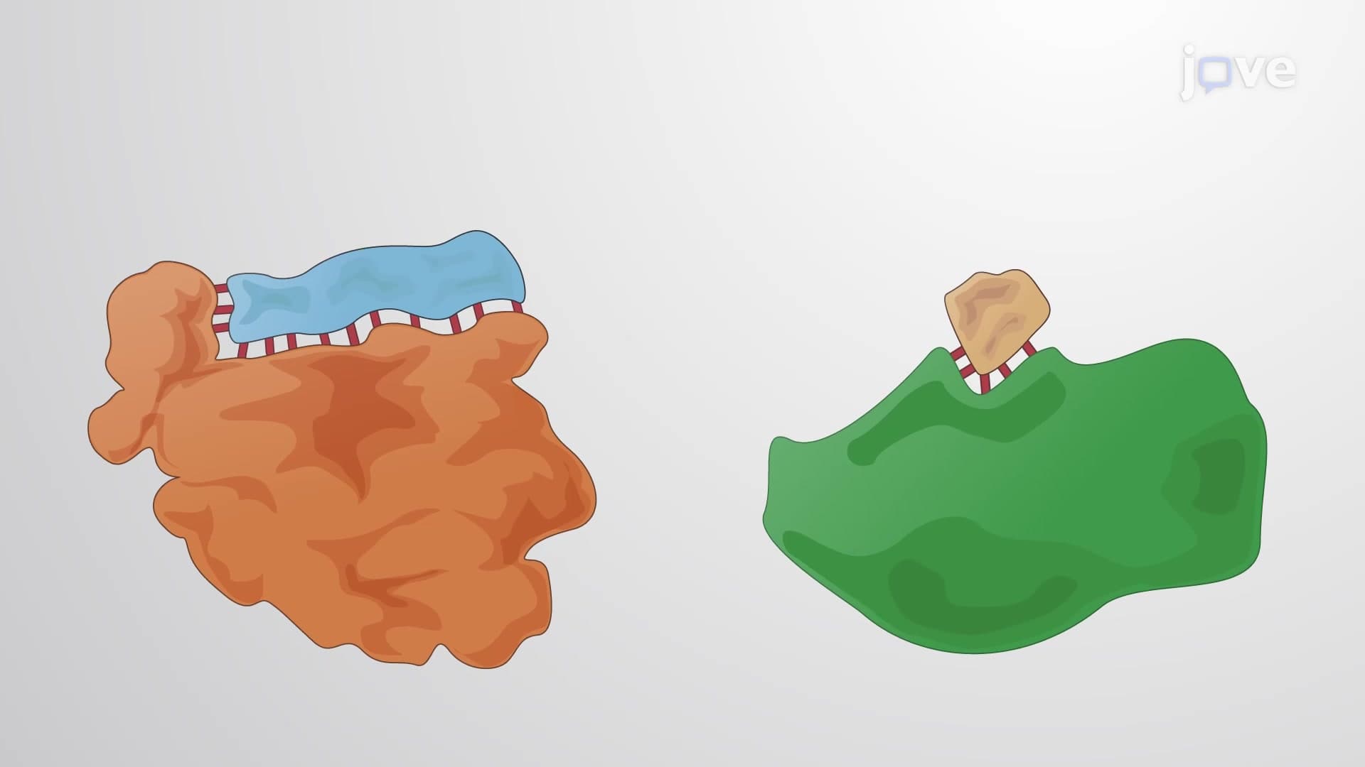
The SH2 domain, found in 115+ proteins, specifically recognizes phosphorylated tyrosine residues-a crucial mechanism for cellular signaling. Meanwhile, SH3 domains bind proline-rich sequences and appear 300 times in the human genome. These domains function like molecular adapters, allowing proteins to interact with specific partners and participate in complex signaling networks.
Domain conservation has profound implications for medicine and biotechnology in the United States. Pharmaceutical companies exploit domain conservation when designing drugs that target protein families. For instance, kinase inhibitors used in cancer treatment often target conserved catalytic domains shared among multiple kinases. Similarly, researchers studying genetic diseases can predict protein function by identifying conserved domains, even in newly discovered genes.
College students preparing for medical school entrance exams should understand that domain conservation explains why mutations in one protein can have effects similar to mutations in seemingly unrelated proteins-they may share conserved functional domains.
Related Micro-courses