Loading video…


 20 Concepts
 36.9K views

Video Summary: What Is Adrenergic Neurons Neurotransmission
Did you know that every time you feel your heart race before a big presentation, specialized nerve cells called adrenergic neurons are firing norepinephrine to activate your body's fight-or-flight response? Adrenergic neurons neurotransmission is the complex process by which these sympathetic nerve fibers synthesize, release, and recycle norepinephrine to communicate with target organs throughout your body. When a UCLA medical student takes the MCAT, their adrenergic system helps maintain focus and alertness during this crucial exam. Watch the full video on JoVE Coach to master this concept with expert-led visuals and step-by-step explanations.
Adrenergic neurons neurotransmission represents one of the most clinically significant signaling mechanisms in human physiology. These specialized postganglionic sympathetic neurons control everything from heart rate regulation during exercise to blood pressure maintenance in emergency situations. For students preparing for the MCAT or advanced placement biology exams, mastering this concept provides essential foundation knowledge for understanding cardiovascular pharmacology and autonomic disorders.
The neurotransmitter synthesis process begins when tyrosine, a dietary amino acid found in foods like turkey and cheese, enters the neuron through sodium-dependent transporters. This amino acid undergoes a precisely controlled four-step transformation. First, tyrosine hydroxylase converts tyrosine to L-DOPA in the rate-limiting step that determines overall norepinephrine production. Next, aromatic L-amino acid decarboxylase transforms L-DOPA into dopamine in the cytoplasm. Dopamine then travels via vesicular monoamine transporters into synaptic vesicles, where dopamine β-hydroxylase completes the final conversion to norepinephrine.
When an action potential reaches the nerve terminal, voltage-gated calcium channels open, allowing calcium ions to flood into the cytoplasm. This calcium surge triggers synaptic vesicles to fuse with the presynaptic membrane through SNARE protein interactions, releasing norepinephrine into the synaptic cleft. The released neurotransmitter then binds to α- or β-adrenergic receptors on target organs, initiating G-protein coupled cascades that generate second messengers like cyclic adenosine monophosphate (cAMP). These molecular signals ultimately produce physiological responses such as increased heart rate, bronchodilation, or vasoconstriction.
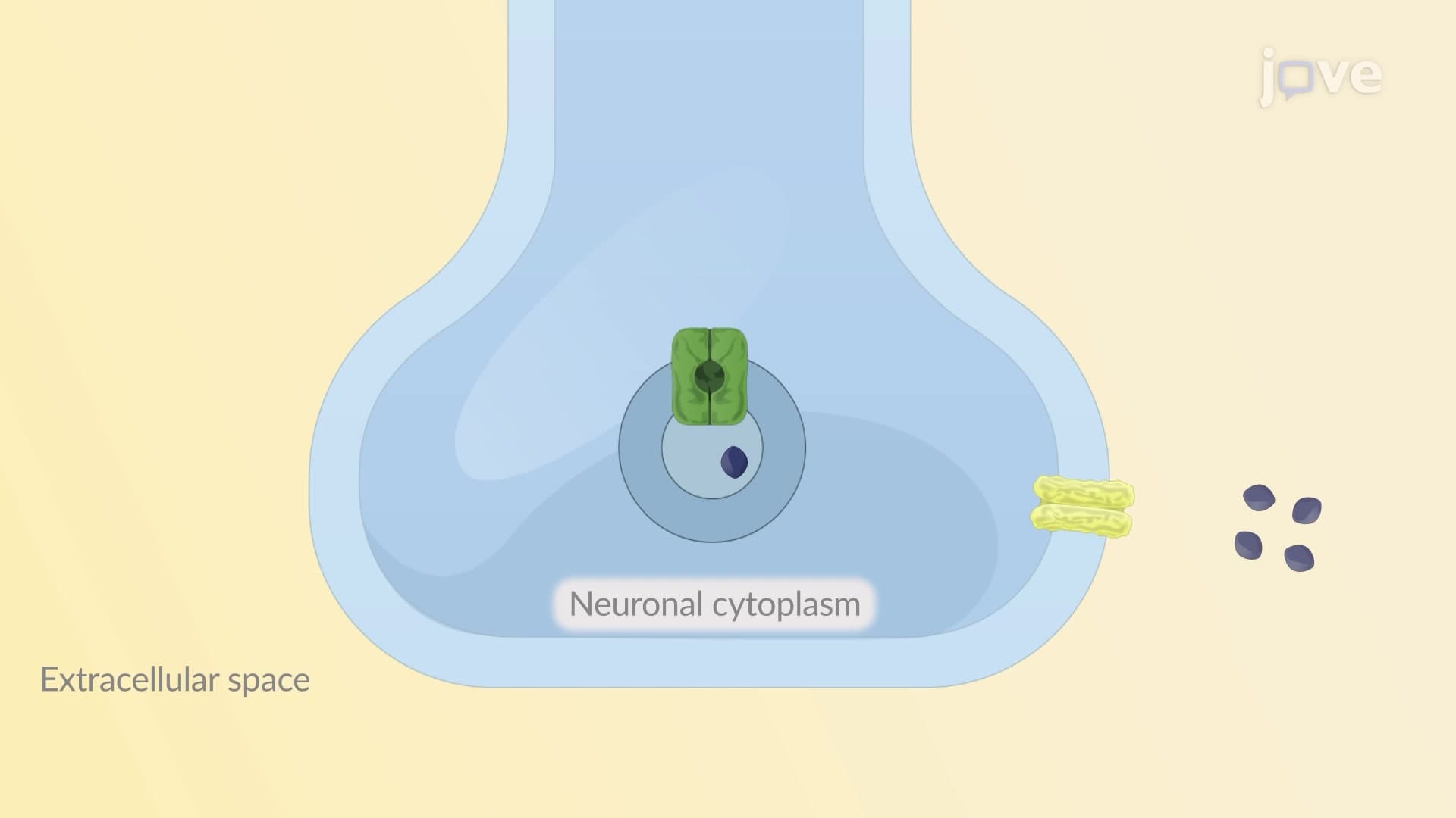
Efficient signal termination prevents overstimulation and maintains precise physiological control. Norepinephrine removal occurs through three primary mechanisms. Some neurotransmitter diffuses away from the synapse into systemic circulation, eventually reaching the kidneys for elimination. Catechol-O-methyltransferase (COMT) enzymes in the synaptic cleft directly metabolize norepinephrine into inactive metabolites. Most importantly, the norepinephrine transporter (NET) rapidly recaptures released neurotransmitter back into the presynaptic terminal. Once inside, norepinephrine either returns to synaptic vesicles for reuse or undergoes degradation by monoamine oxidase (MAO) enzymes. Understanding these pathways helps explain why medications like MAO inhibitors and NET blockers effectively treat depression and attention disorders by prolonging neurotransmitter availability.
Related Micro-courses