Loading video…


 19 Concepts
 132.6K views

Video Summary: What Is Compact Bone
Did you know that compact bone structure is so strong it can withstand forces up to 180 MPa-roughly equivalent to the pressure needed to crush a car? This incredible strength comes from the precisely organized architecture of compact bone tissue, the dense outer layer that forms the shaft of every long bone in your body, from your femur to the bones in baseball players' arms at major league stadiums across America. Understanding what is compact bone in skeletal anatomy reveals how your skeleton achieves both strength and efficiency through its microscopic organization. Watch the full video on JoVE Coach to master this concept with expert-led visuals and step-by-step explanations.
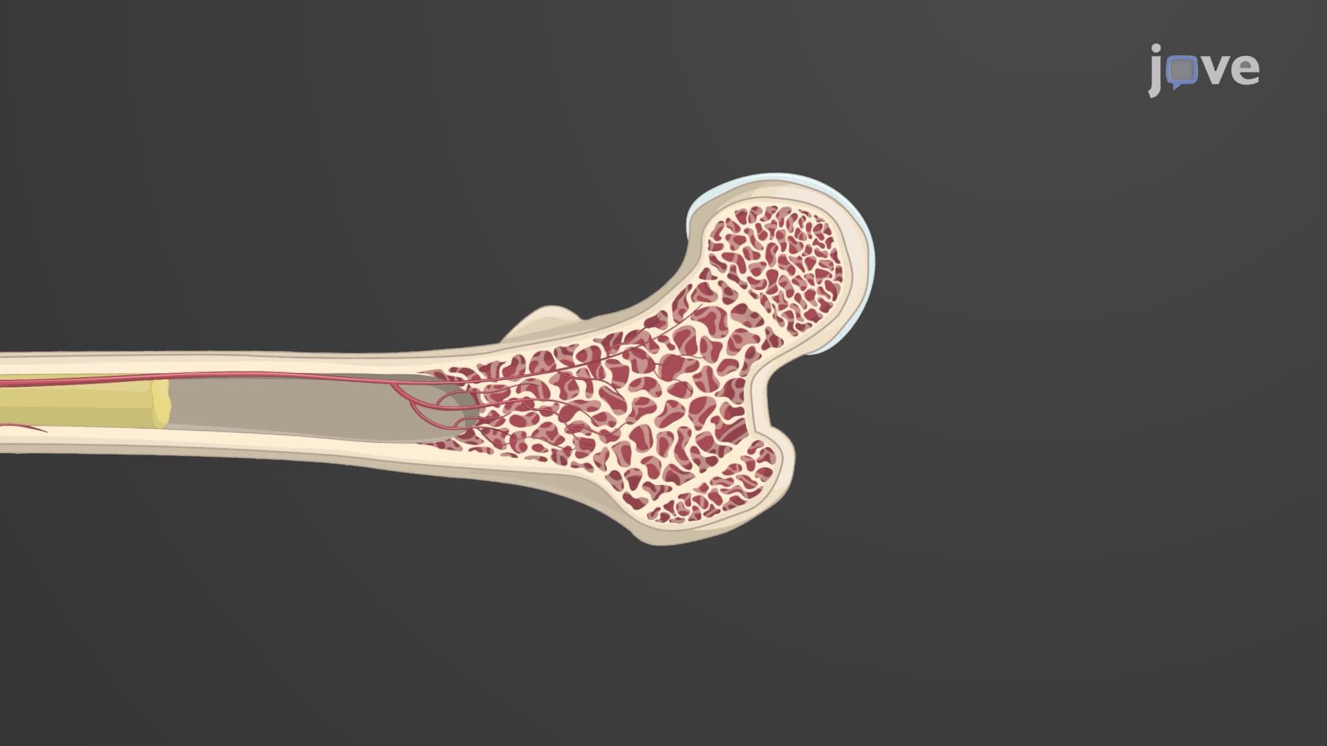
Compact bone represents one of nature's most sophisticated engineering solutions, combining maximum strength with minimal weight. Unlike the spongy interior of bones, compact bone tissue explained reveals a dense, organized structure that forms the outer shell of all bones and comprises up to 80% of the adult skeleton's total bone mass.
The structural brilliance of compact bone lies in its osteons-cylindrical units measuring approximately 200-250 micrometers in diameter that run parallel to the bone's long axis. Think of osteons like the steel reinforcement columns in skyscrapers across American cities like Chicago or New York. Each osteon contains 4-20 concentric rings called lamellae, with collagen fibers oriented in alternating directions between layers. This alternating pattern is crucial for the bone osteon structure, as it prevents catastrophic failure under twisting forces-similar to how plywood's alternating grain prevents splitting.
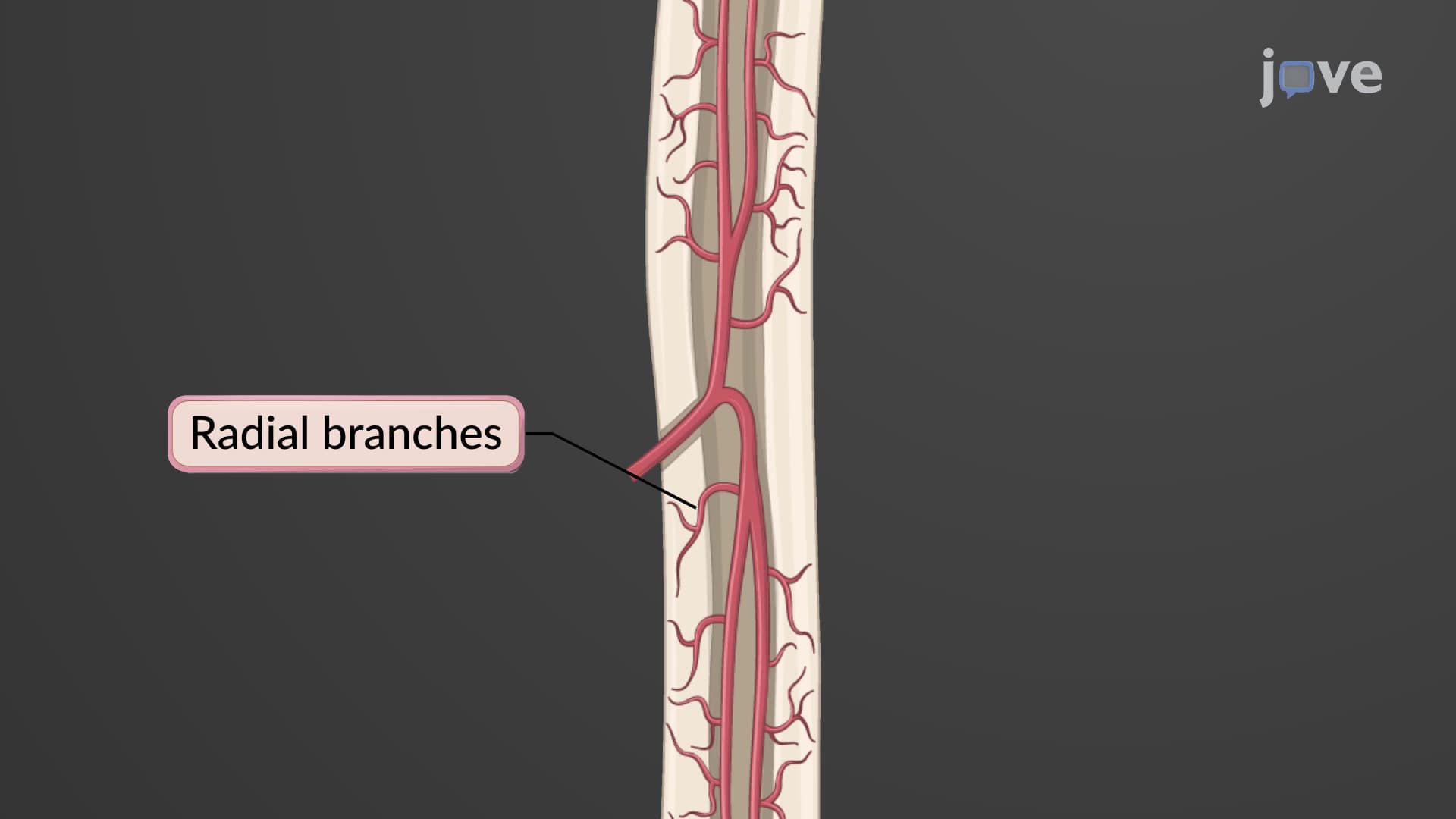
At the center of each osteon lies the Haversian canal, containing blood vessels, lymphatic vessels, and nerves essential for bone maintenance. These canals connect to the bone's main vascular supply through perpendicular Volkmann canals, creating a three-dimensional network that ensures every bone cell receives nutrients. This system is particularly important for athletes-studies of NFL players show that compact cortical bone matrix responds to training by increasing vascular density, supporting the high metabolic demands of bone remodeling.
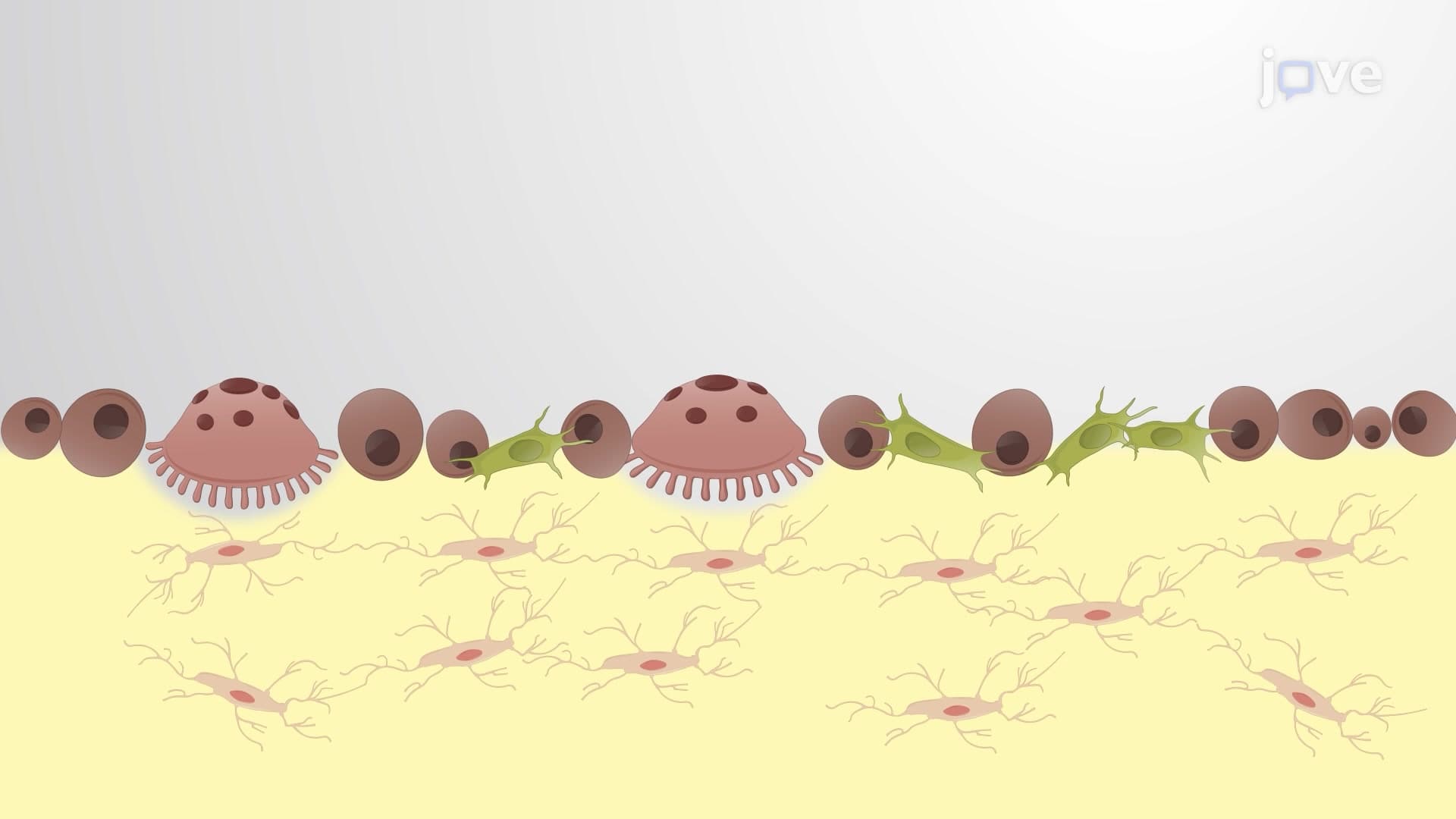
Between the lamellae lie small spaces called lacunae, housing mature bone cells called osteocytes. These cells communicate through tiny channels called canaliculi, forming a network that can detect mechanical stress and coordinate bone remodeling. When you see X-rays at American hospitals, the white areas represent this dense compact bone tissue-a testament to its mineral density and organized structure.
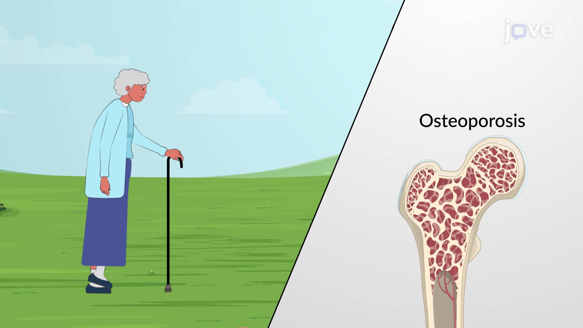
Understanding compact bone structure is essential for students preparing for the MCAT, AP Biology, or college anatomy courses. Medical students studying for the USMLE frequently encounter questions about osteon organization, particularly in orthopedic and pathology contexts. This knowledge directly applies to understanding fracture healing, osteoporosis development, and why certain bones are more prone to specific types of injuries-concepts that appear regularly on nursing entrance exams like the HESI A2 and TEAS.
Related Micro-courses