Loading video…


 19 Concepts
 132.6K views

Video Summary: What Is Spongy Bone
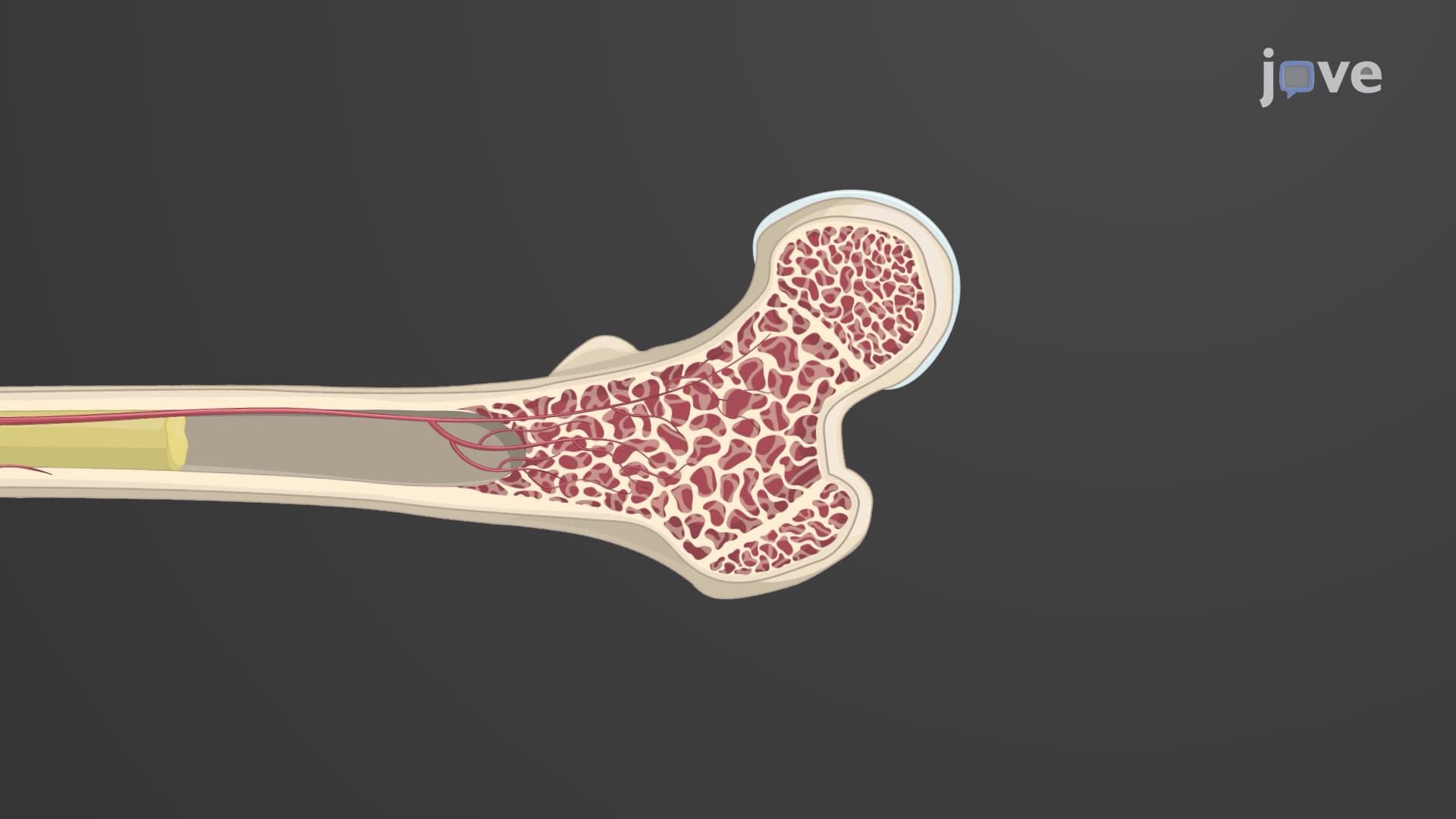
Ever wonder why your bones don't shatter when you jump off a diving board at your local YMCA? The secret lies in spongy bone structure, a remarkable honeycomb-like tissue that makes up about 20% of your skeleton. This lightweight yet incredibly strong bone tissue forms the inner core of bones, particularly in areas like the ends of your femur and humerus. Understanding what is spongy bone reveals how nature engineered the perfect balance between strength and weight in human anatomy. Watch the full video on JoVE Coach to master this concept with expert-led visuals and step-by-step explanations.
Spongy bone structure represents one of nature's most elegant engineering solutions in human anatomy. Unlike the dense, solid appearance of compact bone that forms the outer shell of bones, spongy bone creates an intricate three-dimensional lattice system. This cancellous bone marrow-filled framework occupies approximately 20% of total bone mass while providing crucial structural support where bones experience the greatest mechanical stress.
The genius of this design becomes apparent when examining bones like the vertebrae in your spine or the ends of long bones such as the femur. These regions must absorb tremendous forces-imagine the impact your spine experiences when landing a basketball shot or the stress your hip bones endure during a marathon. The trabeculae spongy bone network distributes these forces across multiple pathways, preventing catastrophic failure that would occur in a solid, brittle structure.
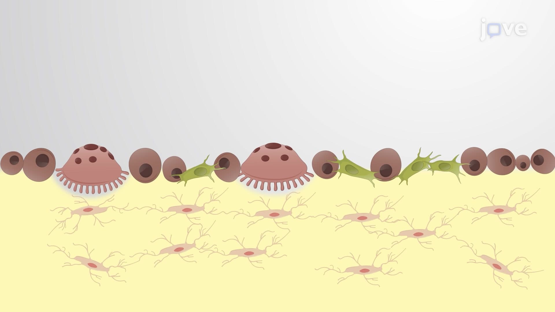
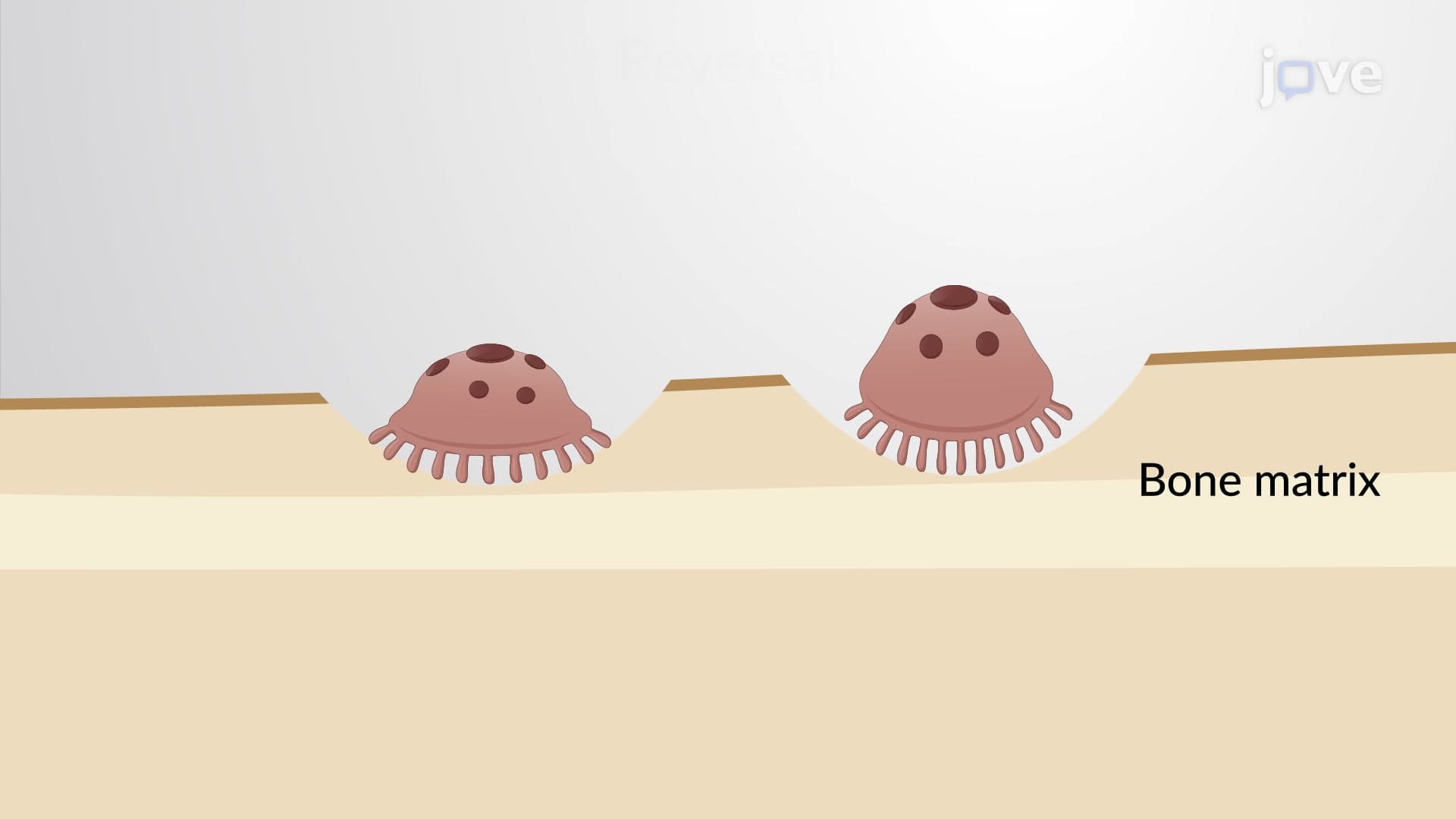
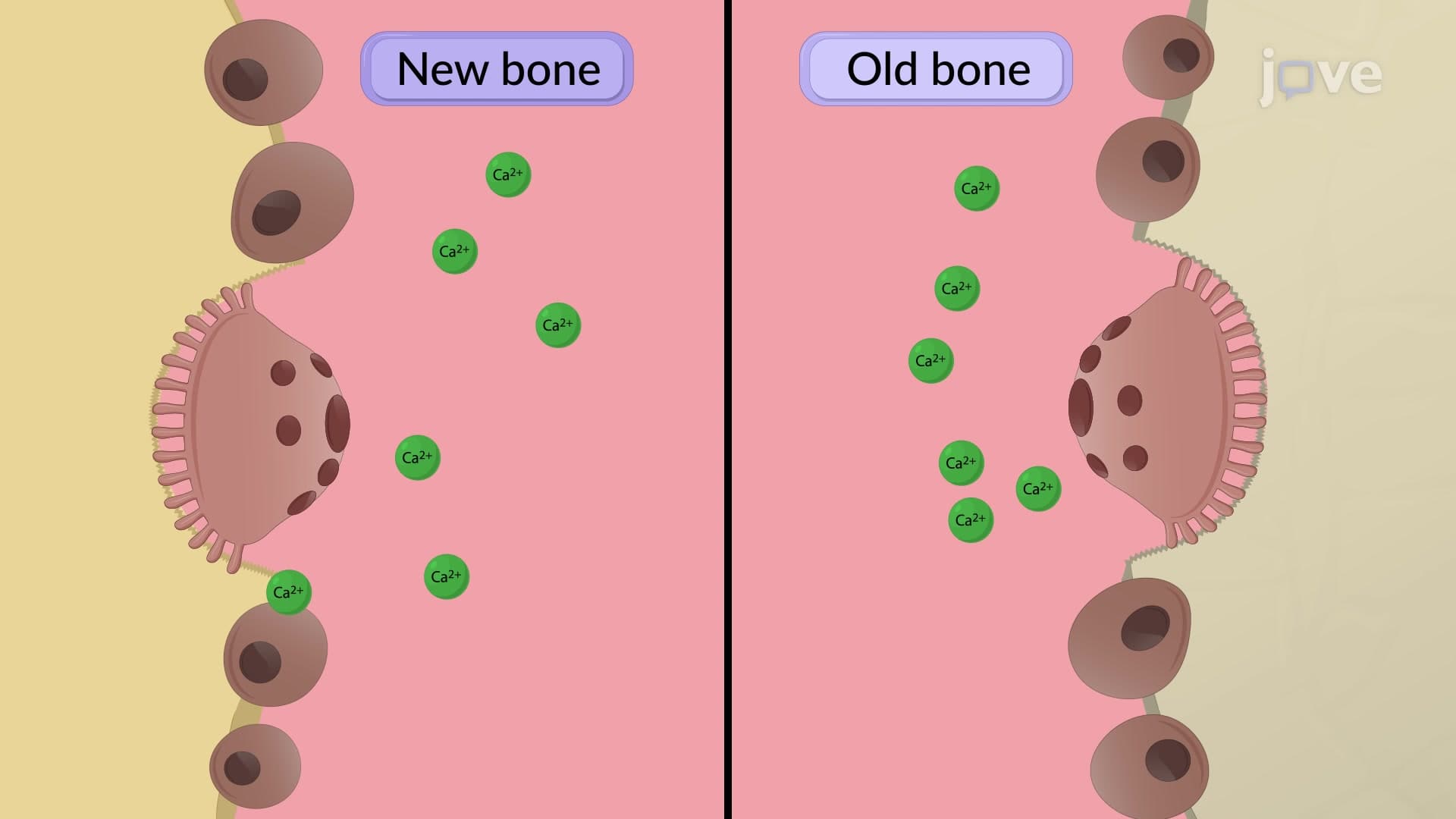
The spongy bone lattice consists of thin, branching structures called trabeculae that align themselves along lines of mechanical stress. This isn't random-your bones continuously remodel throughout life, with osteoblasts and osteoclasts working together to strengthen areas under frequent stress while removing bone tissue from less-used regions. This adaptive process explains why weight-bearing exercise strengthens bones and why astronauts experience bone loss in microgravity.
Each trabecula contains the same basic components as compact bone: concentric lamellae surrounding central spaces, osteocytes housed in lacunae, and an interconnected network of canaliculi. However, the spongy bone internal structure differs significantly in organization. Rather than forming complete osteons, the lamellae create incomplete rings that maximize surface area for nutrient exchange while maintaining structural integrity.
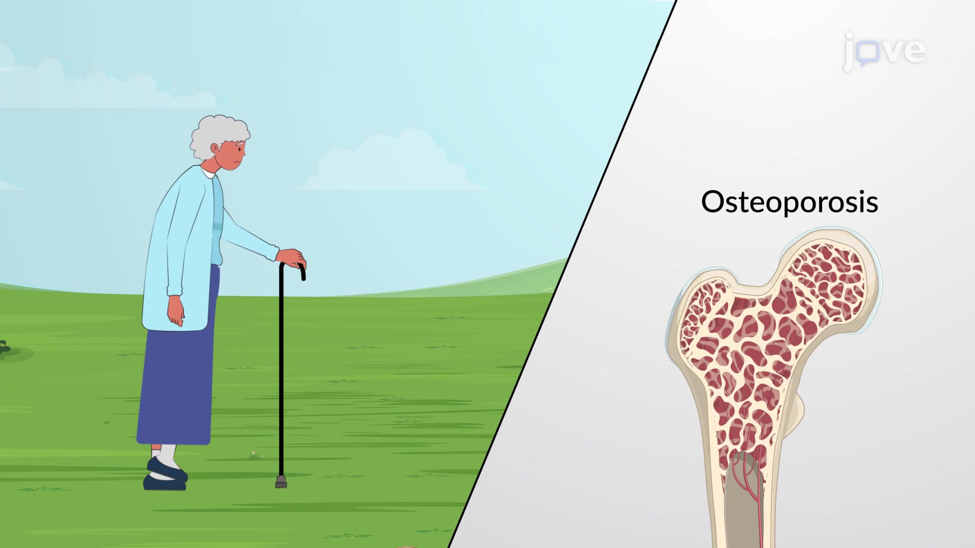
For students preparing for the MCAT or AP Biology exams, understanding spongy bone structure proves essential for questions about bone physiology, calcium homeostasis, and fracture mechanics. Medical students studying for the USMLE frequently encounter scenarios involving osteoporosis, where red marrow trabeculae become thin and disconnected, leading to increased fracture risk.
The spongy bone tissue explained concept also connects to broader physiological processes. The red bone marrow housed within trabecular spaces serves as the primary site of hematopoiesis-blood cell production-in adults. This makes spongy bone not just a structural component but also a vital organ system supporting immune function and oxygen transport throughout the body.
Related Micro-courses