Loading video…


 10 Concepts
 37.3K views

Video Summary: What are Functional Classification of Joints
Ever wonder why your shoulder can rotate 360 degrees while your skull bones never budge? The functional joint classification system explains this fascinating difference by categorizing joints based on their mobility into three distinct types. In human anatomy, synarthrosis immovable joints provide stability, amphiarthrosis slightly movable joints offer limited flexibility, and diarthrosis freely movable joints enable complex movements like those seen in NBA players' spectacular dunks. Watch the full video on JoVE Coach to master this concept with expert-led visuals and step-by-step explanations.
The functional joint classification system organizes joints based on their degree of movement rather than their structural composition. This classification method proves invaluable for understanding how different body regions balance stability with mobility to serve specific physiological functions. Unlike structural classification, which focuses on tissue types, functional classification directly relates to biomechanical performance and clinical relevance.
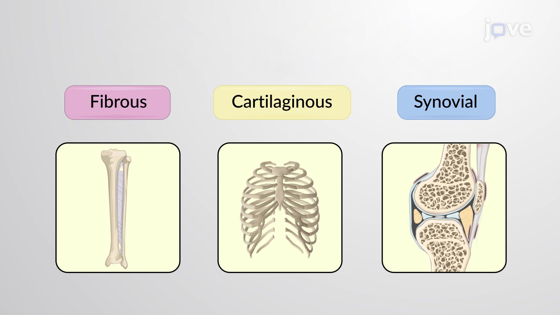
Synarthrosis immovable joints form the body's structural foundation, providing maximum stability where movement would compromise protection or function. These joints utilize dense fibrous connective tissue or cartilage to create essentially permanent connections between bones. Classic examples include the sutures between skull bones, which protect the brain while allowing for growth during childhood, and the gomphoses connecting teeth to their sockets in the maxilla and mandible.
The immobile nature of synarthroses makes them crucial for maintaining structural integrity in weight-bearing and protective roles. In clinical settings, understanding these joints helps explain why certain fractures, like those affecting skull sutures, require specialized treatment approaches. Medical students preparing for the MCAT often encounter questions about how synarthroses contribute to overall skeletal stability.
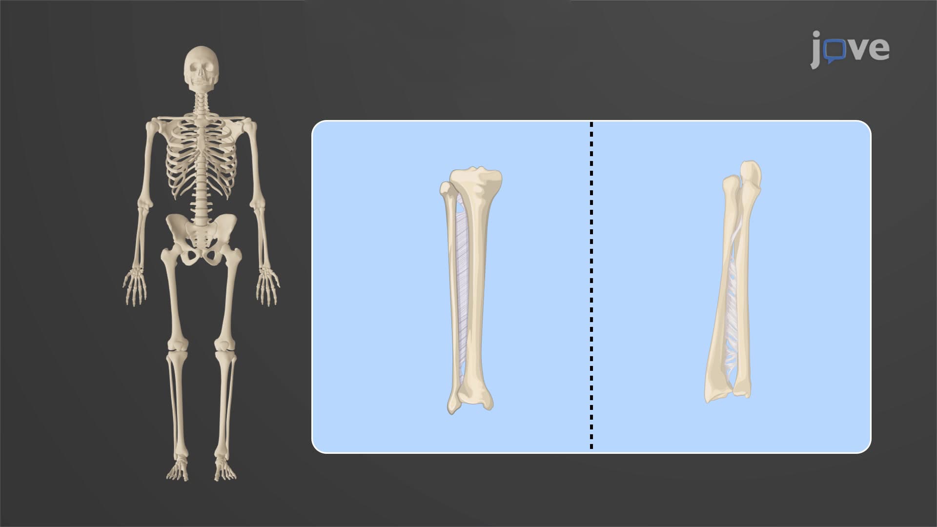
Amphiarthrosis slightly movable joints represent an evolutionary compromise between stability and flexibility. These joints permit limited movement while maintaining substantial structural support, making them ideal for areas requiring both functions. The fibrous membrane connecting the tibia and fibula (interosseous membrane) exemplifies this classification, allowing slight movement during walking while maintaining leg stability.
Another prime example includes the pubic symphysis, where cartilaginous tissue permits slight movement during childbirth while normally maintaining pelvic stability. Physical therapy students studying for licensing exams like the NPTE frequently analyze how amphiarthroses contribute to functional movement patterns while preventing excessive motion that could cause injury.
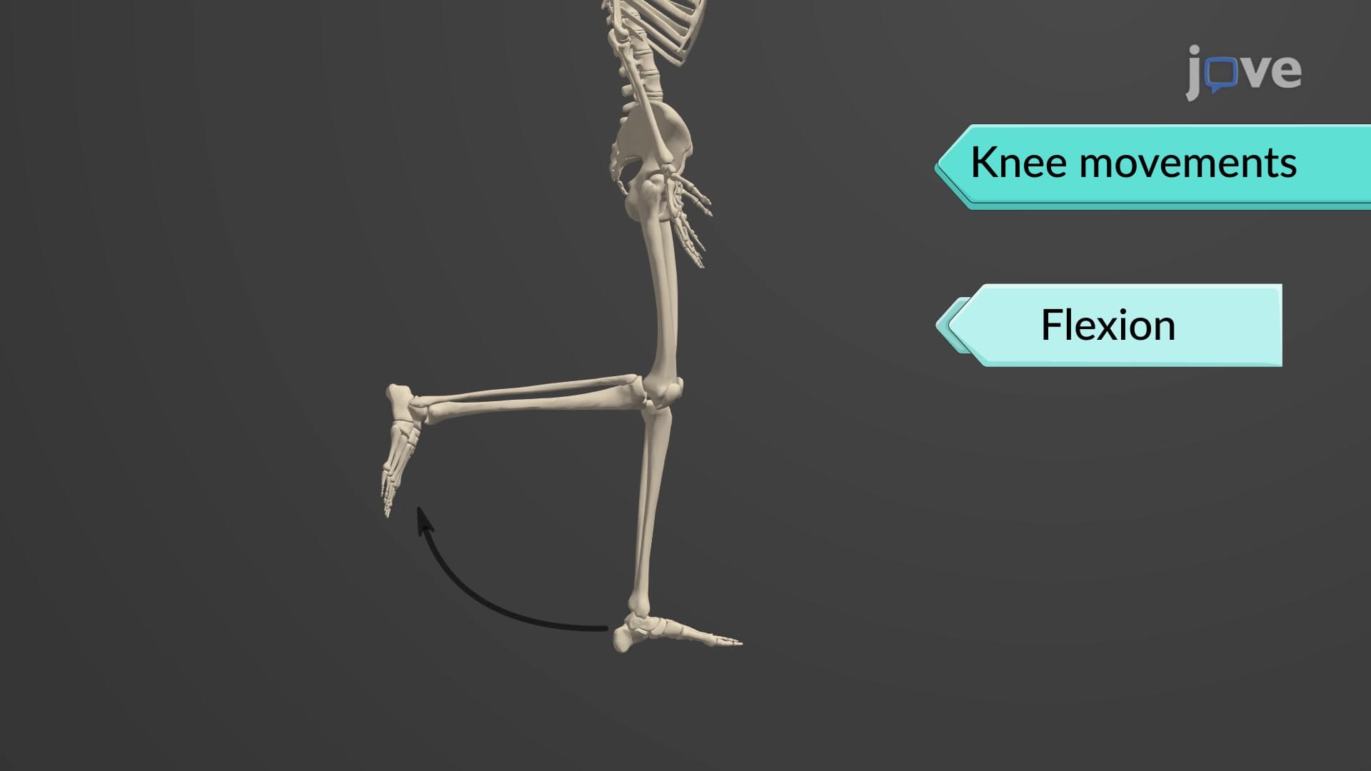
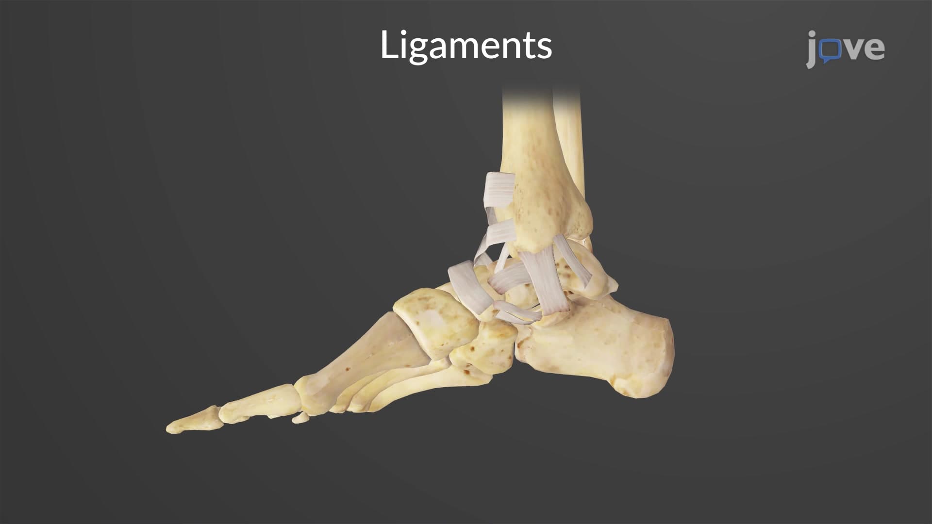
Diarthrosis freely movable joints enable the complex movements that define human mobility and dexterity. These synovial joints feature fluid-filled cavities that reduce friction and permit extensive range of motion. However, this mobility comes at the cost of stability, making diarthroses more susceptible to injuries like dislocations and ligament tears commonly seen in sports medicine.
Examples include the glenohumeral (shoulder) joint, which offers the greatest range of motion in the body, and the knee joint, which combines significant mobility with weight-bearing capacity. Athletic trainers and orthopedic residents studying for board examinations must thoroughly understand how diarthroses function under stress and their common injury patterns. The trade-off between mobility and stability in these joints explains why athletes like gymnasts and football players experience different injury patterns based on their sport-specific demands.
Related Micro-courses