Loading video…


 10 Concepts
 37.3K views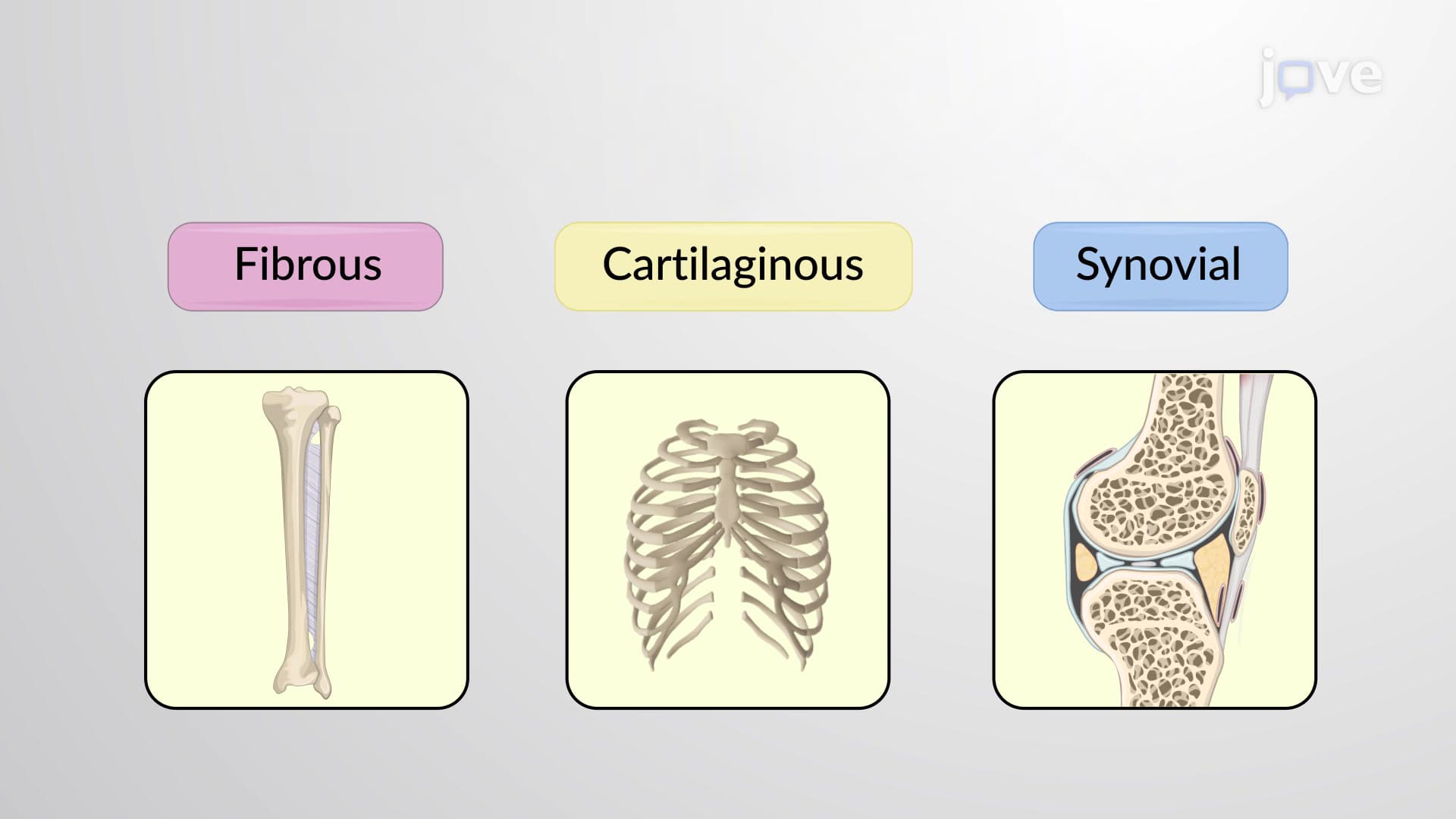

Video Summary: What are Structural Classification of Joints
Ever wonder why your skull feels rock-solid while your shoulder moves freely in all directions? The answer lies in structural joint classification, which categorizes joints based on their connective tissue composition and mobility. From the immovable sutures protecting your brain to the freely moving ball-and-socket joint in your shoulder, understanding what structural classification of joints means reveals how anatomy directly impacts function in the human body. Watch the full video on JoVE Coach to master this concept with expert-led visuals and step-by-step explanations.
The structural classification of joints provides a systematic approach to understanding how different joints function based on their anatomical composition. Unlike functional classification, which focuses on movement capability, structural classification examines the actual materials that connect bones and the presence or absence of joint cavities. This classification system is fundamental to anatomy and physiology courses at both high school AP Biology level and undergraduate programs.
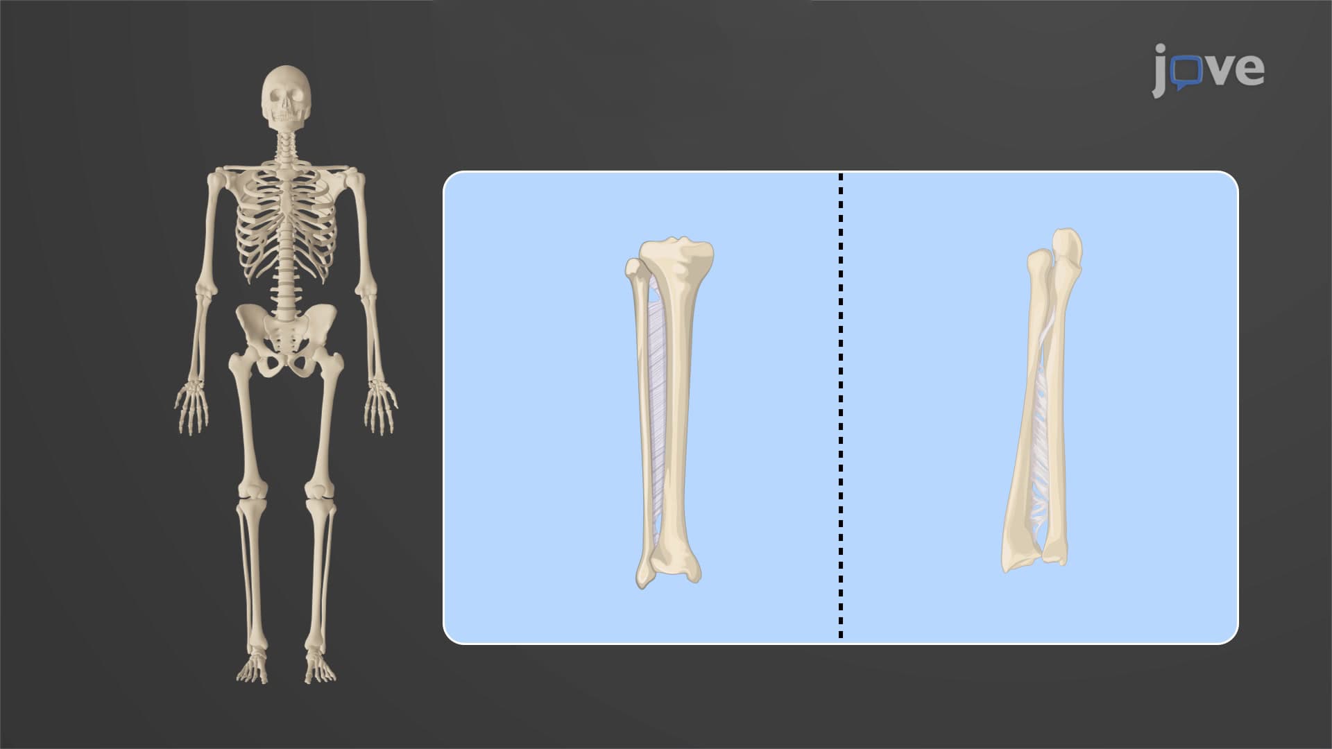
Fibrous joints represent the most restrictive structural category, characterized by dense irregular connective tissue rich in collagen fibers. The length and density of these fibers directly correlate with joint mobility. Sutures in the skull exemplify immovable fibrous joints, where short, tightly packed collagen fibers create an interlocking pattern that protects the brain while allowing for slight expansion during growth. In contrast, the syndesmosis joint between the radius and ulna in the forearm demonstrates slightly movable fibrous joints, where longer connective tissue fibers permit limited rotation during pronation and supination movements.
Students preparing for the MCAT or AP Biology exams should note that fibrous joints are particularly important in questions about skeletal development and injury mechanisms. For instance, understanding how skull sutures fuse over time helps explain why pediatric skull fractures differ significantly from adult injuries.
Cartilaginous joints utilize either hyaline cartilage or fibrocartilage to connect bones, creating joints that balance stability with controlled movement. Primary cartilaginous joints (synchondroses) feature hyaline cartilage and are typically temporary structures that ossify with age, such as the epiphyseal plates in growing bones. Secondary cartilaginous joints (symphyses) contain fibrocartilage discs that provide shock absorption and limited movement.
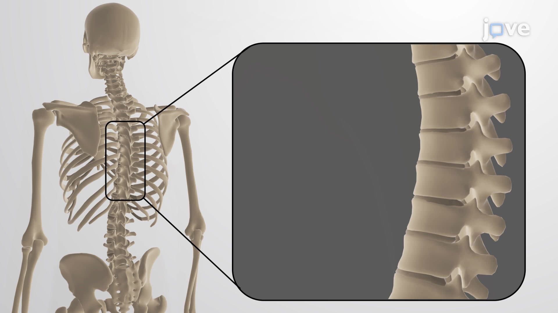
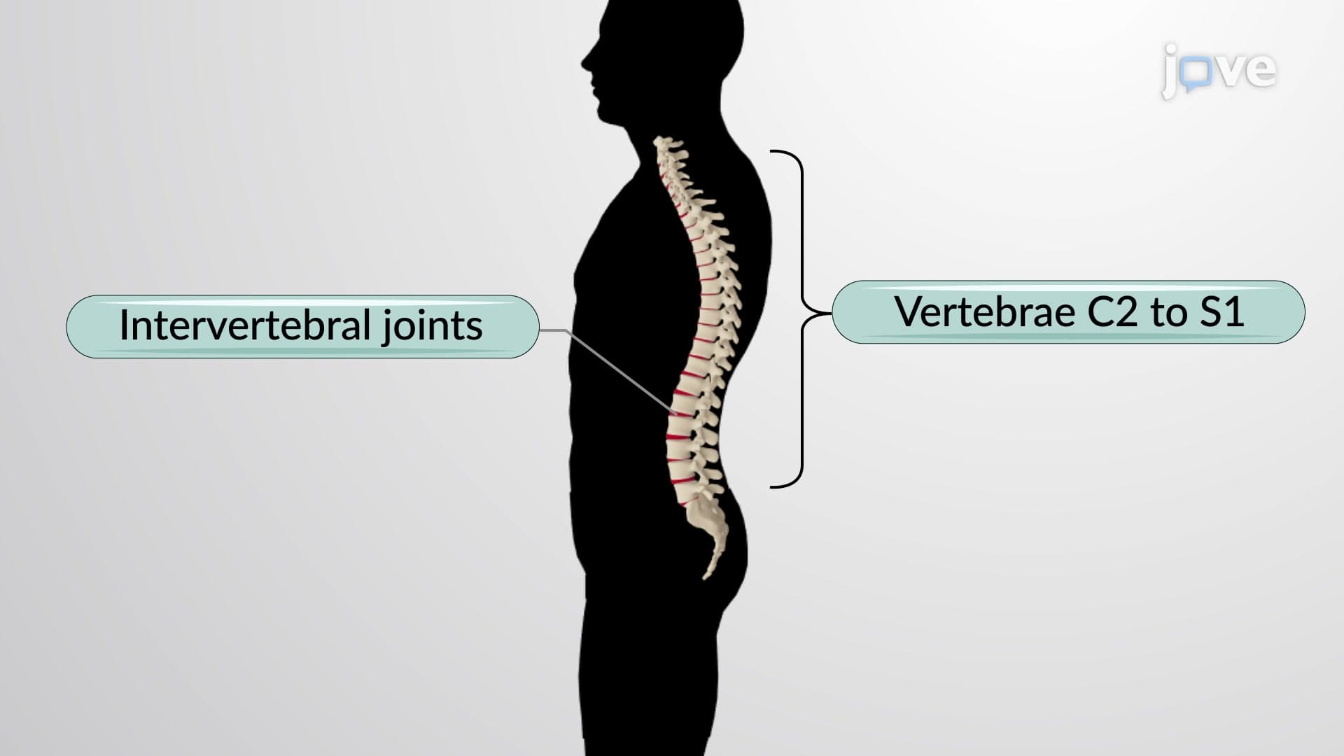
The intervertebral discs serve as excellent examples for understanding this concept. These fibrocartilaginous structures allow slight movement between individual vertebrae while maintaining spinal stability. When students encounter spine-related questions on nursing exams like the NCLEX or HESI A2, understanding cartilaginous joint structure helps explain both normal spinal mechanics and common pathologies like herniated discs.
Synovial joints represent the most structurally complex category, featuring joint cavities filled with synovial fluid and articular cartilage covering bone surfaces. The joint capsule, composed of an outer fibrous layer and inner synovial membrane, creates a sealed environment that facilitates smooth movement while providing structural support.
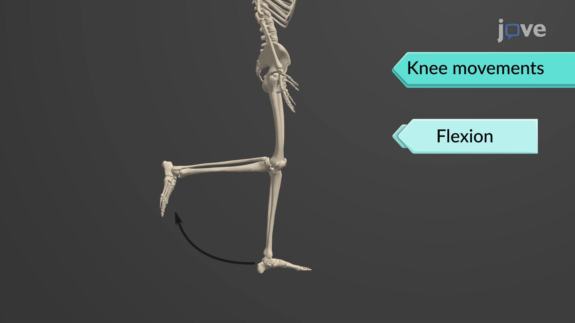
The knee joint exemplifies synovial joint complexity, incorporating multiple structural features including menisci, ligaments, and bursae. For pre-med students, understanding synovial joint anatomy is crucial for MCAT success and forms the foundation for comprehending orthopedic medicine concepts in medical school.
Related Micro-courses