Loading subject page…


37,301 views

Video Summary: What Is Ankle Joint
Ever wonder why basketball players like Stephen Curry can pivot so smoothly on the court? The ankle joint anatomy structure forms a remarkable hinge mechanism that enables precise foot movements essential for athletic performance. This talocrural joint allows dorsiflexion and plantarflexion while preventing dangerous sideways motion through its unique bone and ligament arrangement. What is ankle joint mechanics become crucial for understanding sports injuries and rehabilitation in American athletics. Watch the full video on JoVE Coach to master this concept with expert-led visuals and step-by-step explanations.
The ankle joint anatomy structure represents one of the most mechanically sophisticated joints in the human body. This talocrural joint serves as the critical link between the leg and foot, bearing the entire body weight during walking, running, and jumping activities. Understanding what is the structure of the ankle joint becomes essential for students pursuing healthcare careers, as ankle injuries account for approximately 85% of all sports-related injuries in American high schools and colleges.
The ankle mortise joint derives its name from its resemblance to a carpentry mortise-and-tenon connection. Three bones create this stable yet mobile structure: the talus (ankle bone), tibia (shin bone), and fibula (smaller leg bone). The talus fits snugly between the medial lateral malleolus ankle projections, creating what anatomists call the "ankle fork." This design prevents dangerous sideways motion while allowing the foot to flex upward (dorsiflexion) and downward (plantarflexion).
Students preparing for the MCAT or AP Biology exams should note that this joint classification as a uniaxial hinge joint limits movement to one plane, unlike ball-and-socket joints. The distal tibia forms the superior surface of the joint, while the medial malleolus of the tibia and lateral malleolus of the fibula create the sides of this bony socket.
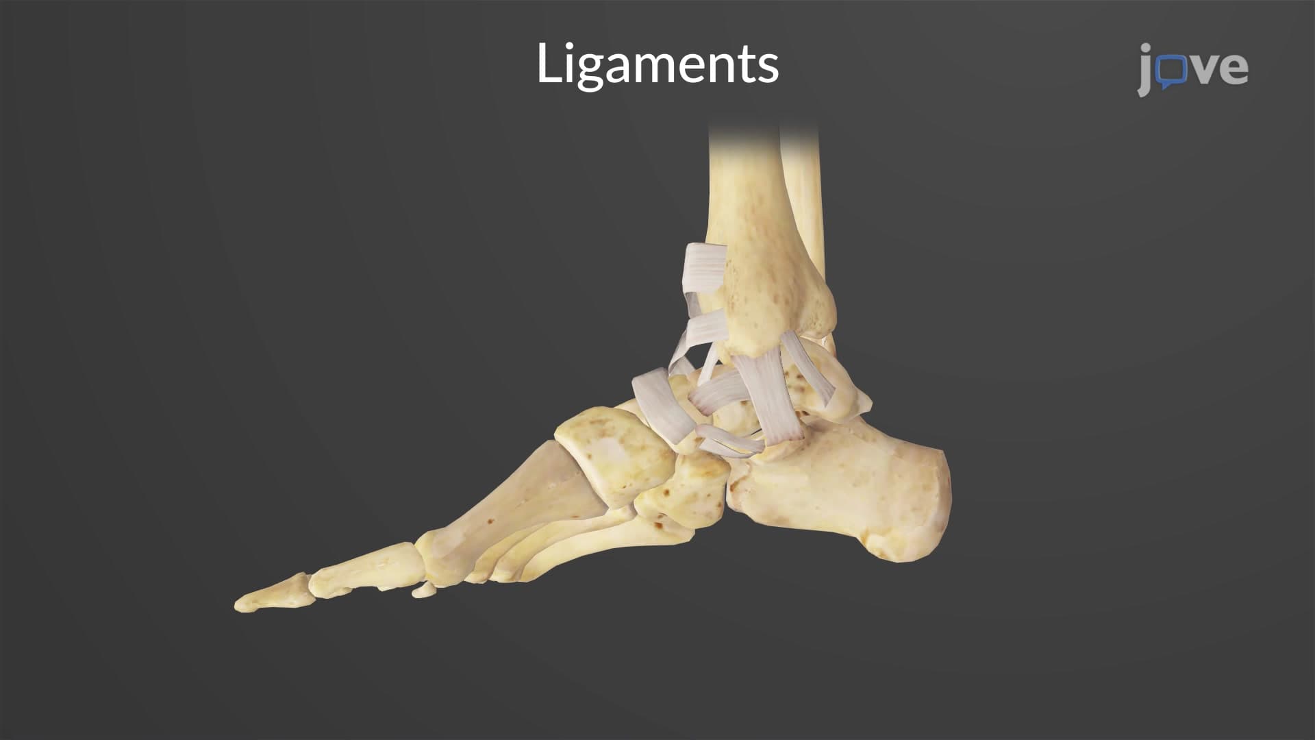
The deltoid lateral ankle ligament system provides crucial joint stability. On the medial side, the broad deltoid ligament prevents excessive eversion (outward rolling) of the foot. This triangular ligament complex connects the medial malleolus to multiple foot bones, creating a strong medial anchor.
Laterally, three distinct ligaments work together: the anterior talofibular ligament (most commonly injured in ankle sprains), posterior talofibular ligament, and calcaneofibular ligament. These structures prevent inversion injuries, which occur when the foot rolls inward excessively. Understanding this anatomy helps explain why lateral ankle sprains are more common than medial injuries in American football and basketball players.
The talocrural joint ankle design includes hyaline cartilage covering all articular surfaces, reducing friction during the estimated 1 million steps Americans take annually. The fibrous joint capsule provides additional stability while allowing necessary movement. This anatomical arrangement makes the ankle joint particularly relevant for nursing students preparing for the NCLEX-RN, as ankle assessment and injury prevention are common clinical scenarios.
Related Micro-courses