Loading video…


 18 Concepts
 14.5K views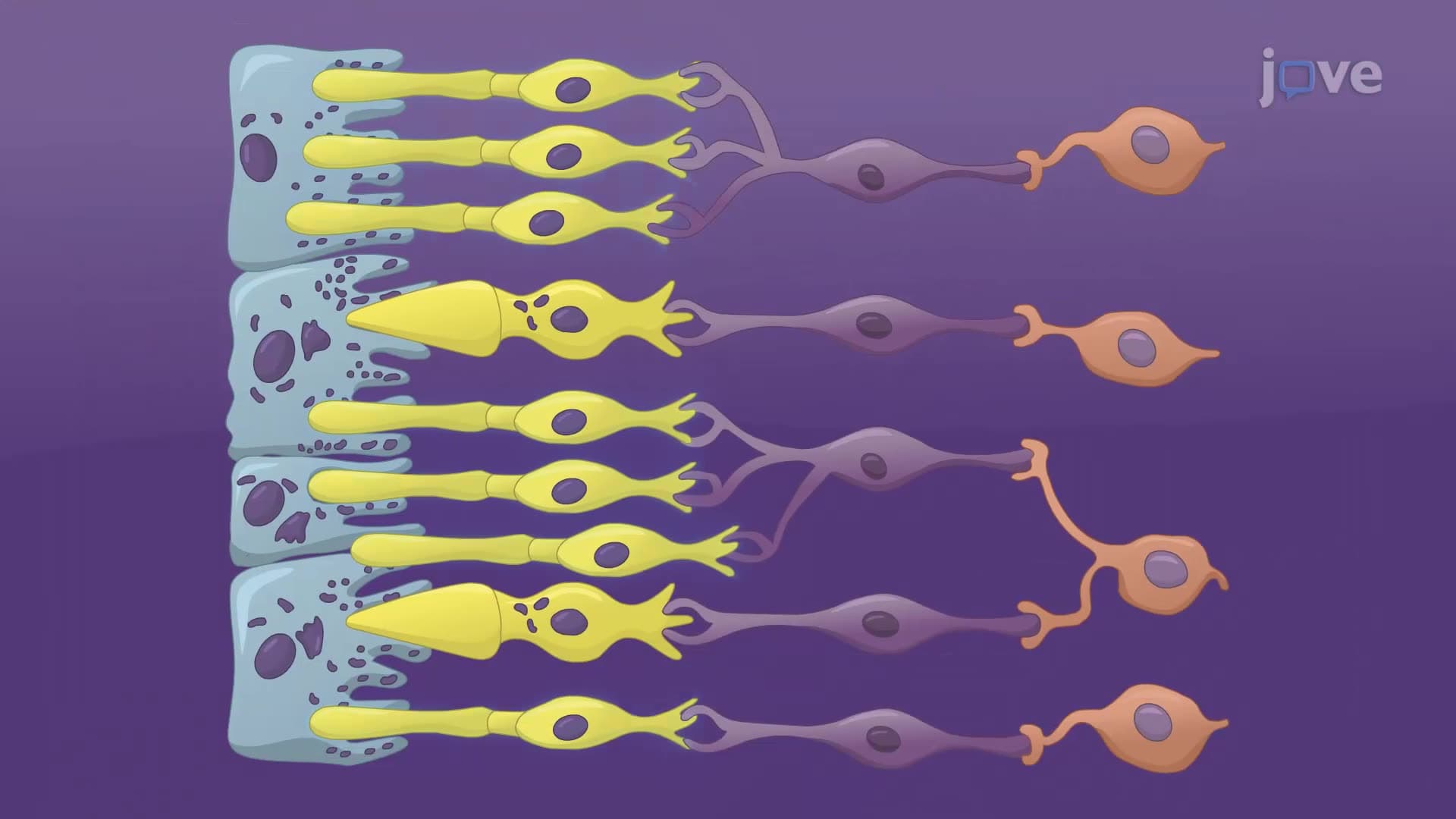

Video Summary: What Is Visual System
Ever wonder why traffic lights use red, yellow, and green instead of other colors? Color vision allows us to distinguish between millions of different hues, making everyday tasks like driving safer and more intuitive. The visual system is our body's sophisticated network that transforms light waves into meaningful visual information, with specialized cells called cones enabling us to perceive the full spectrum of colors around us. Watch the full video on JoVE Coach to master this concept with expert-led visuals and step-by-step explanations.
The visual system represents one of biology's most remarkable sensory mechanisms, transforming electromagnetic radiation into the rich tapestry of colors we experience daily. This complex process begins when light photons enter the eye and culminates in conscious color perception within the brain's visual cortex. Color vision specifically refers to our ability to distinguish between different wavelengths of light, typically ranging from 380 to 700 nanometers in the visible spectrum.
Color vision definition encompasses the biological process by which specialized photoreceptor cells called cones detect and respond to different wavelengths of light. Unlike rods, which function primarily in low-light conditions and provide black-and-white vision, cones operate optimally in bright light and contain photopigments sensitive to specific wavelength ranges. Humans possess three types of cones: S-cones (short wavelength/blue), M-cones (medium wavelength/green), and L-cones (long wavelength/red). This trichromatic system enables us to perceive approximately 10 million distinct colors.
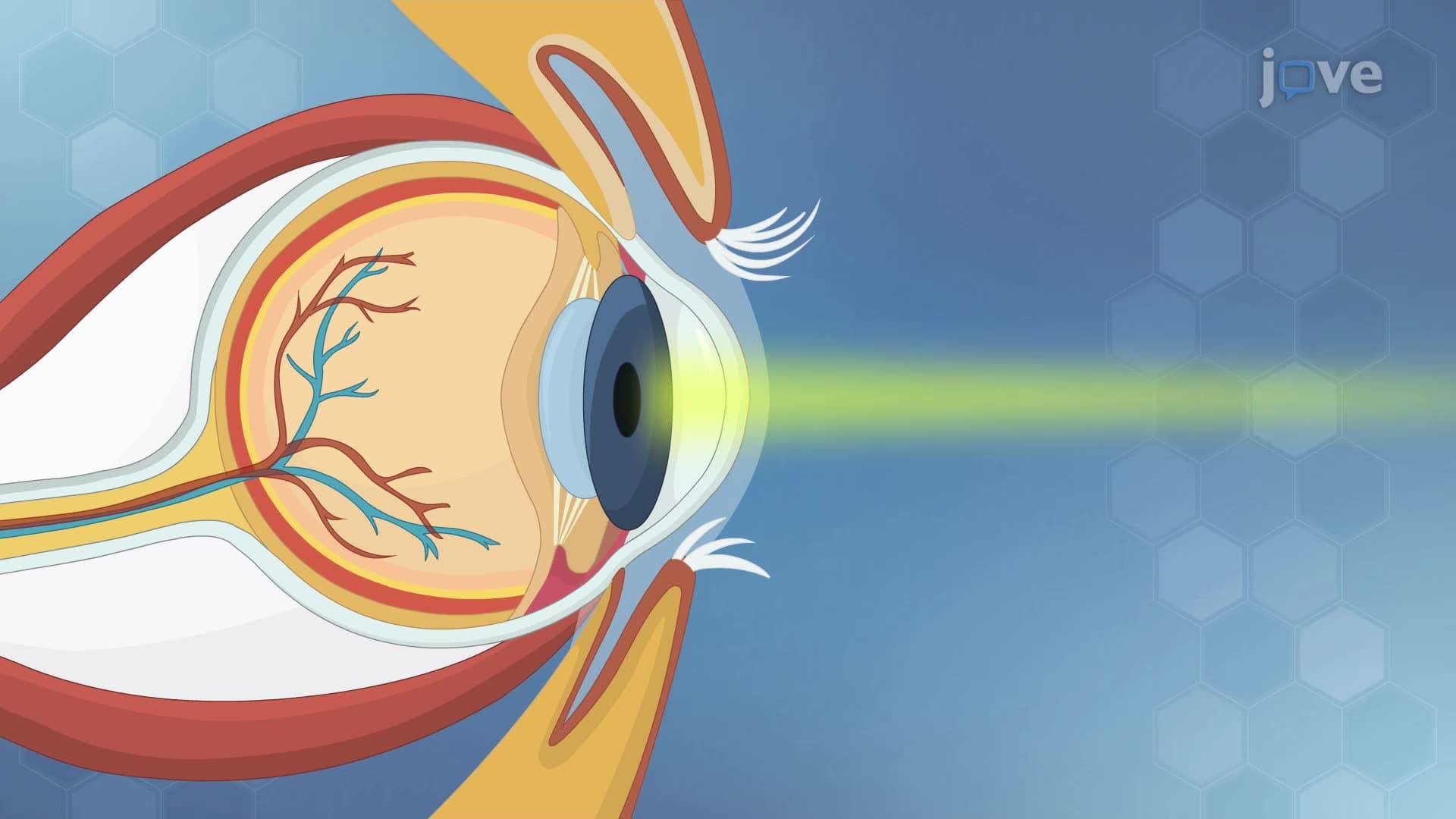
When studying what is color vision in detail, students must understand that color perception results from complex neural processing rather than simple wavelength detection. The retina contains approximately 6-7 million cone cells concentrated in the fovea, the eye's central region responsible for sharp, detailed vision. These cells convert light energy into electrical signals through a process called phototransduction, where photons trigger conformational changes in photopigment molecules.
The color vision concept extends beyond initial photoreceptor activation to include sophisticated neural processing mechanisms. Bipolar cells receive signals from multiple photoreceptors and perform initial signal processing before transmitting information to ganglion cells. These ganglion cells, whose axons form the optic nerve, carry visual information to the lateral geniculate nucleus (LGN) in the thalamus, which acts as a relay station before signals reach the primary visual cortex (V1) in the occipital lobe.
Feature detector neurons in V1 respond to specific visual attributes, including color boundaries, orientation, and spatial frequency. This parallel processing system allows simultaneous analysis of multiple visual features, with specialized neural pathways dedicated to color processing (the parvocellular pathway) operating alongside those processing motion and brightness (the magnocellular pathway).
Understanding color vision proves essential for students pursuing careers in optometry, ophthalmology, neuroscience, or psychology. Color vision deficiencies, commonly called color blindness, affect approximately 8% of men and 0.5% of women in the United States, making this topic clinically relevant. The Ishihara color test, commonly used in medical examinations and driver's license applications, assesses color vision function by presenting numbers embedded within colored dot patterns.
For standardized exam preparation, students should focus on the anatomical pathway from cornea to visual cortex, understanding how cone cell responses create color opponent processing, and recognizing how parallel processing enables binding of visual features into cohesive perceptions.
Related Micro-courses