Loading subject page…

- Analytical Chemistry
- Principles of Nuclear Magnetic Resonance
- Atomic Nuclei: Types of Nuclear Relaxation
Micro-course: Principles of Nuclear Magnetic Resonance
24,278 views
Learn key concepts and earn your certificate.


Atomic Nuclei: Types of Nuclear Relaxation

00:00
01:28
Video Summary: Atomic Nuclei Types of Nuclear Relaxation Explained
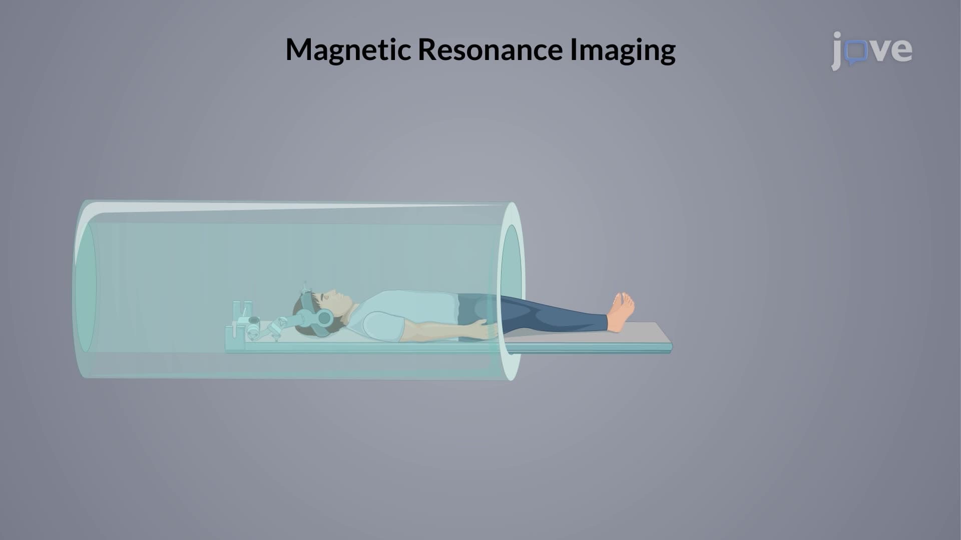
Ever wonder why MRI machines at hospitals like Johns Hopkins can capture crystal-clear images of your brain? The secret lies in understanding the types of nuclear relaxation that occur when atomic nuclei return to their ground state after magnetic excitation. Nuclear magnetic resonance relies on two distinct relaxation mechanisms-spin-lattice (T1) and spin-spin (T2) relaxation-each with unique time constants that determine signal quality and imaging contrast. Watch the full video on JoVE Coach to master this concept with expert-led visuals and step-by-step explanations.
- Understand the fundamental principles behind atomic nuclei types of nuclear relaxation and their role in NMR spectroscopy
- Identify the two primary relaxation mechanisms: spin-lattice (longitudinal) and spin-spin (transverse) relaxation
- Compare T1 and T2 relaxation time constants and explain why T2 is typically shorter than T1
- Analyze how molecular size, temperature, and nuclear environment affect relaxation rates
- Evaluate the relationship between relaxation rates and spectral line broadening in NMR
- Apply knowledge of relaxation mechanisms to predict signal behavior in different molecular systems
- Recognize the importance of optimal relaxation times (0.1-10 seconds) for obtaining quality NMR spectra
- Explore how relaxation phenomena enable practical applications like medical MRI imaging
Understanding Atomic Nuclei Types of Nuclear Relaxation
Nuclear relaxation represents the fundamental process by which excited atomic nuclei return to their equilibrium state after absorbing electromagnetic energy. This phenomenon follows first-order exponential decay kinetics and occurs through two distinct pathways that are essential for NMR spectroscopy and medical imaging applications.
Spin-Lattice Relaxation: The T1 Mechanism
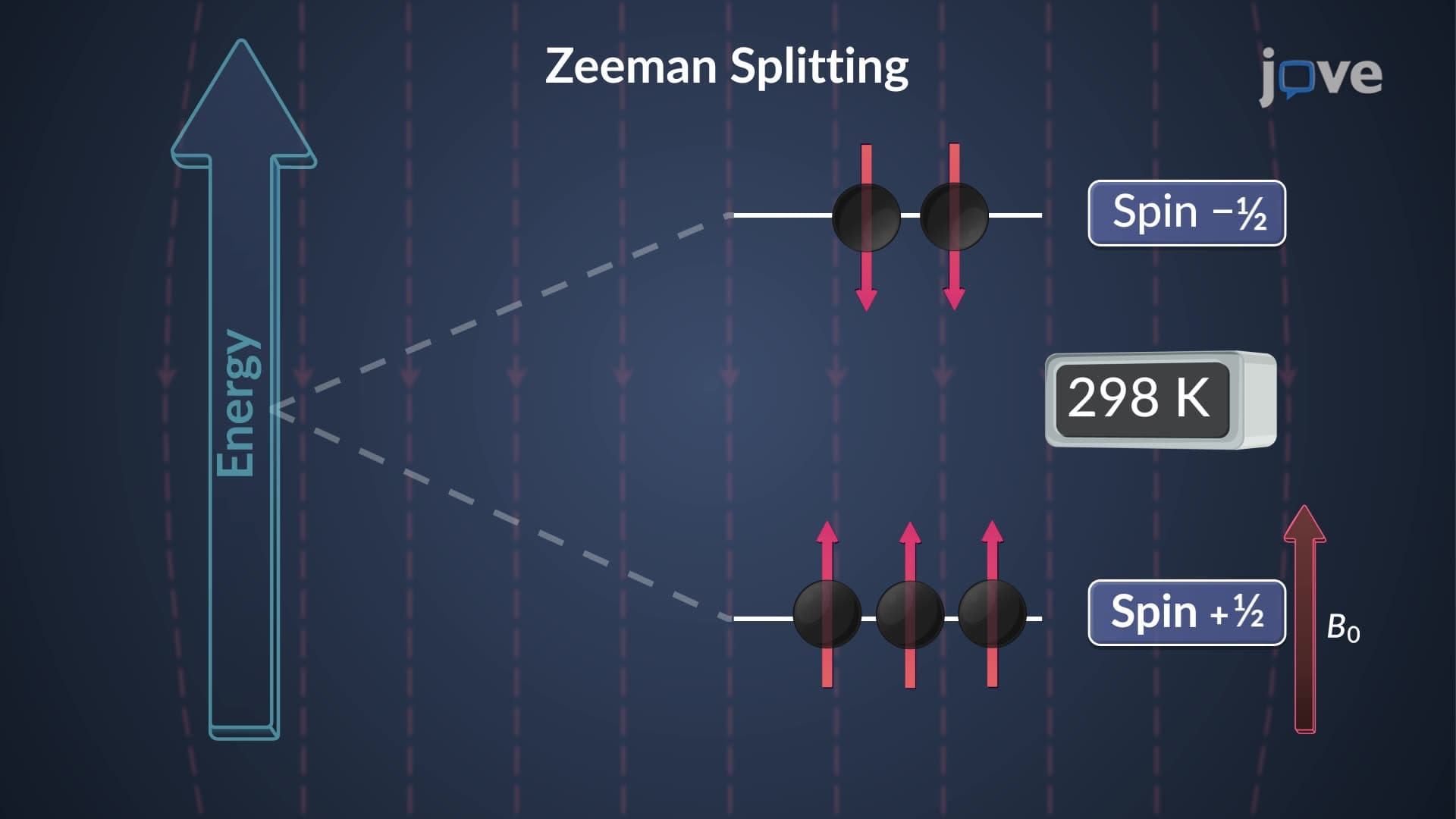
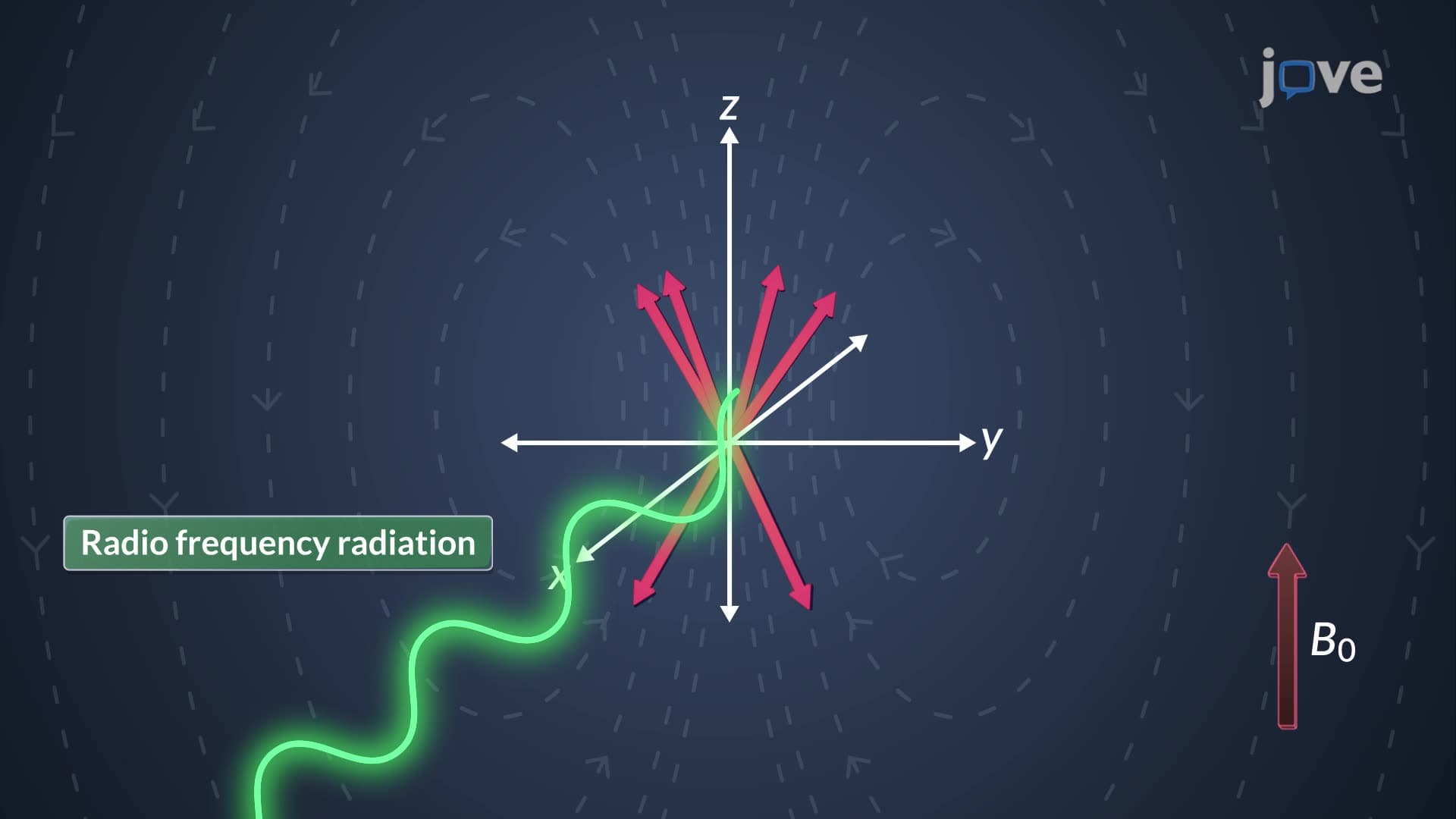
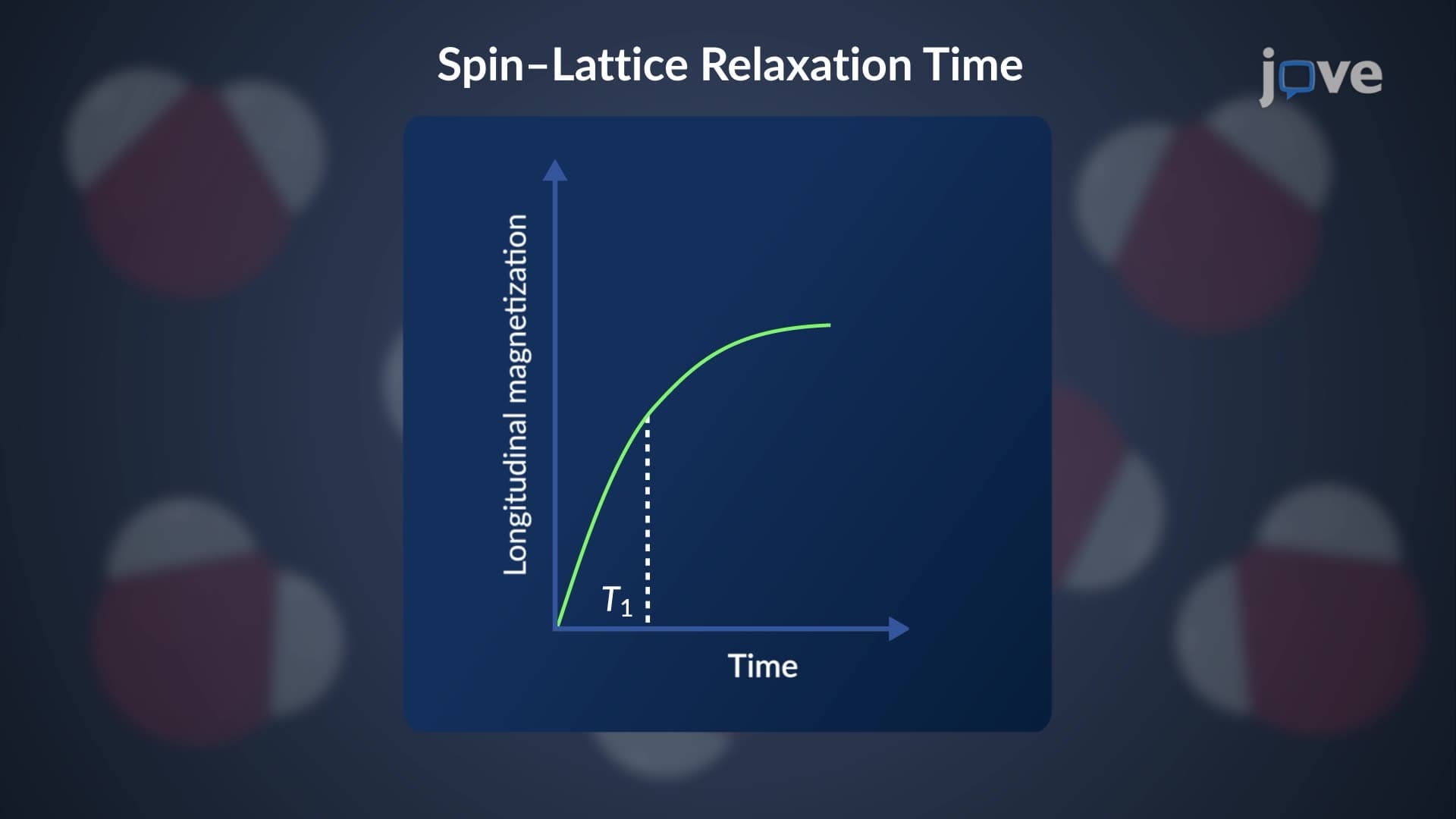
Longitudinal relaxation T1, also called spin-lattice relaxation, describes how excited nuclei transfer energy to their molecular surroundings. This process primarily occurs through magnetic dipole-dipole interactions, where the excited nucleus releases energy to nearby magnetic dipoles-most commonly tumbling protons in the molecular environment. The spin lattice relaxation mechanism restores the Boltzmann distribution of nuclear spin states, effectively "cooling" the nuclear spin system back to thermal equilibrium.
The T1 relaxation time constant represents the average time required for 63% of excited nuclei to return to the ground state. For liquid samples commonly analyzed in US research laboratories at institutions like MIT and Stanford, T1 values typically range from 0.01 to 100 seconds. These values depend critically on nuclear type, molecular position, molecular size, and temperature-factors that students encounter in AP Chemistry and college-level physical chemistry courses.
Spin-Spin Relaxation: The T2 Mechanism
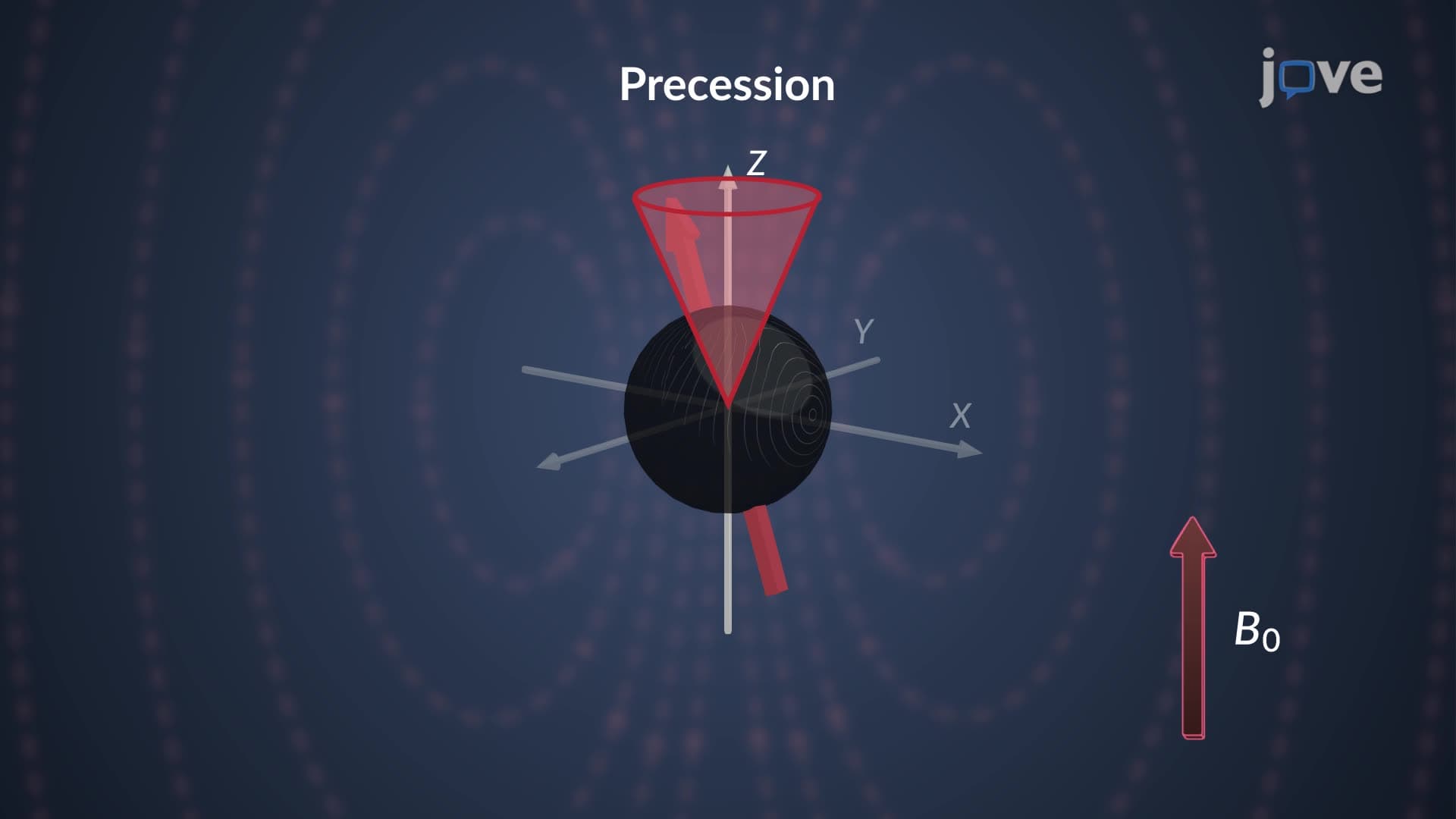
Transverse relaxation T2, known as spin spin relaxation NMR, occurs through a fundamentally different mechanism. Rather than energy transfer to the surroundings, T2 relaxation results from magnetic interactions between neighboring precessing nuclei. These spin-spin interactions cause dephasing in the transverse plane, where individual nuclear magnetic moments lose their synchronized precession.
The relaxation time NMR spectroscopy parameter T2 is invariably shorter than T1 because transverse dephasing can occur without energy loss to the environment. This distinction becomes crucial when students analyze NMR spectra in organic chemistry labs or prepare for the MCAT's chemistry sections.
Practical Implications and Applications
Understanding T1 T2 relaxation NMR types proves essential for optimizing experimental conditions and interpreting results. High relaxation rates cause spectral line broadening, reducing resolution and signal quality. Conversely, extremely long relaxation times can lead to signal saturation, preventing adequate signal detection.
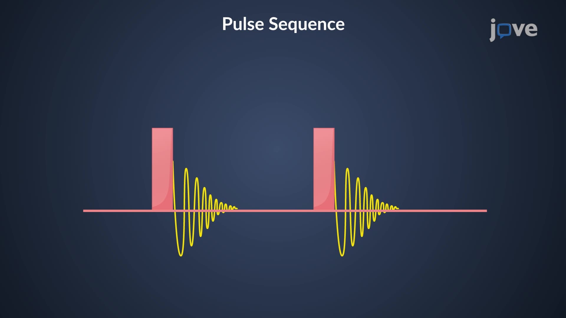
The ideal nuclear half-life of 0.1-10 seconds represents a practical compromise between signal intensity and spectral resolution. This principle guides pulse sequence design in modern NMR spectrometers used across US pharmaceutical companies and academic research centers, making this knowledge valuable for students pursuing careers in chemistry, biochemistry, or materials science.
Frequently Asked Questions
The two primary types are spin-lattice (T1 or longitudinal) relaxation and spin-spin (T2 or transverse) relaxation. T1 relaxation involves energy transfer to the molecular environment, while T2 relaxation results from magnetic interactions between neighboring nuclei causing signal dephasing.
T1 relaxation restores thermal equilibrium by transferring energy to surrounding molecules, while T2 relaxation causes signal decay through nuclear spin dephasing without energy loss. T2 is typically shorter than T1 because dephasing can occur without requiring energy dissipation to the environment.
MCAT questions often test understanding of relaxation mechanisms in NMR spectroscopy and MRI applications. Students should know that T1 determines recovery time between pulses, T2 affects signal decay rates, and both influence image contrast in medical imaging scenarios.
While not explicitly tested, understanding relaxation helps explain spectroscopic techniques used in advanced chemistry labs. This knowledge provides foundation for college physical chemistry and demonstrates how quantum mechanical principles apply to real analytical instruments.
MRI machines in hospitals like Cleveland Clinic exploit T1 and T2 relaxation differences between tissues to create image contrast. Different tissues have characteristic relaxation times, allowing radiologists to distinguish between healthy and diseased tissue based on signal intensity patterns.
Basic understanding requires only exponential decay concepts from high school algebra and chemistry. The mathematical treatment involves first-order kinetics similar to radioactive decay, making it accessible to students with solid chemistry fundamentals.
Focus on understanding the physical mechanisms rather than memorizing equations. Create concept maps linking molecular motion, magnetic interactions, and relaxation pathways. Practice interpreting how molecular size and temperature affect T1 and T2 values.
Consider studying pulse sequences in multidimensional NMR, relaxation in solid-state systems, and advanced MRI contrast mechanisms. These topics appear in graduate-level spectroscopy courses and professional applications in pharmaceutical research.
Related Micro-courses