Loading subject page…


24,278 views


Video Summary: Diamagnetic Shielding of Nuclei Local Diamagnetic Currents
Ever wonder why hydrogen atoms in water show up differently on MRI scans than those in fat tissue? The answer lies in diamagnetic shielding nuclei mechanisms. When magnetic fields interact with molecules, electrons create protective currents around atomic nuclei, much like how MRI machines detect different tissue types in US hospitals. This diamagnetic shielding of nuclei: local diamagnetic currents phenomenon explains why identical atoms can produce distinct NMR signals based on their electronic environments. Watch the full video on JoVE Coach to master this concept with expert-led visuals and step-by-step explanations.
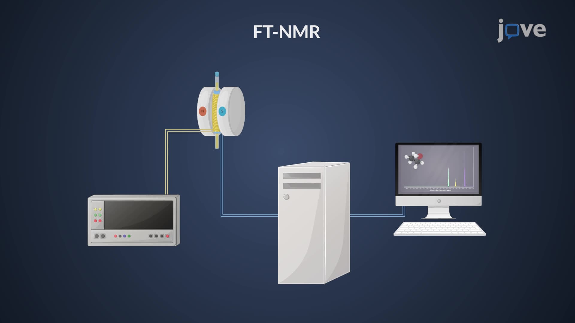
When placed in a strong magnetic field, molecules don't simply absorb energy uniformly. Instead, the electrons surrounding each nucleus respond by creating tiny circular currents that oppose the applied field-a phenomenon central to Nuclear Magnetic Resonance (NMR) spectroscopy used in research labs across US universities and medical facilities.
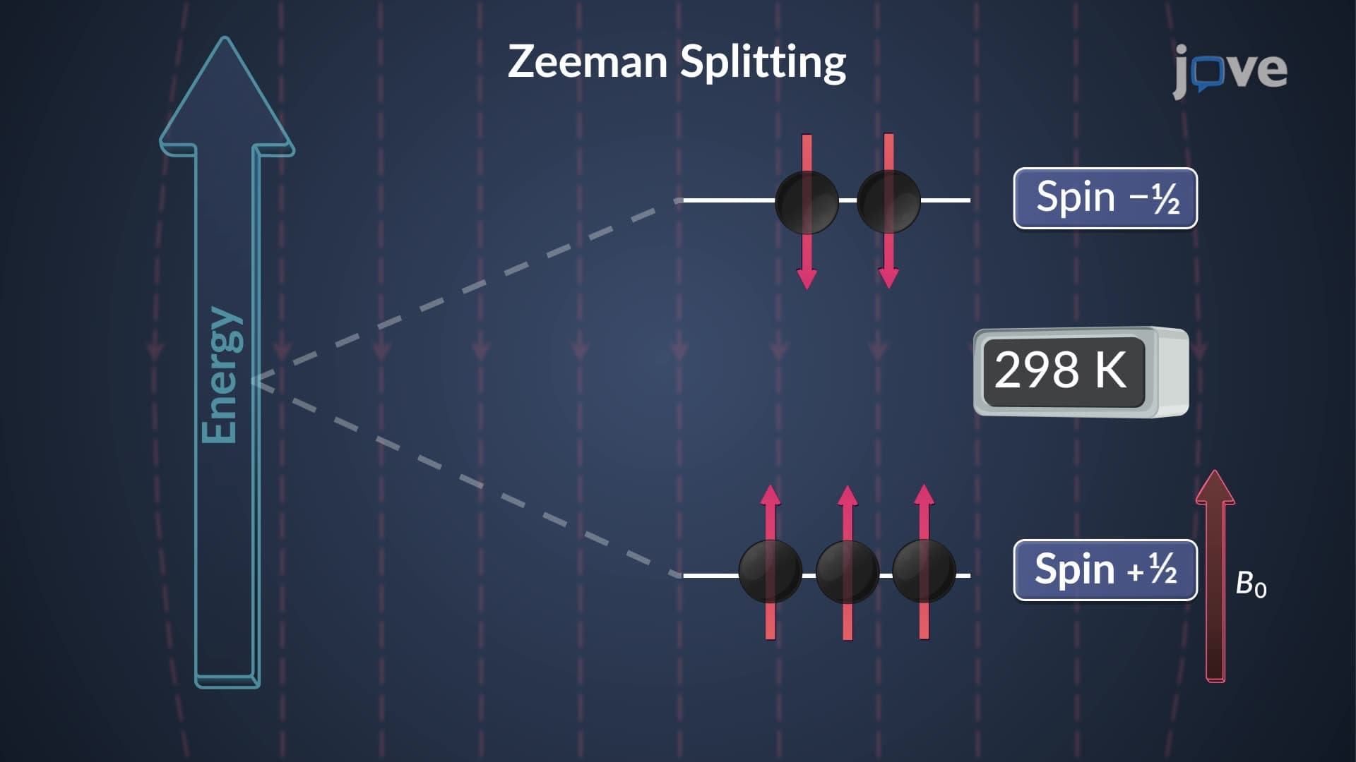
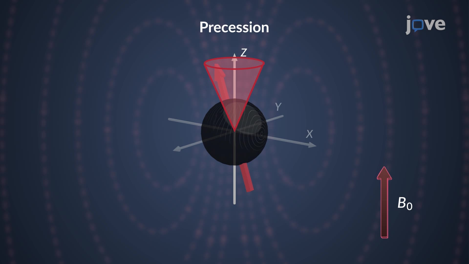
Think of electrons as microscopic shields protecting nuclei from external magnetic fields. When an external field B₀ is applied, electrons begin circulating around nuclei, generating their own magnetic field (Blocal) that opposes the original field. This creates an effective field (Beffective = B₀ - Blocal) that's weaker than the applied field. The strength of this protection depends entirely on electron density-areas with more electrons provide better shielding.
This concept frequently appears on AP Chemistry exams and college organic chemistry courses, where students must predict NMR chemical shifts based on molecular structure. For example, protons near electronegative atoms like oxygen appear "downfield" (higher chemical shift) because they're less shielded.
Different positions within the same molecule experience vastly different shielding effects. Consider ethanol (C₂H₅OH), commonly studied in US undergraduate chemistry labs. The methyl protons (CH₃) are well-shielded by surrounding electrons and appear upfield in NMR spectra. Meanwhile, the hydroxyl proton (OH) experiences minimal shielding due to oxygen's electronegativity pulling electron density away, causing it to appear downfield.
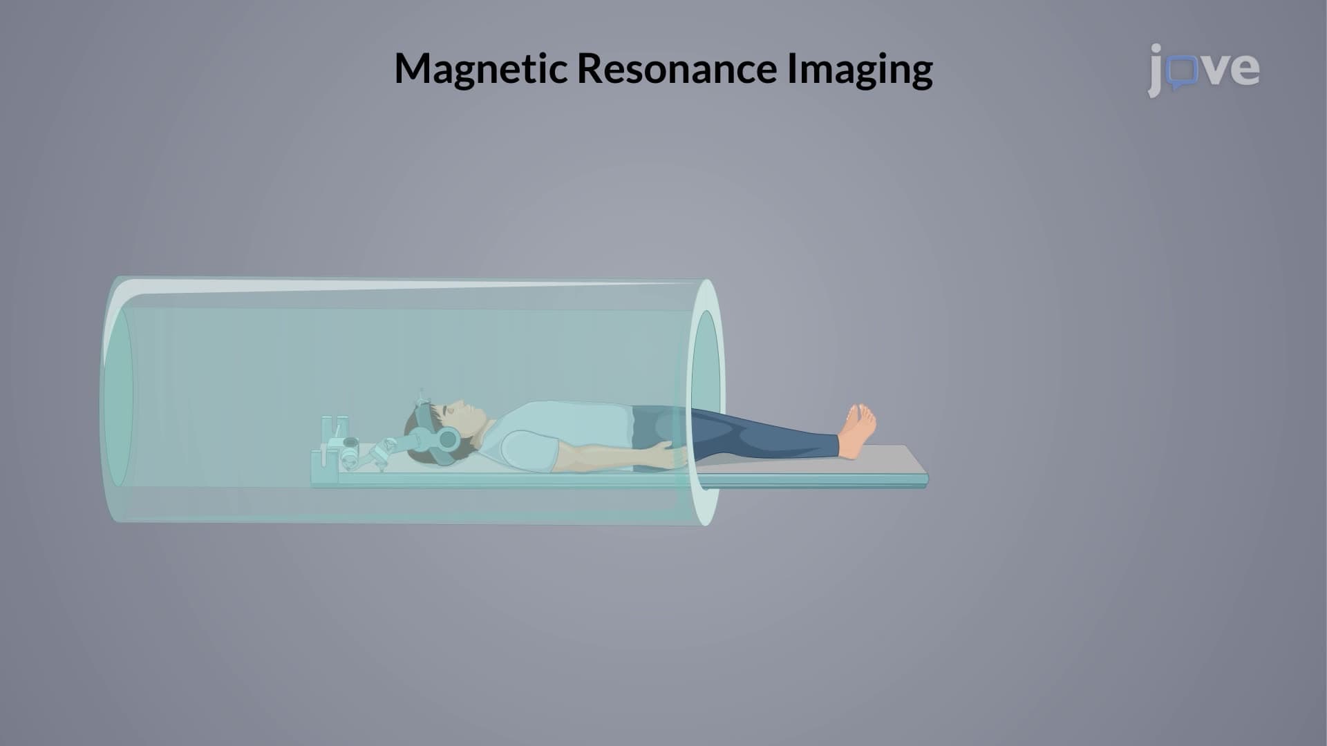
This shielding principle enables MRI technology used in US hospitals. Different tissues contain molecules with varying electron densities around hydrogen nuclei, creating distinct signal patterns that allow doctors to differentiate between healthy and diseased tissue. Similarly, pharmaceutical companies use NMR spectroscopy to verify drug compound structures, relying on predictable shielding patterns to confirm molecular identity.
Understanding diamagnetic shielding helps students excel on standardized tests like the MCAT, where they must interpret spectroscopic data and predict molecular behavior in biological systems.
Related Micro-courses