Loading subject page…


24,278 views

Video Summary: What Is Atomic Nuclei Magnetic Resonance
Ever wondered how MRI machines at hospitals like Johns Hopkins can see inside your body without surgery? Atomic nuclei magnetic resonance occurs when nuclear spins absorb radiofrequency energy at their specific Larmor frequency, causing them to flip between energy states and create detectable magnetic signals. This fundamental phenomenon enables modern medical imaging and chemical analysis across the United States. Understanding what is atomic nuclei magnetic resonance reveals how tiny nuclear spins can be manipulated to provide life-saving diagnostic information. Watch the full video on JoVE Coach to master this concept with expert-led visuals and step-by-step explanations.
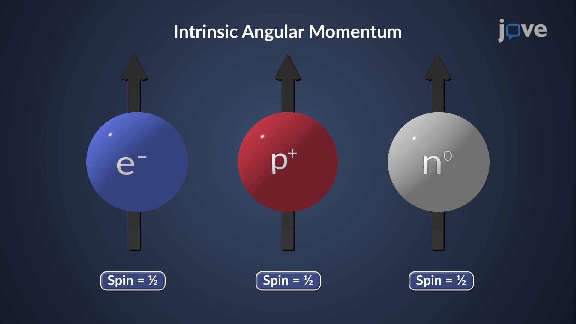
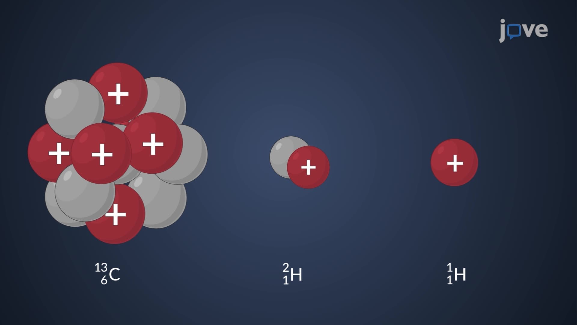
Atomic nuclei magnetic resonance represents one of the most elegant examples of quantum mechanics meeting practical application. At its core, this phenomenon occurs when atomic nuclei with non-zero spin (like hydrogen-1, carbon-13, and phosphorus-31) are placed in a strong external magnetic field and exposed to radiofrequency radiation at precisely the right frequency.
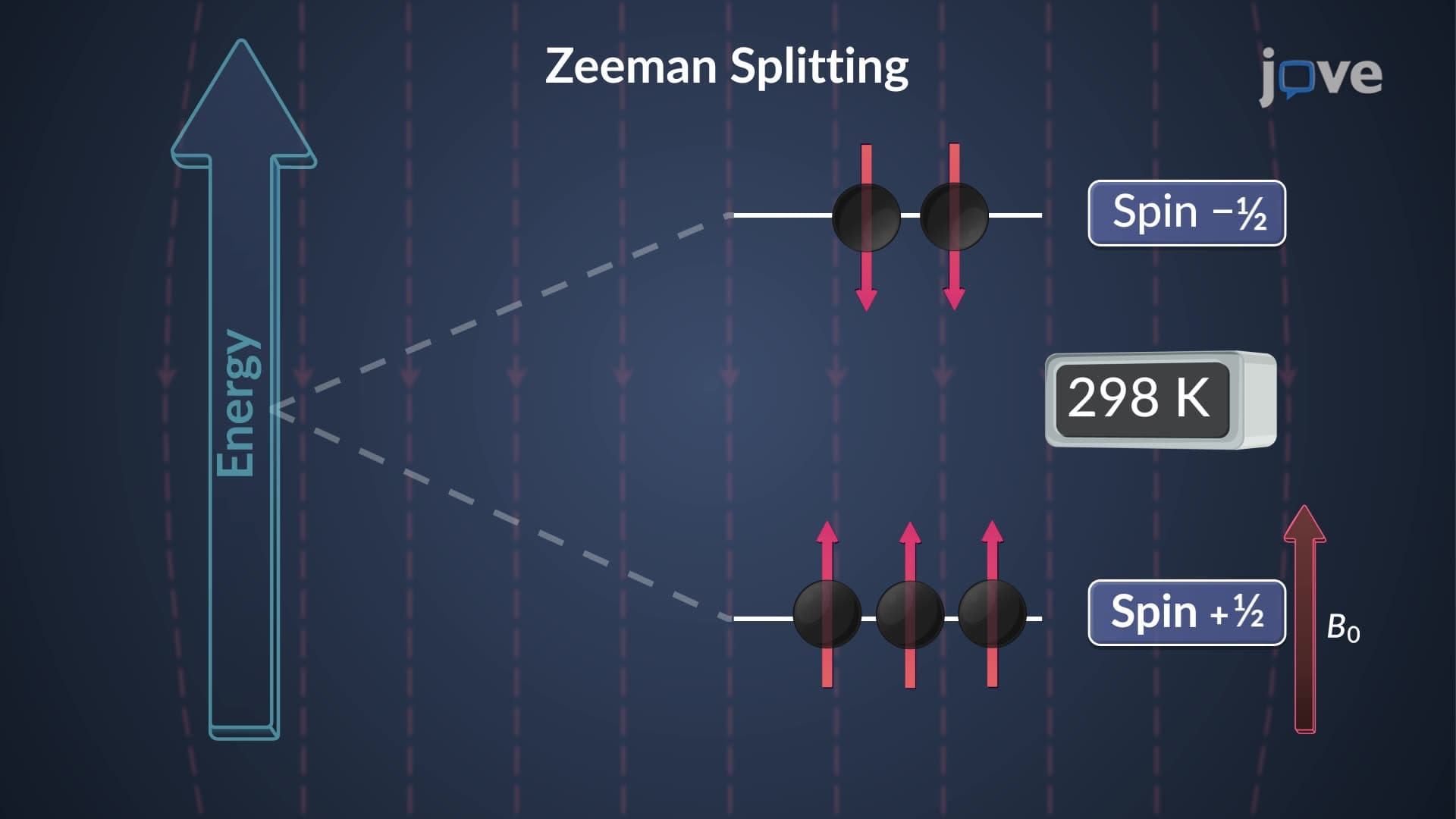
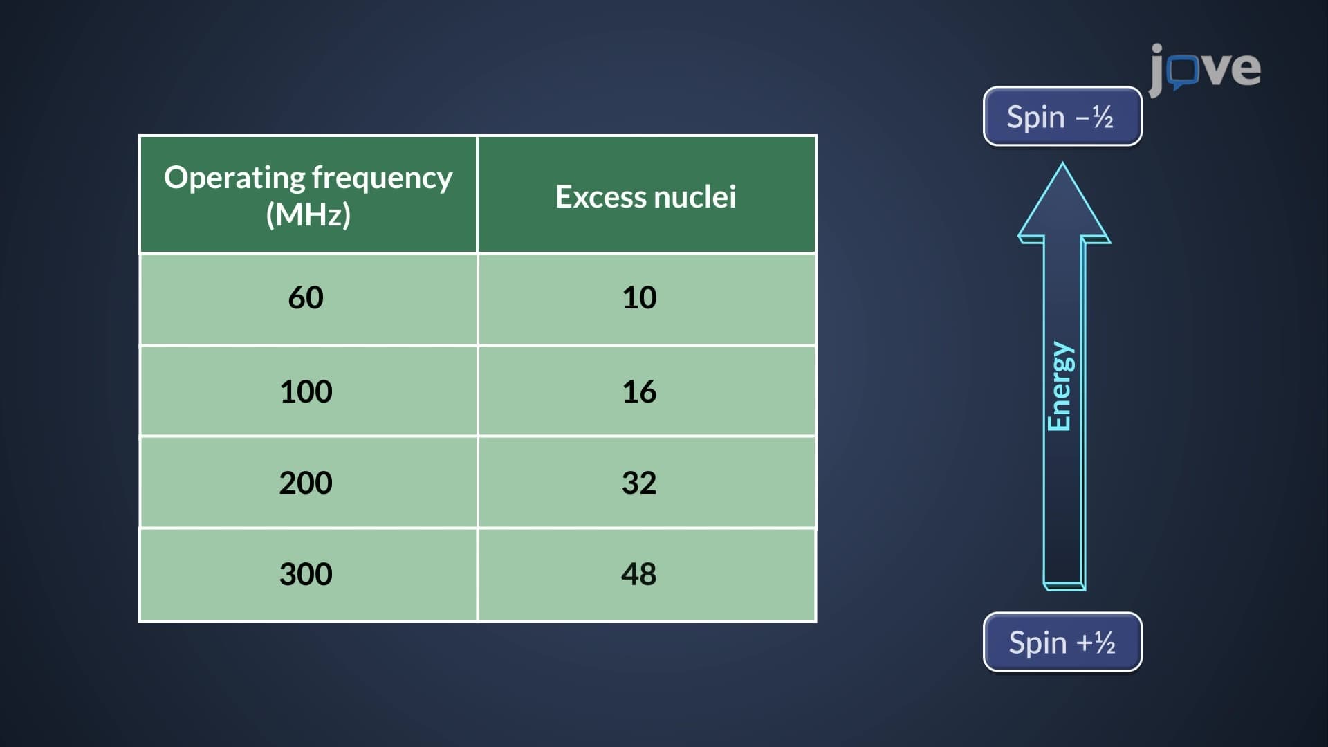
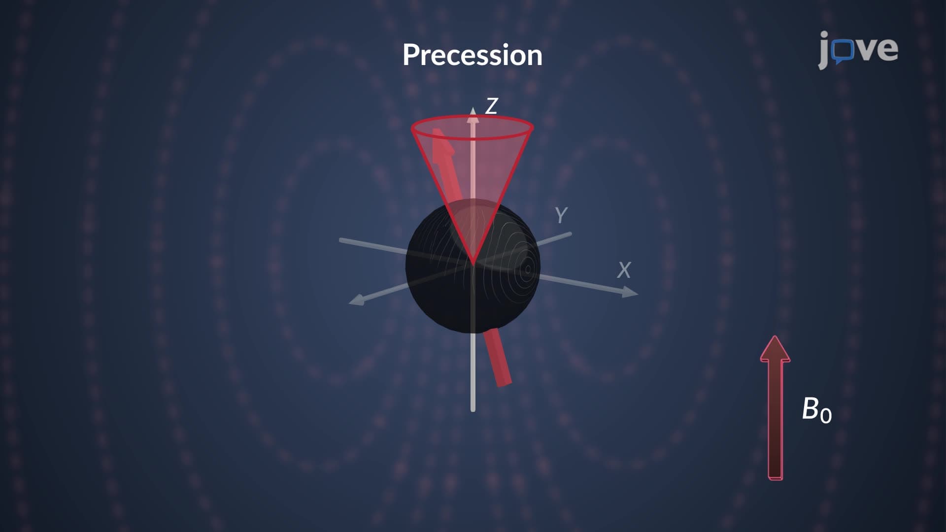
The process begins with nuclear spin alignment. In the presence of a magnetic field B₀, nuclear spins adopt one of two orientations: aligned with the field (lower energy) or against it (higher energy). While the distribution appears random, slightly more spins occupy the lower energy state, creating a small but measurable population difference-typically just a few parts per million at room temperature.
The resonance frequency nuclear spin depends entirely on the Larmor frequency, calculated as ω = γB₀, where γ is the gyromagnetic ratio specific to each nucleus type. For hydrogen nuclei in a 1.5 Tesla MRI scanner (common in US hospitals), this frequency equals approximately 64 MHz-right in the FM radio band. This precise frequency matching enables selective excitation of specific nuclear species.
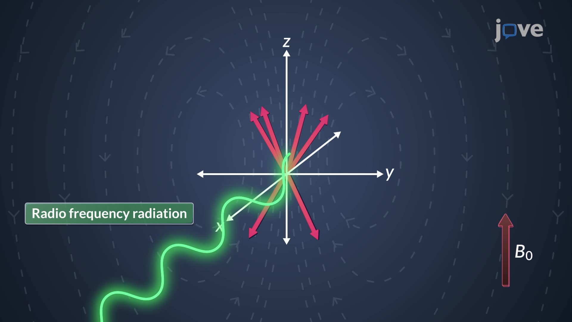
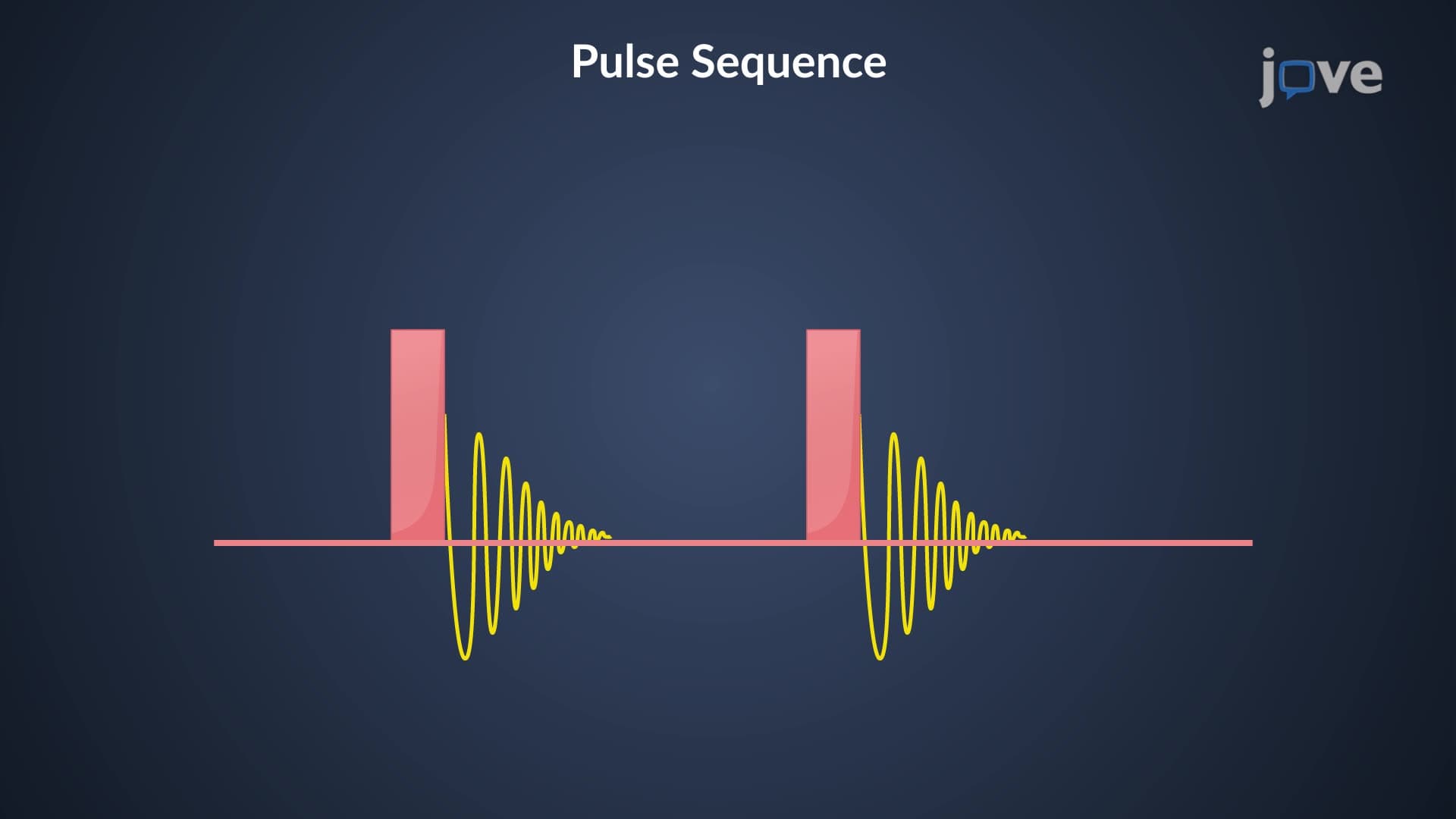
When RF pulse magnetic resonance occurs, the applied radiofrequency energy matches the Larmor frequency exactly. This energy absorption nuclei experience causes nuclear spins to "flip" from the lower energy state to the higher energy state. The net effect shifts the bulk magnetization vector away from its equilibrium position along the z-axis toward the xy-plane.
Resonance absorption NMR involves a carefully orchestrated energy exchange. The RF pulse duration and strength determine how far the magnetization vector tips-90-degree pulses are common in NMR spectroscopy, while MRI often uses different pulse angles for contrast optimization. After the pulse ends, the excited nuclear spins begin relaxing back to equilibrium, releasing the absorbed energy as detectable electromagnetic signals.
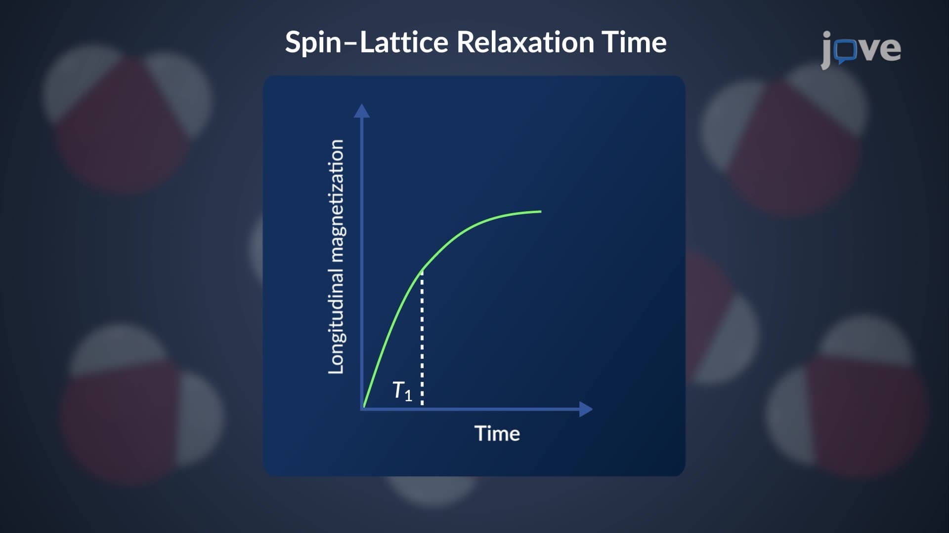
This relaxation process occurs through two main mechanisms: T1 (spin-lattice) relaxation, where energy transfers to surrounding molecules, and T2 (spin-spin) relaxation, where spins lose coherence through mutual interactions. These relaxation times vary dramatically between different tissues and chemical environments, providing the contrast mechanisms exploited in medical imaging and chemical analysis.
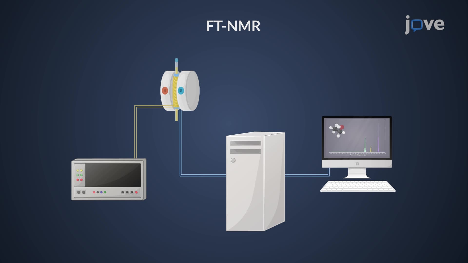
US chemistry students encounter these principles in organic chemistry labs using benchtop NMR spectrometers, while pre-med students preparing for the MCAT must understand how magnetic resonance enables non-invasive medical diagnostics. AP Chemistry courses increasingly include NMR spectroscopy as a molecular identification tool, making this concept essential for college-bound students.
Related Micro-courses