Loading subject page…
- Analytical Chemistry
- Molecular Vibrational Spectroscopy
- IR Frequency Region: X–H Stretching
Micro-course: Molecular Vibrational Spectroscopy
28,168 views
Learn key concepts and earn your certificate.



IR Frequency Region: X–H Stretching

00:00
01:24
Video Summary: Ir Frequency Region X H Stretching Explained
Ever wonder how forensic scientists identify unknown substances in crime labs? The XH stretching IR region holds the key-specific frequency ranges between 2850-3600 cm⁻¹ where C-H, N-H, and O-H bonds create unique spectral fingerprints. The FBI's chemistry labs use these exact X H stretching frequency IR patterns to analyze drug samples and trace evidence. Understanding IR Frequency Region X H Stretching Explained reveals how molecular vibrations translate into diagnostic peaks that distinguish between different functional groups. Watch the full video on JoVE Coach to master this concept with expert-led visuals and step-by-step explanations.
- Understand the IR Frequency Region X H Stretching Explained concept and identify the diagnostic 2850-3600 cm⁻¹ range where single bonds create characteristic absorption patterns
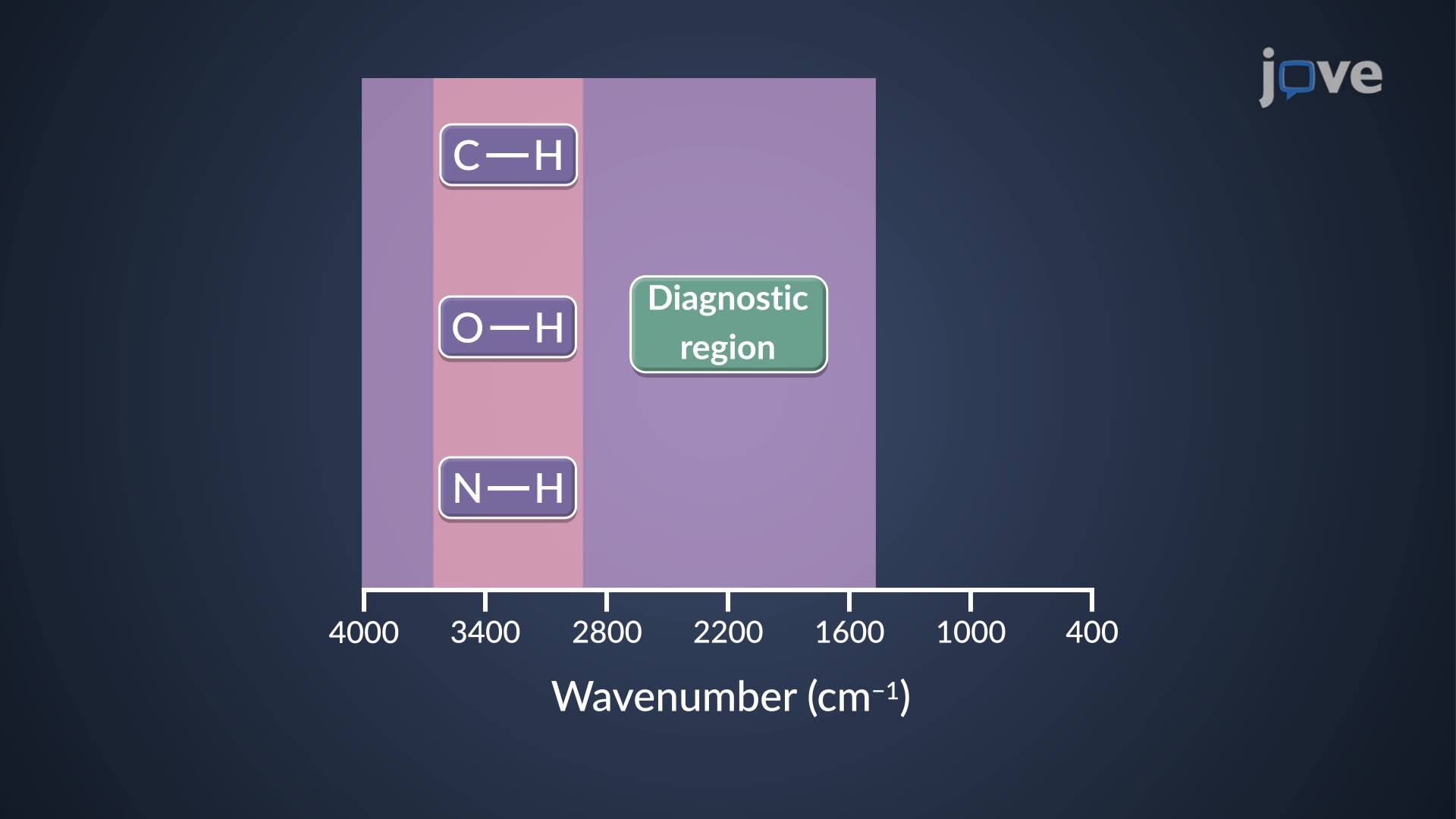
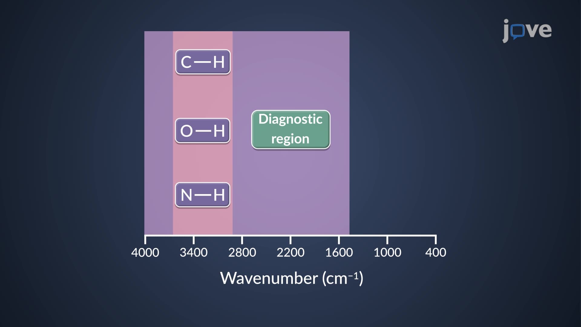
- Identify specific wavenumber ranges for C-H stretching (2850-3000 cm⁻¹), N-H stretching (3300-3500 cm⁻¹), and O-H stretching (3200-3600 cm⁻¹) in infrared spectra
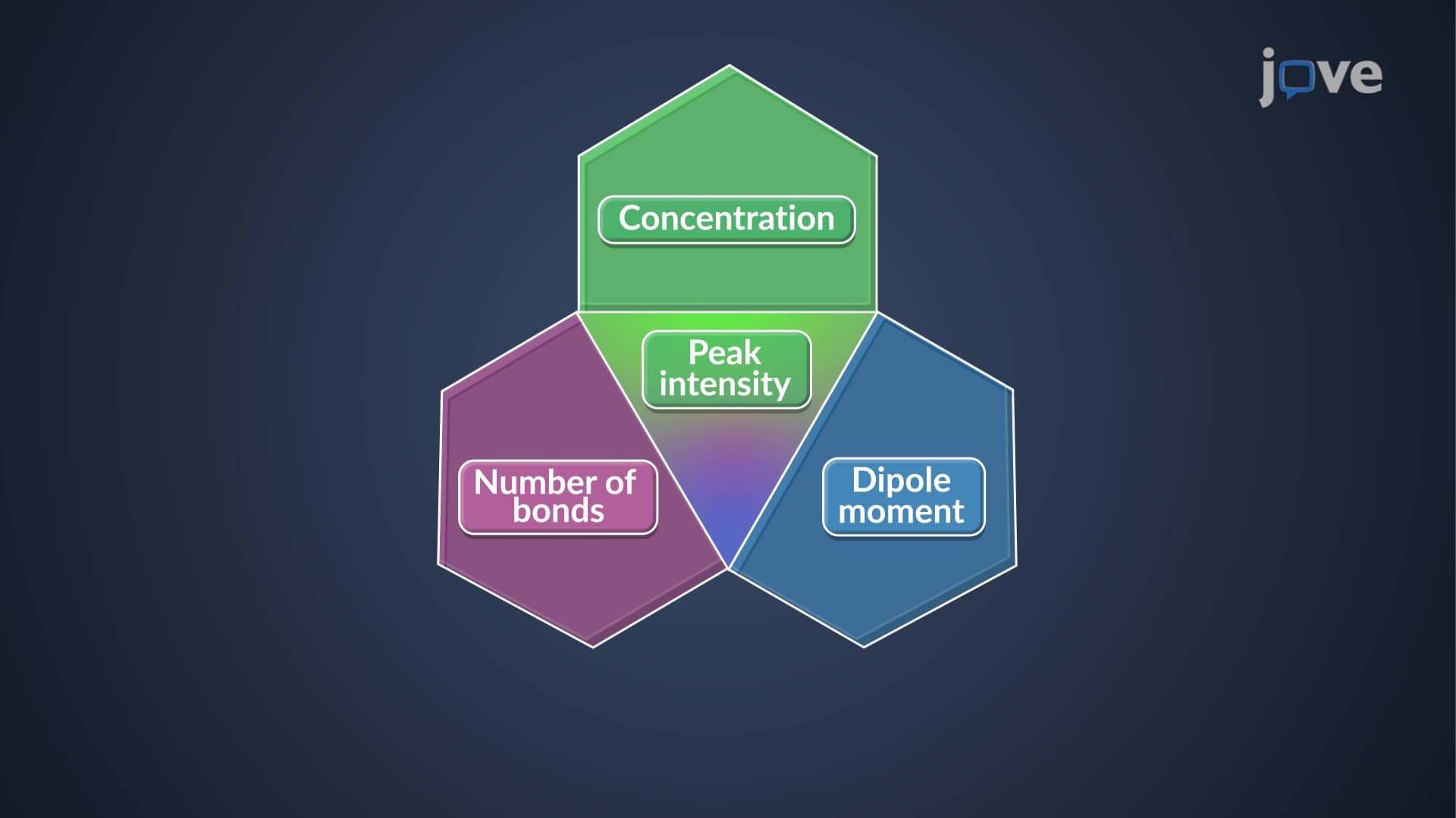
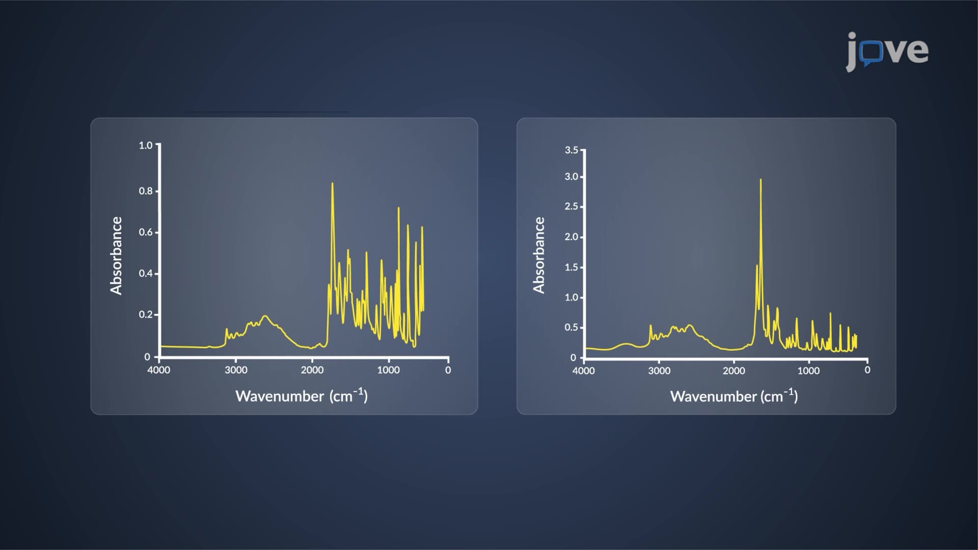
- Analyze peak characteristics including intensity, shape, and splitting patterns to distinguish between different X-H functional groups
- Compare hydrogen-bonded versus non-hydrogen-bonded O-H stretching vibrations based on peak width and frequency position
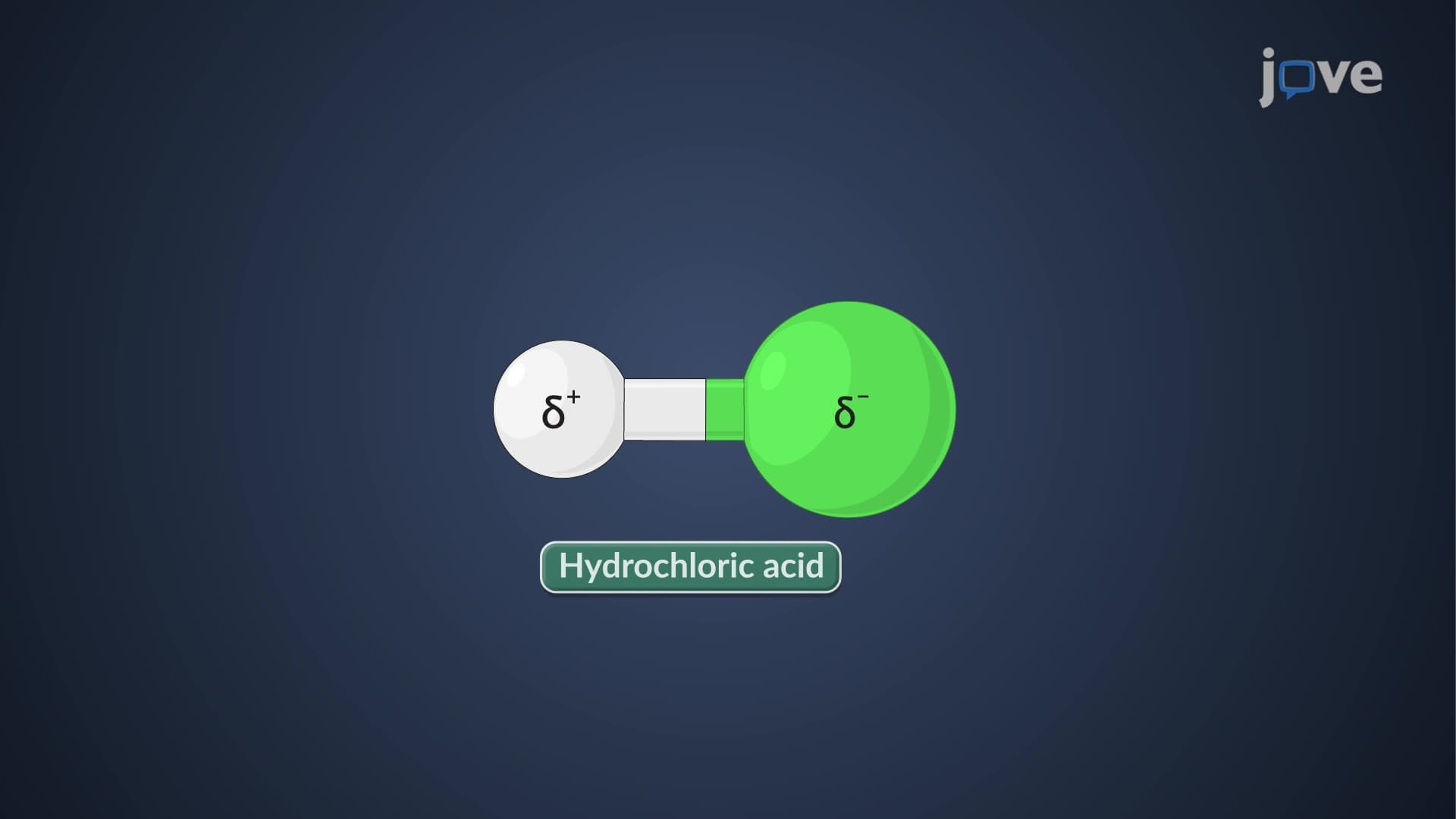
- Evaluate the relationship between dipole moment changes and absorption intensity for various X-H bonds
- Apply spectral interpretation skills to identify primary amines through symmetric and asymmetric N-H stretching patterns
- Recognize how molecular environment affects peak appearance, particularly hydrogen bonding effects on O-H stretching
- Explore the diagnostic value of the X-H stretching region for structural elucidation in organic chemistry applications
Understanding IR Frequency Region X H Stretching Fundamentals
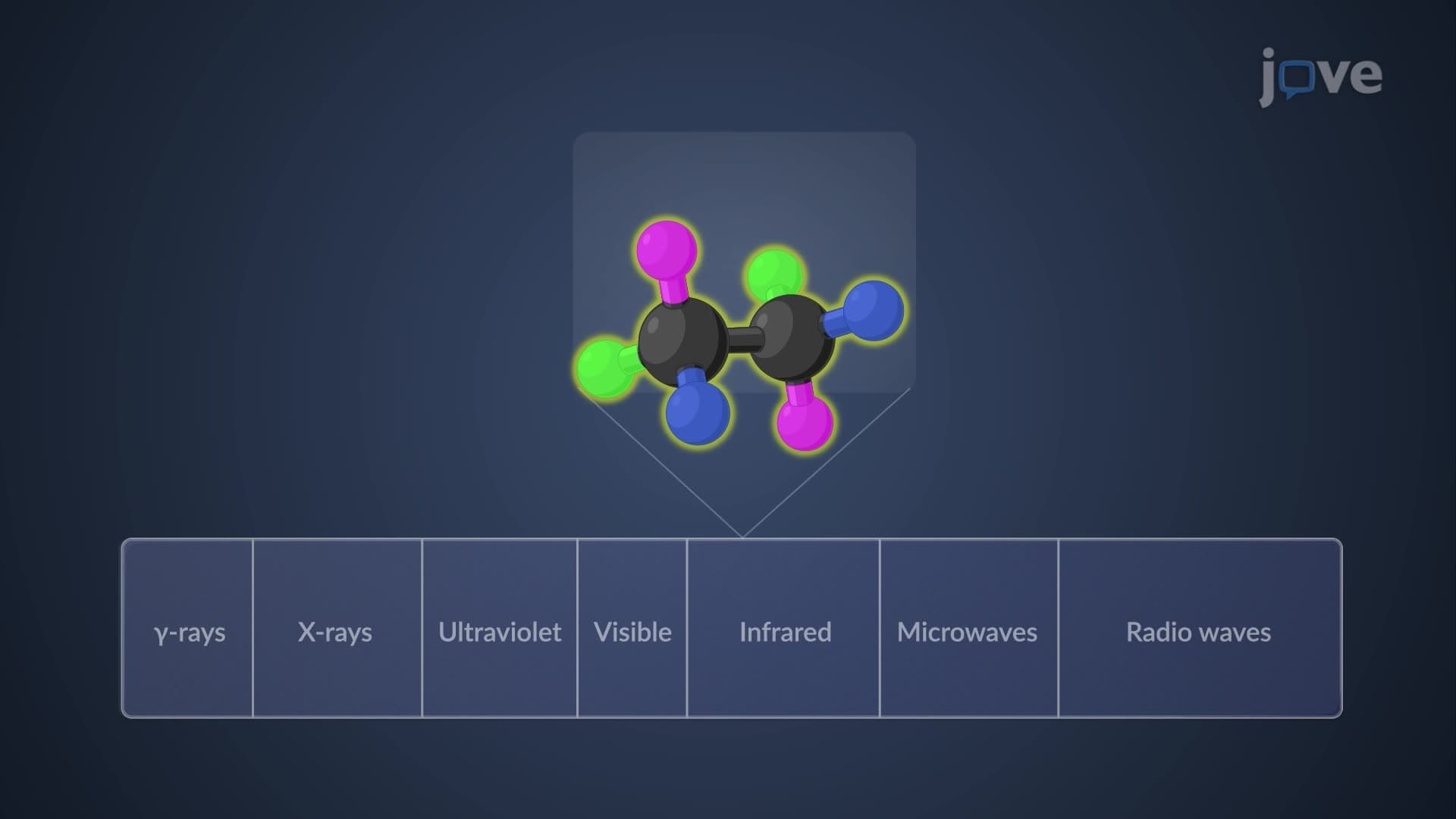
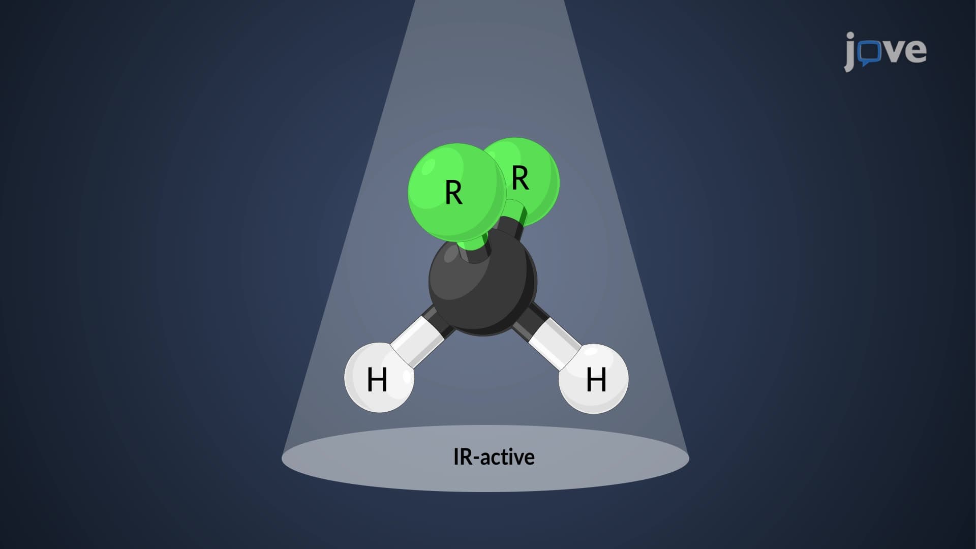
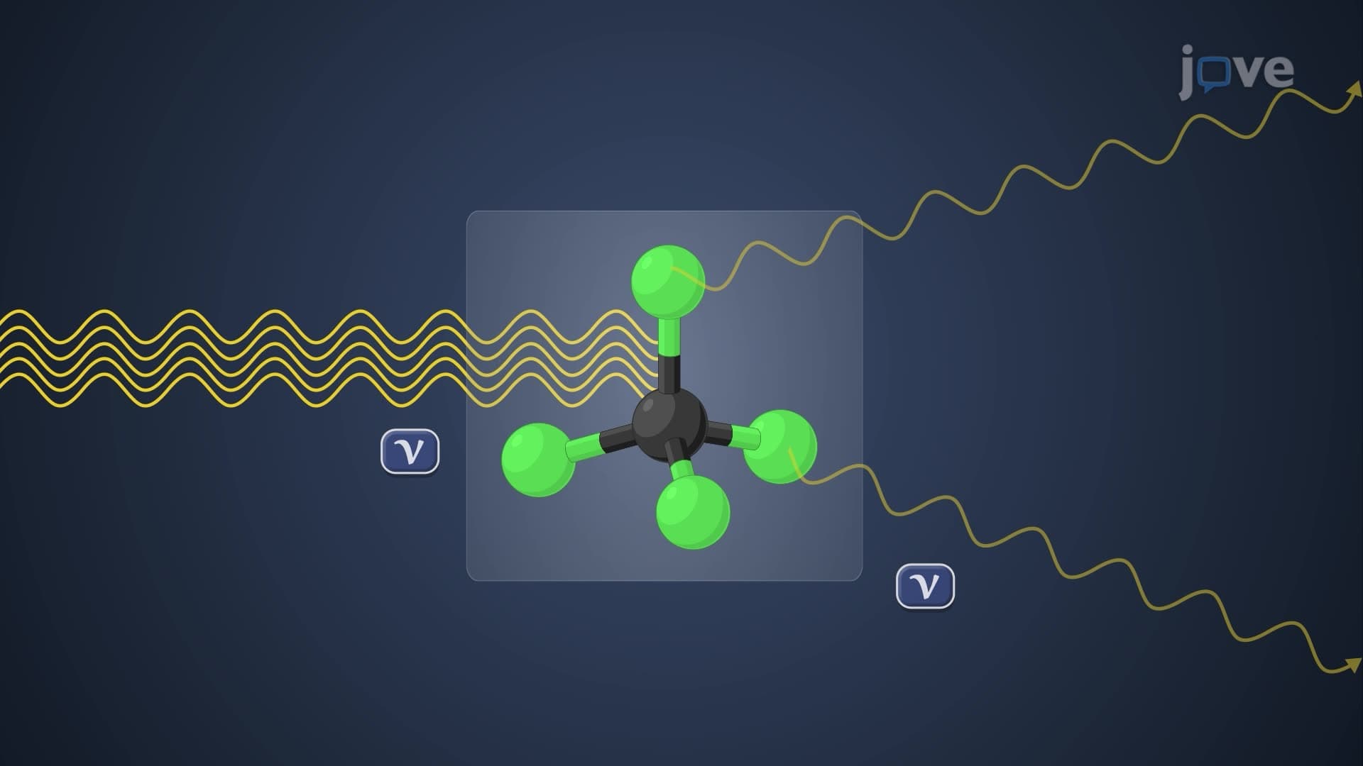
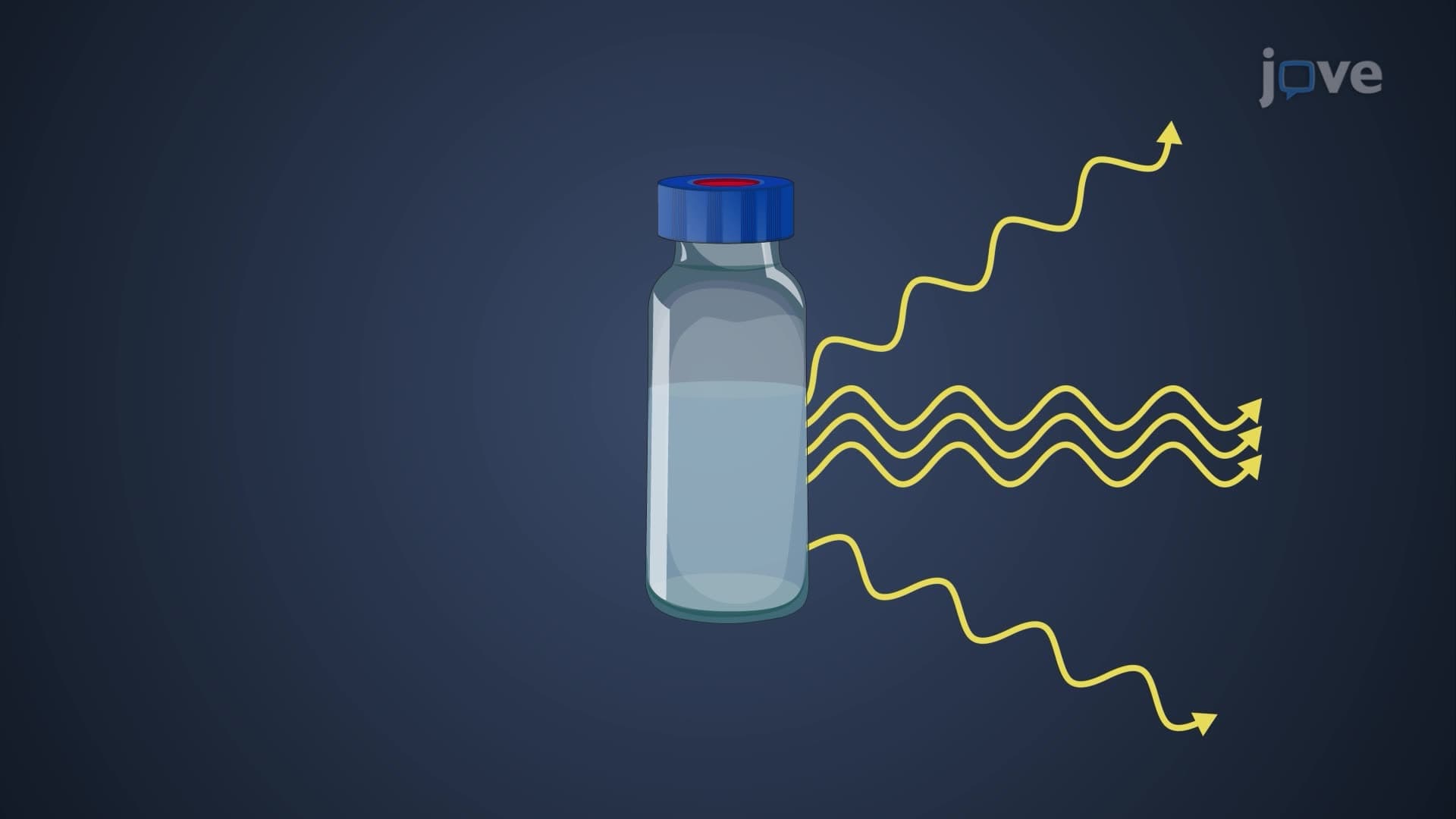
The XH stretching IR region represents one of the most diagnostically valuable areas in infrared spectroscopy, spanning 2850-3600 cm⁻¹. This region captures the vibrational frequencies of single bonds between hydrogen and heavier atoms like carbon, nitrogen, and oxygen. When molecules absorb infrared radiation, these X-H bonds stretch and contract at characteristic frequencies, creating unique spectral signatures that chemists use for molecular identification.
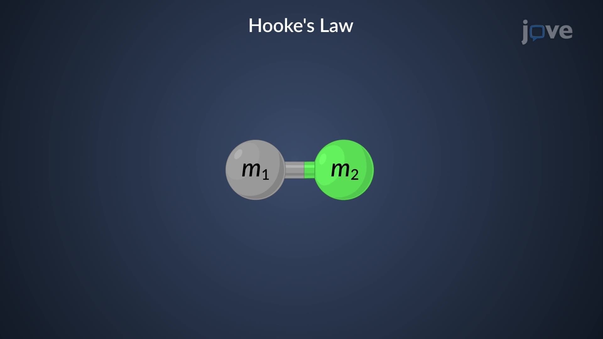
The fundamental principle underlying X H stretching frequency IR patterns lies in Hooke's law, where bond strength and reduced mass determine vibrational frequency. Stronger bonds and lighter atoms produce higher frequency absorptions, explaining why O-H bonds typically appear at higher wavenumbers than C-H bonds.
C-H Stretching Characteristics and Applications
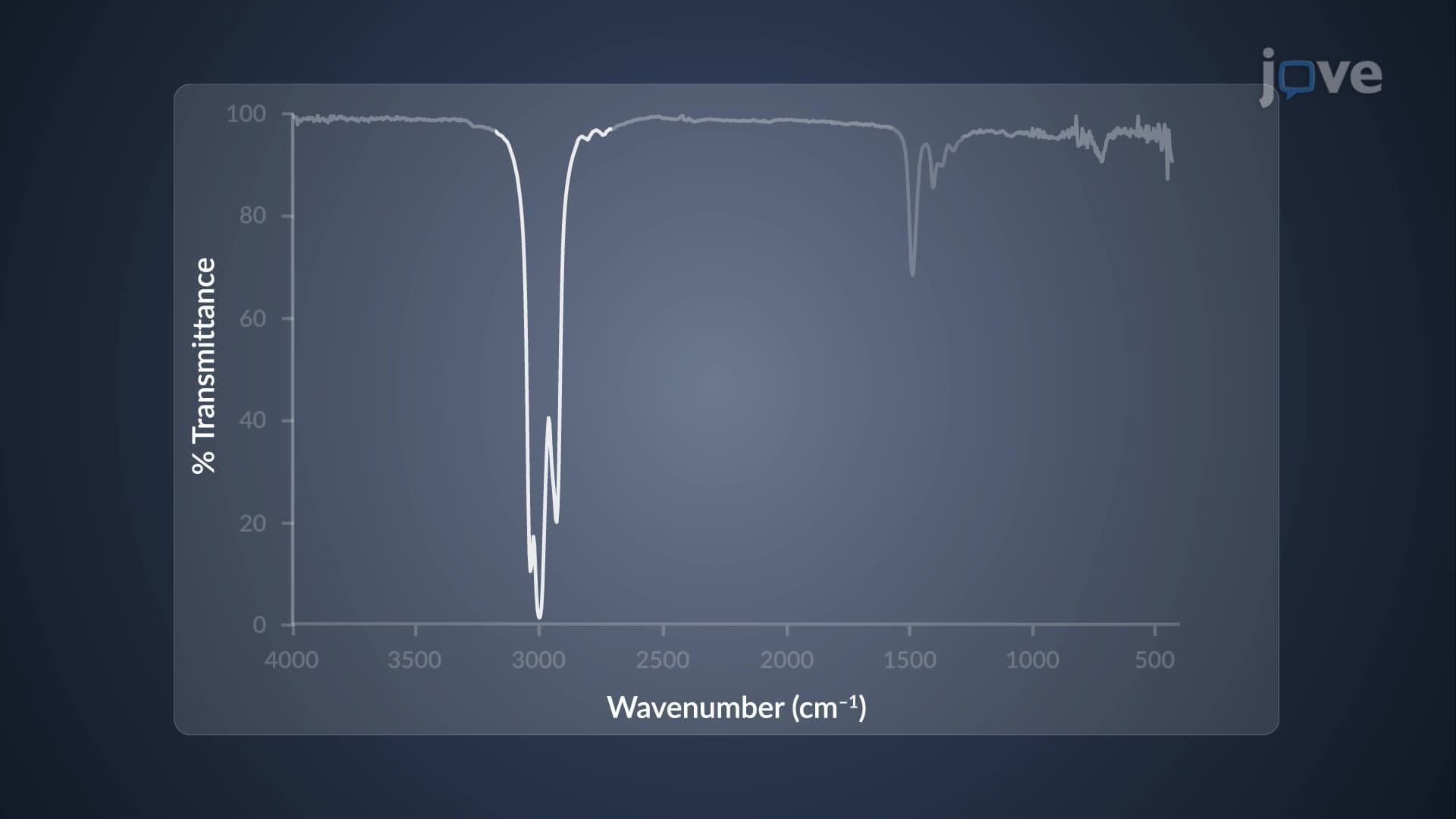
Carbon-hydrogen stretching vibrations dominate the 2850-3000 cm⁻¹ region, exhibiting complex splitting patterns particularly evident in alkanes. Methyl groups produce asymmetric stretching around 2960 cm⁻¹ and symmetric stretching near 2870 cm⁻¹, while methylene groups show characteristic peaks around 2930 cm⁻¹ and 2850 cm⁻¹.
These CH stretching IR region patterns prove invaluable in pharmaceutical analysis. FDA laboratories routinely use these fingerprints to verify drug purity and identify contaminants. For AP Chemistry students, mastering C-H peak interpretation often determines success on free-response questions involving spectral analysis.
N-H and O-H Stretching: Distinguishing Features
NH stretching IR absorptions appear as sharp, relatively weak bands between 3300-3500 cm⁻¹. Primary amines like methylamine exhibit two distinct peaks due to symmetric and asymmetric stretching modes, while secondary amines show single peaks. This multiplicity pattern serves as a diagnostic tool for amine classification.
OH stretching IR frequency patterns vary dramatically based on hydrogen bonding. Free O-H groups produce sharp, intense peaks near 3600 cm⁻¹, while hydrogen-bonded alcohols create broad absorptions spanning 3200-3500 cm⁻¹. This distinction proves crucial in environmental chemistry, where EPA laboratories analyze water contaminants and identify hydrogen-bonding interactions in complex mixtures.
Practical Applications in US Academic and Professional Settings
Medical schools incorporate XH bond stretch wavenumber interpretation into biochemistry curricula, particularly for MCAT preparation. Students learn to identify functional groups in biomolecules, understanding how hydrogen bonding affects protein structure through O-H and N-H stretching analysis.
Industrial applications extend from quality control in pharmaceutical manufacturing to forensic analysis in criminal investigations. Understanding hydrogen stretching IR absorption patterns enables chemists to differentiate between structural isomers and identify unknown compounds with remarkable precision, making this knowledge essential for careers in analytical chemistry, forensic science, and pharmaceutical research.
Frequently Asked Questions
The XH stretching region spans 2850-3600 cm⁻¹ and contains characteristic absorptions from C-H, N-H, and O-H bonds. This region provides crucial structural information because different X-H bonds produce unique frequency patterns, peak shapes, and intensities. These distinct spectral fingerprints allow chemists to identify functional groups and distinguish between similar molecules with remarkable accuracy.
AP Chemistry and college exams frequently test XH stretching interpretation through spectral analysis problems. Students must identify functional groups, explain peak multiplicity in primary amines, and distinguish hydrogen-bonded from free O-H groups. MCAT questions often combine IR spectroscopy with biomolecule structure, requiring knowledge of how hydrogen bonding affects peak appearance in proteins and nucleic acids.
Focus on key wavenumber ranges: C-H stretching (2850-3000 cm⁻¹), N-H stretching (3300-3500 cm⁻¹), and O-H stretching (3200-3600 cm⁻¹). Remember that primary amines show two N-H peaks, hydrogen-bonded O-H groups produce broad absorptions, and C-H peaks exhibit multiple splitting patterns. Understanding these patterns helps answer MCAT questions about biomolecule identification and functional group analysis.
FBI and state crime laboratories use XH stretching patterns to identify drugs, explosives, and trace evidence. Each substance produces unique C-H, N-H, and O-H fingerprints that help distinguish between different compounds. For example, cocaine shows characteristic C-H stretching patterns different from other stimulants, while explosive materials exhibit specific N-H and O-H combinations that aid in criminal prosecutions.
XH stretching concepts are perfectly accessible to high school students with basic chemistry knowledge. The key is understanding that different bonds vibrate at different frequencies, creating unique patterns. AP Chemistry students regularly master these concepts, and many honors chemistry courses introduce IR spectroscopy basics. Starting with simple examples like distinguishing alcohols from alkanes builds confidence for more complex applications.
Create visual associations and practice with real spectra rather than rote memorization. Remember "CON" order: Carbon-H lowest (2850-3000), Oxygen-H middle (3200-3500), Nitrogen-H highest (3300-3500). Use practice problems from your textbook and online resources to reinforce pattern recognition. Focus on peak shapes and intensities, not just wavenumber ranges, since these characteristics often determine exam answers.
XH stretching knowledge provides essential foundation for physical chemistry, analytical chemistry, and biochemistry courses. Graduate programs expect students to interpret complex spectra confidently, while pharmaceutical and materials science careers require daily spectroscopic analysis. Mastering these fundamentals in undergraduate courses prepares students for research opportunities and professional laboratory work.
Advanced applications include coupling patterns, overtone analysis, and time-resolved IR spectroscopy. Students can explore how molecular environment affects peak positions, study isotope effects on vibrational frequencies, and investigate two-dimensional IR techniques. These concepts appear in graduate-level physical chemistry and are essential for careers in spectroscopy research and advanced analytical chemistry.
Related Micro-courses

17 Concepts

15 Concepts