Loading subject page…

- Analytical Chemistry
- Molecular Vibrational Spectroscopy
- IR Spectrum Peak Splitting: Symmetric vs Asymmetric Vibrations
Micro-course: Molecular Vibrational Spectroscopy
28,168 views
Learn key concepts and earn your certificate.
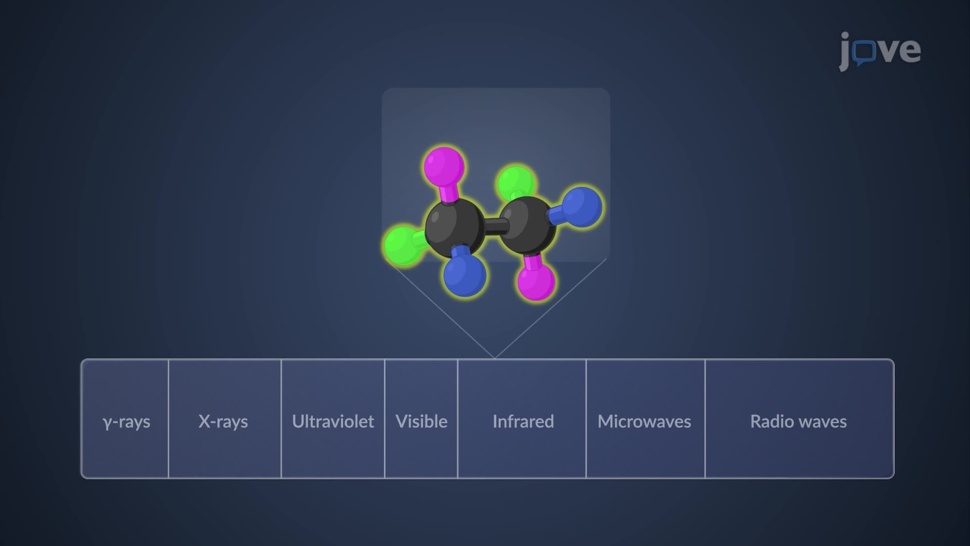


IR Spectrum Peak Splitting: Symmetric vs Asymmetric Vibrations


00:00
01:08
Video Summary: Ir Spectrum Peak Splitting Symmetric Explained
Ever wonder why your chemistry lab's infrared spectrometer shows multiple peaks for what seems like a single bond? IR peak splitting symmetric phenomena occur when molecules contain identical atoms that can vibrate in coordinated patterns. For instance, when pharmaceutical companies analyze aspirin's structure, they observe distinct peaks for N-H stretching in amine groups rather than single absorptions. This splitting reveals whether bonds stretch together (symmetric) or opposite each other (asymmetric), providing crucial molecular fingerprint information. Watch the full video on JoVE Coach to master this concept with expert-led visuals and step-by-step explanations.
- Understand how IR spectrum peak splitting symmetric patterns arise from multiple identical bonds within polyatomic groups
- Identify the difference between symmetric and asymmetric stretching modes in molecules containing two or more equivalent atoms
- Analyze why primary amines (RNH₂) produce two distinct N-H stretching peaks while secondary amines (R₂NH) show only one
- Compare frequency ranges for symmetric versus asymmetric stretching, recognizing that asymmetric modes typically appear at higher frequencies
- Evaluate how bond equivalence through delocalization affects peak patterns in groups like NO₂
- Apply knowledge of stretching patterns to predict IR spectral features for various polyatomic functional groups
- Recognize the relationship between molecular symmetry and the number of observable IR absorption bands
- Explore how symmetric splitting IR spectrum peaks provide structural information for compound identification
Understanding IR Spectrum Peak Splitting Symmetric Fundamentals
IR spectrum peak splitting symmetric behavior emerges from a fundamental principle of molecular vibrations: when molecules contain two or more identical bonds, these bonds can vibrate in different coordinated patterns. This phenomenon is crucial for structural determination in analytical chemistry, appearing frequently on AP Chemistry exams and college organic chemistry courses.
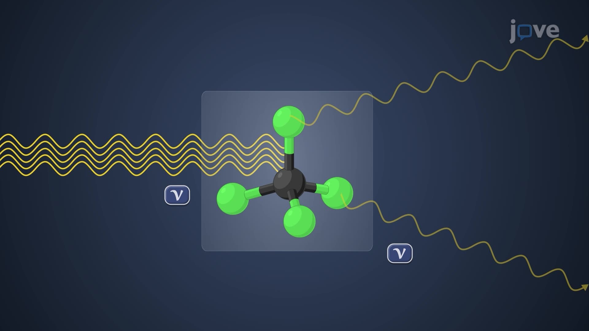
The key lies in understanding vibrational modes. When identical bonds exist within a molecule, they don't vibrate independently. Instead, they couple together, creating distinct vibrational patterns that absorb infrared radiation at different frequencies. This coupling explains what causes peak splitting in IR spectra - it's the mathematical combination of individual bond vibrations into collective molecular movements.
Primary Amines: A Classic Example of Symmetric vs Asymmetric Stretching
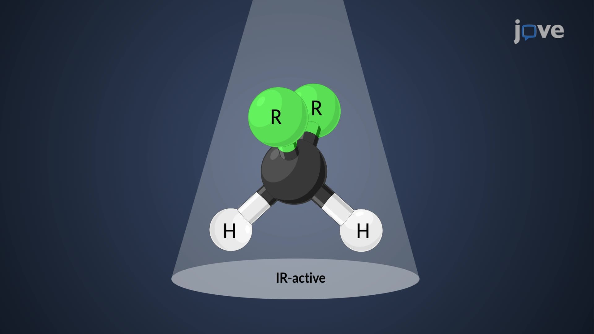
Primary amines (RNH₂) provide an excellent illustration of this concept. These molecules contain two equivalent N-H bonds that can stretch in two distinct ways. In symmetric stretching, both N-H bonds elongate and contract simultaneously, maintaining the molecule's overall symmetry. Conversely, asymmetric stretching involves one N-H bond stretching while the other contracts, creating an asymmetric deformation.
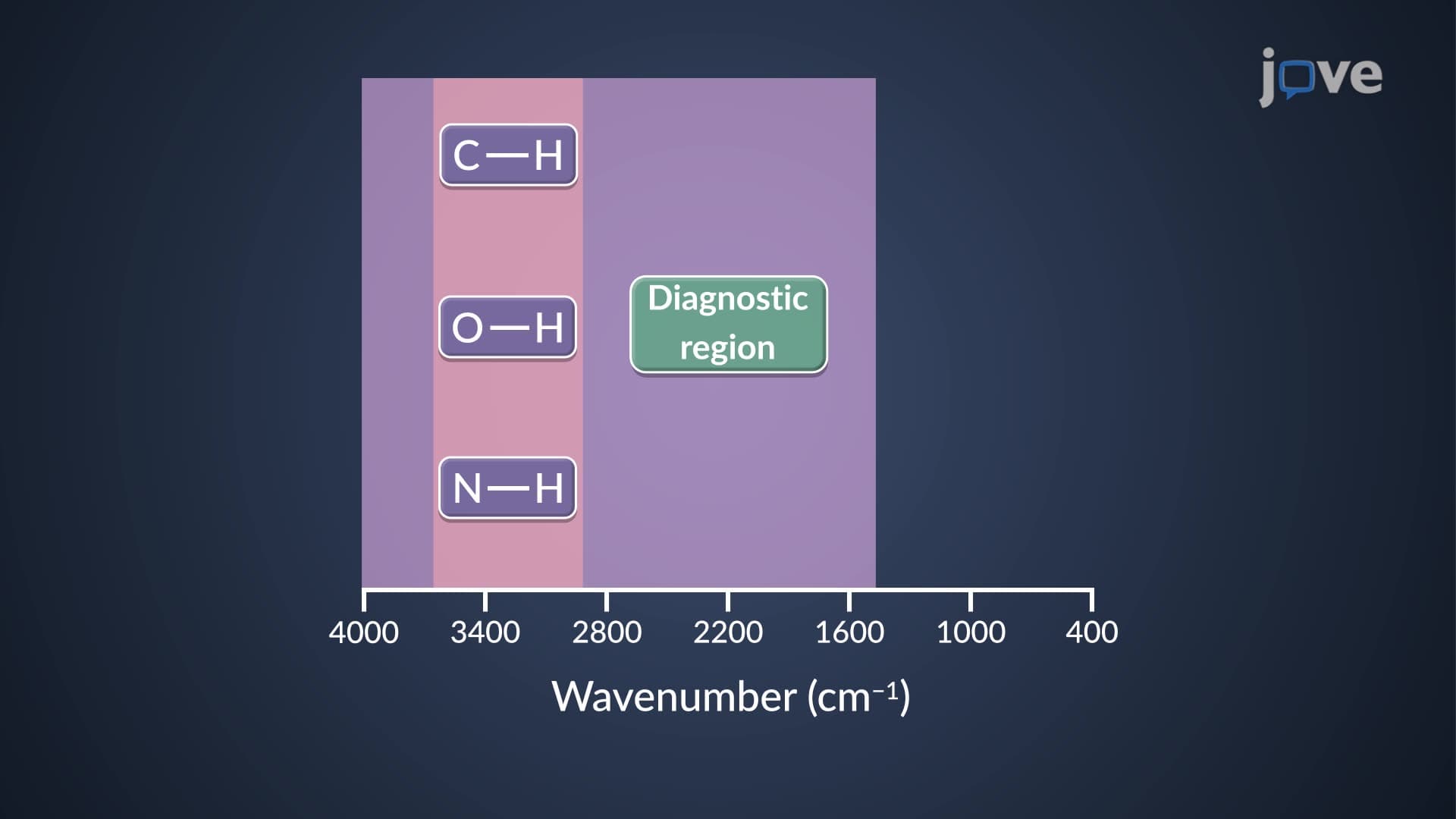
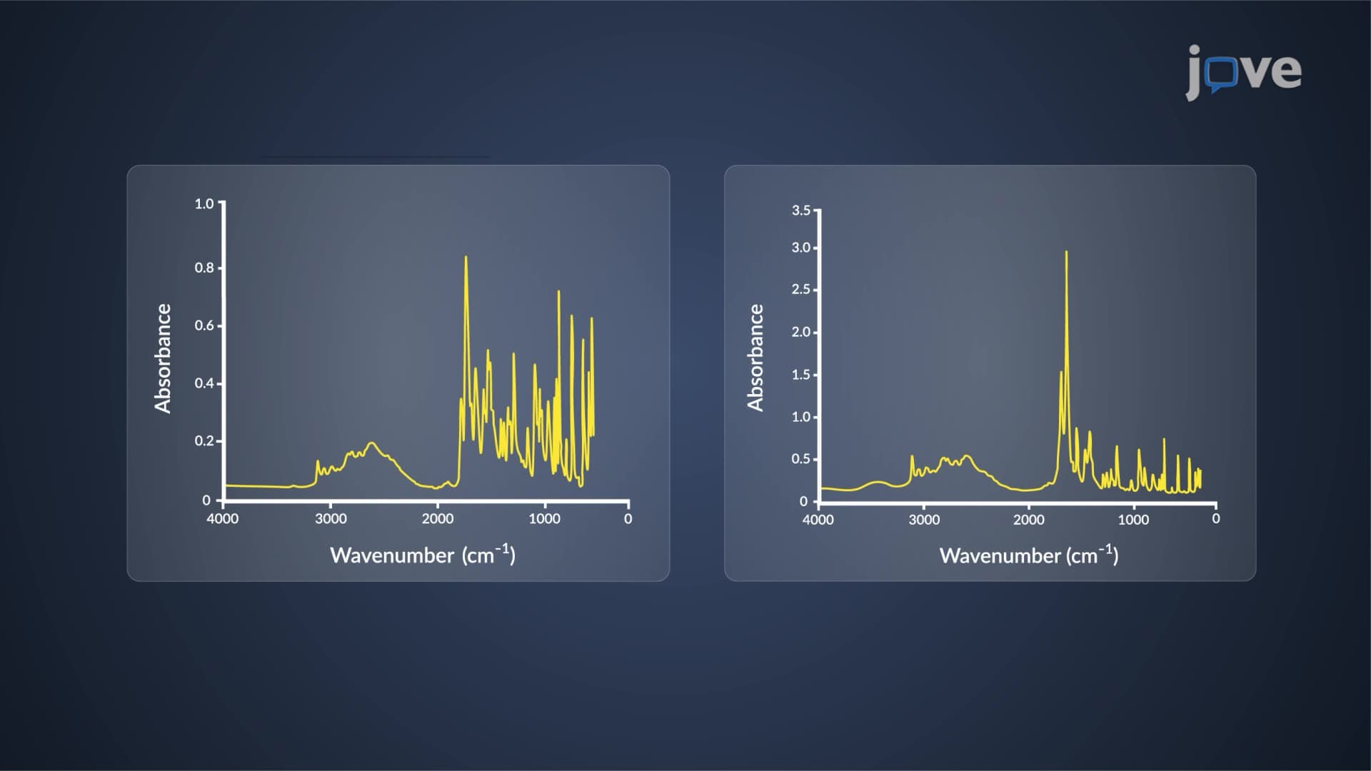
These different stretching modes absorb infrared radiation at distinct frequencies, typically appearing in the 3300-3500 cm⁻¹ range. The asymmetric stretch consistently appears at higher frequency because it requires more energy to break the molecule's symmetry. This pattern helps pharmaceutical chemists identify primary amine groups in drug compounds, a skill tested extensively on MCAT examinations.
Secondary Amines and the Single Peak Phenomenon
Secondary amines (R₂NH) demonstrate the importance of bond count in determining spectral patterns. With only one N-H bond present, no coupling can occur between identical stretching vibrations. Consequently, these molecules produce a single, sharp absorption peak around 3300 cm⁻¹, contrasting sharply with the doublet pattern of primary amines.
NO₂ Groups: Delocalization and Equivalent Bonds
Nitro groups (NO₂) present an interesting case study in symmetric antisymmetric IR behavior. Although the two N-O bonds might appear structurally different, electron delocalization creates equivalent bonds throughout the group. This equivalence enables both symmetric and asymmetric stretching modes, producing characteristic peak splitting patterns that help forensic scientists identify explosive compounds in criminal investigations.
This delocalization concept frequently appears in college-level physical chemistry courses, where students learn to predict spectral behavior based on molecular orbital theory and resonance structures.
Frequently Asked Questions
IR spectrum peak splitting symmetric refers to the phenomenon where single functional groups produce multiple absorption peaks due to different vibrational modes of equivalent bonds. This occurs when molecules contain two or more identical bonds that can vibrate together (symmetric) or in opposition (asymmetric), each mode absorbing at distinct frequencies. The splitting provides valuable structural information for molecular identification.
Yes, AP Chemistry frequently includes IR spectroscopy interpretation questions, including peak splitting patterns. You'll typically see problems asking you to distinguish between primary and secondary amines based on their N-H stretching patterns, or identify functional groups from characteristic splitting. Focus on memorizing key frequency ranges and understanding why equivalent bonds produce multiple peaks.
MCAT passages often include IR spectra requiring you to identify functional groups based on splitting patterns. Recognizing that primary amines show doublet N-H peaks while secondary amines show singlets helps eliminate answer choices quickly. The exam particularly emphasizes relating molecular structure to spectroscopic evidence, making peak splitting knowledge essential for biochemistry and organic chemistry sections.
Peak splitting in pharmaceutical IR spectra results from vibrational coupling between equivalent bonds within functional groups. When drug molecules contain groups like NH₂, NO₂, or SO₂, their identical bonds create symmetric and asymmetric stretching modes that absorb at different frequencies. Pharmaceutical companies use this splitting to verify drug purity and identify synthetic intermediates during manufacturing quality control.
No advanced mathematics is required to grasp IR peak splitting fundamentals. The concept relies on visualizing how identical bonds can move together or in opposition, similar to synchronized swimming versus opposite movements. While quantum mechanical calculations explain the exact frequencies, high school chemistry focuses on pattern recognition and functional group identification using established frequency tables.
Create comparison charts showing splitting patterns for different functional groups, focusing on primary vs secondary amines as your foundation example. Practice with real spectra, covering the molecule names and trying to predict peak patterns before checking answers. Use flashcards for key frequency ranges and remember that asymmetric stretches typically appear at higher frequencies than symmetric ones.
NMR spectroscopy exhibits analogous splitting through spin-spin coupling, where neighboring hydrogen atoms cause peak multiplication. Raman spectroscopy also shows vibrational splitting patterns complementary to IR. Understanding IR peak splitting provides excellent preparation for these advanced techniques typically encountered in upper-level chemistry courses and graduate programs.
IR peak splitting fundamentals prepare you for molecular orbital theory, group theory applications in chemistry, and advanced spectroscopic methods like 2D-IR spectroscopy. Physical chemistry courses extensively use these concepts when discussing vibrational energy states and selection rules, while analytical chemistry applies them to complex mixture analysis and structural determination of unknown compounds.
Related Micro-courses

17 Concepts

15 Concepts