Loading subject page…

- Anatomy and Physiology
- Bone Tissue and the Skeletal System
- Bone Formation by Endochondral Ossification
Micro-course: Bone Tissue and the Skeletal System
132,600 views
Learn key concepts and earn your certificate.









Bone Formation by Endochondral Ossification


00:00
01:24
Video Summary: Bone Formation by Endochondral Ossification Explained
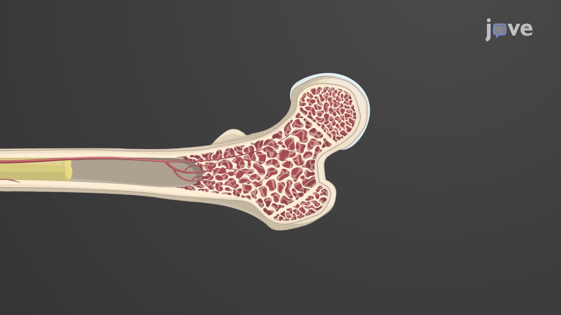
Did you know that a newborn's "bones" are actually made of flexible cartilage? The endochondral ossification process transforms this cartilage template into the rigid bones that support a grown adult's 180-pound frame. This remarkable transformation occurs in developing long bones like the femur in your thigh, where cartilage to bone ossification creates the sturdy framework needed for movement and protection. Understanding Bone Formation By Endochondral Ossification Explained reveals how your skeleton literally rebuilds itself from soft cartilage into hard bone during development. Watch the full video on JoVE Coach to master this concept with expert-led visuals and step-by-step explanations.
- Understand the complete process of Bone Formation By Endochondral Ossification Explained and identify the key stages from cartilage template to mature bone tissue
- Recognize how the perichondrium transforms into periosteum and forms the initial bone collar around the cartilaginous model's diaphysis
- Analyze the role of chondrocyte enlargement and calcification in creating the primary ossification center during embryonic development
- Compare the timing of diaphyseal ossification (12th week of gestation) versus epiphyseal ossification (after birth) in long bone formation
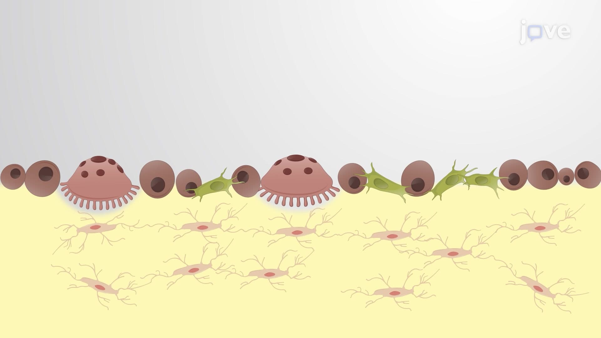
- Identify how osteogenic cells differentiate into osteoblasts and deposit new bone matrix within the cavities left by dying chondrocytes
- Evaluate the function of osteoclasts in forming the medullary cavity through central matrix degradation
- Apply knowledge of nutrient vessel infiltration and its role in supporting bone cell survival during the ossification process
- Explore the relationship between calcification, nutrient diffusion limitations, and chondrocyte death in cartilage replacement
Understanding Bone Formation By Endochondral Ossification
The endochondral ossification process represents one of nature's most sophisticated construction projects, transforming a flexible cartilage blueprint into the rigid bone architecture that defines our skeletal system. Unlike intramembranous ossification, which creates flat bones directly from connective tissue, cartilage to bone ossification follows a precise sequence that replaces an entire cartilaginous framework with mineralized bone tissue.
The Cartilage Template Foundation
During embryonic development, cartilage template ossification begins with hyaline cartilage forming the basic shape of future bones. This cartilaginous model, surrounded by perichondrium, serves as the architectural blueprint for bones like the femur, tibia, and humerus. The process demonstrates remarkable biological efficiency-rather than building bones from scratch, the body uses pre-existing cartilage as a scaffold for bone construction.
Primary Ossification Center Development
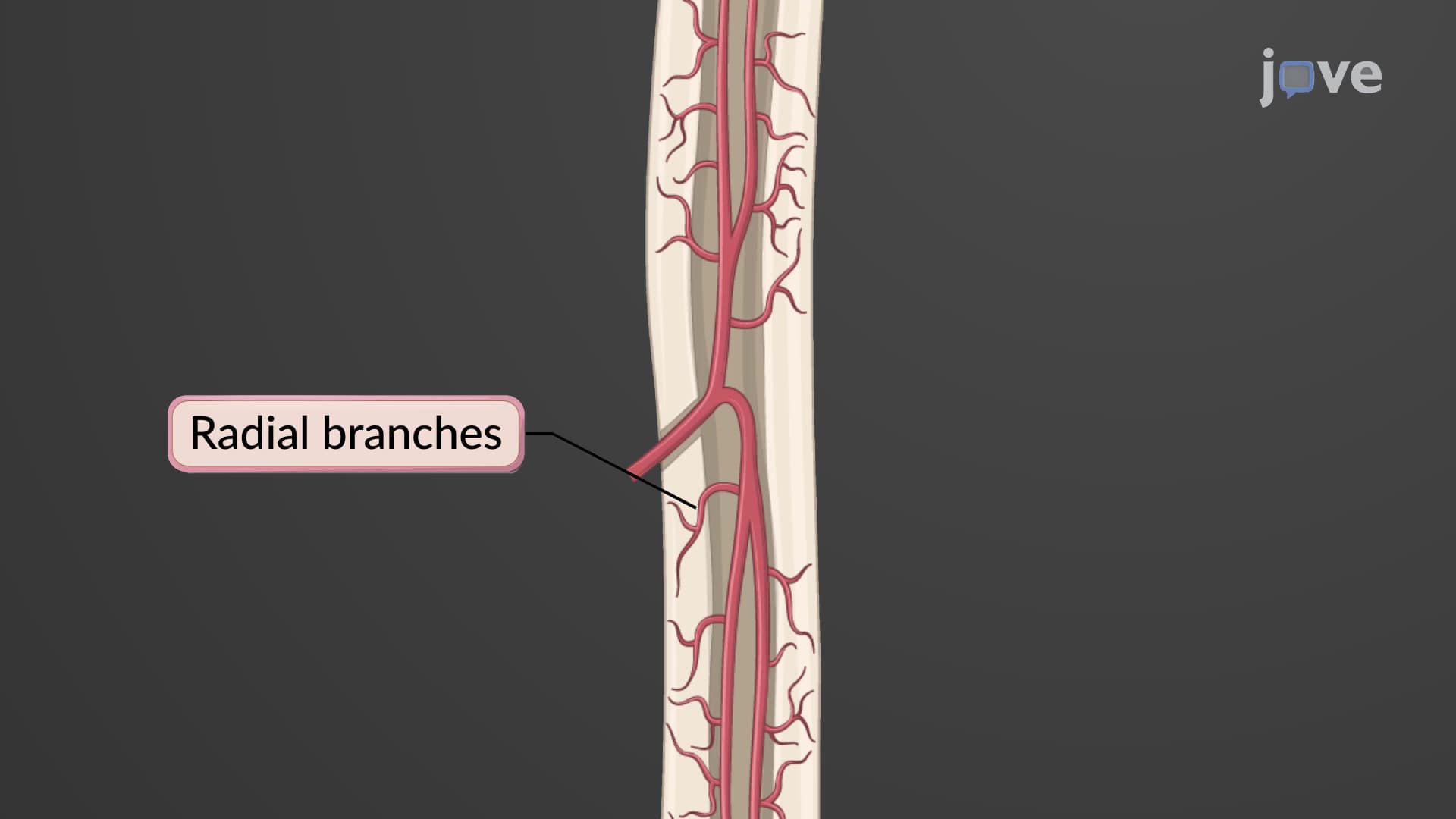
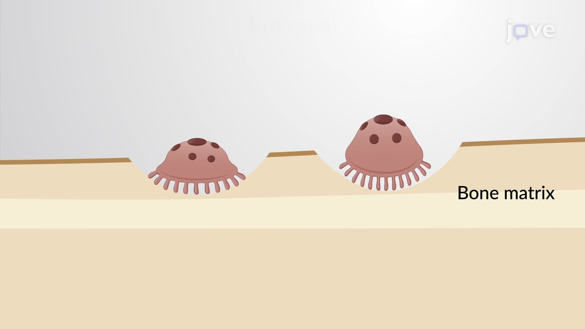
The transformation accelerates when perichondrium evolves into periosteum, immediately forming a bone collar around the diaphysis. Simultaneously, central chondrocytes enlarge dramatically and trigger calcification of surrounding matrix. This calcification creates a critical biological bottleneck: nutrients can no longer diffuse through the hardened matrix, causing chondrocyte death and creating hollow cavities. Primary secondary ossification centers emerge as osteogenic cells, blood vessels, and nerves infiltrate these spaces, with osteoblasts depositing fresh bone matrix while osteoclasts carve out the medullary cavity.
Clinical and Academic Applications
Understanding how endochondral ossification replaces cartilage with bone proves essential for MCAT preparation, AP Biology exams, and college anatomy courses. Students studying for the USMLE must grasp this concept to understand growth disorders like achondroplasia, where cartilage formation defects disrupt normal bone development. In clinical settings, orthopedic surgeons rely on this knowledge when treating pediatric fractures, knowing that epiphyseal plate bone growth continues until approximately age 18-21, affecting treatment decisions for young patients at institutions like Boston Children's Hospital or Cincinnati Children's.
The long bone endochondral process also explains why growth hormone deficiencies during childhood result in proportional dwarfism-the cartilage template formation remains intact, but the ossification process slows significantly. This knowledge helps healthcare providers at facilities like the Mayo Clinic and Cleveland Clinic design targeted treatments for growth disorders.
Frequently Asked Questions
Endochondral ossification is the process where cartilage templates are replaced by bone tissue during development, primarily forming long bones like the femur and humerus. Unlike intramembranous ossification which creates flat bones directly, this process uses pre-existing cartilage as a scaffold. It's the primary mechanism responsible for longitudinal bone growth and occurs in most bones of the appendicular skeleton.
MCAT questions often focus on the cellular mechanisms, timing sequences, and hormonal regulation of the process, particularly growth hormone and thyroid hormone effects. AP Biology exams typically test the stages of ossification, differences between primary and secondary ossification centers, and the role of growth plates. Students should understand the molecular signals that control chondrocyte differentiation and osteoblast activity.
Disrupted endochondral ossification can cause conditions like achondroplasia (dwarfism), where cartilage formation defects lead to shortened limbs but normal trunk development. Growth hormone deficiencies slow the entire process, resulting in proportional but reduced stature. These conditions are treated at specialized centers like Boston Children's Hospital using growth hormone therapy and surgical interventions.
The process begins during embryonic development around the 8th week of gestation, with most diaphyses ossified by the 12th week. However, epiphyseal ossification doesn't start until after birth and continues through adolescence. Growth plates typically close between ages 14-18 in females and 16-21 in males, marking the end of longitudinal bone growth.
No advanced chemistry background is required-basic understanding of calcium phosphate mineralization and protein synthesis is sufficient. Focus on the biological sequence: cartilage formation, calcification, cell death, vascular invasion, and bone deposition. The process is more about cellular biology and tissue development than complex biochemical reactions.
Use the acronym "CCIVO": Cartilage template, Calcification, Invasion (vascular), and Ossification. Create a timeline starting with perichondrium transformation, then chondrocyte enlargement, calcification-induced cell death, vascular invasion, and finally osteoblast/osteoclast activity. Drawing the process while studying helps visualize the spatial relationships between different cell types and structures.
This knowledge is fundamental for orthopedics, pediatrics, and endocrinology specialties. Orthopedic surgeons use it when treating pediatric fractures near growth plates, while pediatric endocrinologists apply it when diagnosing growth disorders. Physical therapists leverage this understanding when working with adolescent athletes, knowing that growth plates remain vulnerable until ossification completes.
Explore bone remodeling and the role of osteoblasts, osteoclasts, and osteocytes in adult bone maintenance. Study hormonal regulation including parathyroid hormone, calcitonin, and vitamin D effects on bone metabolism. Understanding fracture healing and bone repair mechanisms builds naturally on endochondral ossification principles.
Related Micro-courses