Loading subject page…


132,600 views


Video Summary: Bone Formation by Intramembranous Ossification Explained
Did you know that your skull bones develop completely differently from your arm and leg bones? The intramembranous ossification process creates flat bones like those protecting your brain through direct transformation of membrane tissue into bone. Unlike the cartilage-to-bone conversion seen in limb development, this fascinating mechanism builds bones directly from mesenchymal membranes-the same process that forms the frontal and parietal bones of newborns in US hospitals. Watch the full video on JoVE Coach to master this concept with expert-led visuals and step-by-step explanations.
The intramembranous ossification process represents one of nature's most elegant construction projects, building flat bones directly from membrane-like tissues. This mechanism primarily creates the bones of your skull, facial bones, and parts of your clavicle and pelvis. Unlike the more complex endochondral process that forms your limb bones through a cartilage intermediate, intramembranous ossification takes a direct approach-transforming mesenchymal membranes straight into bone tissue.
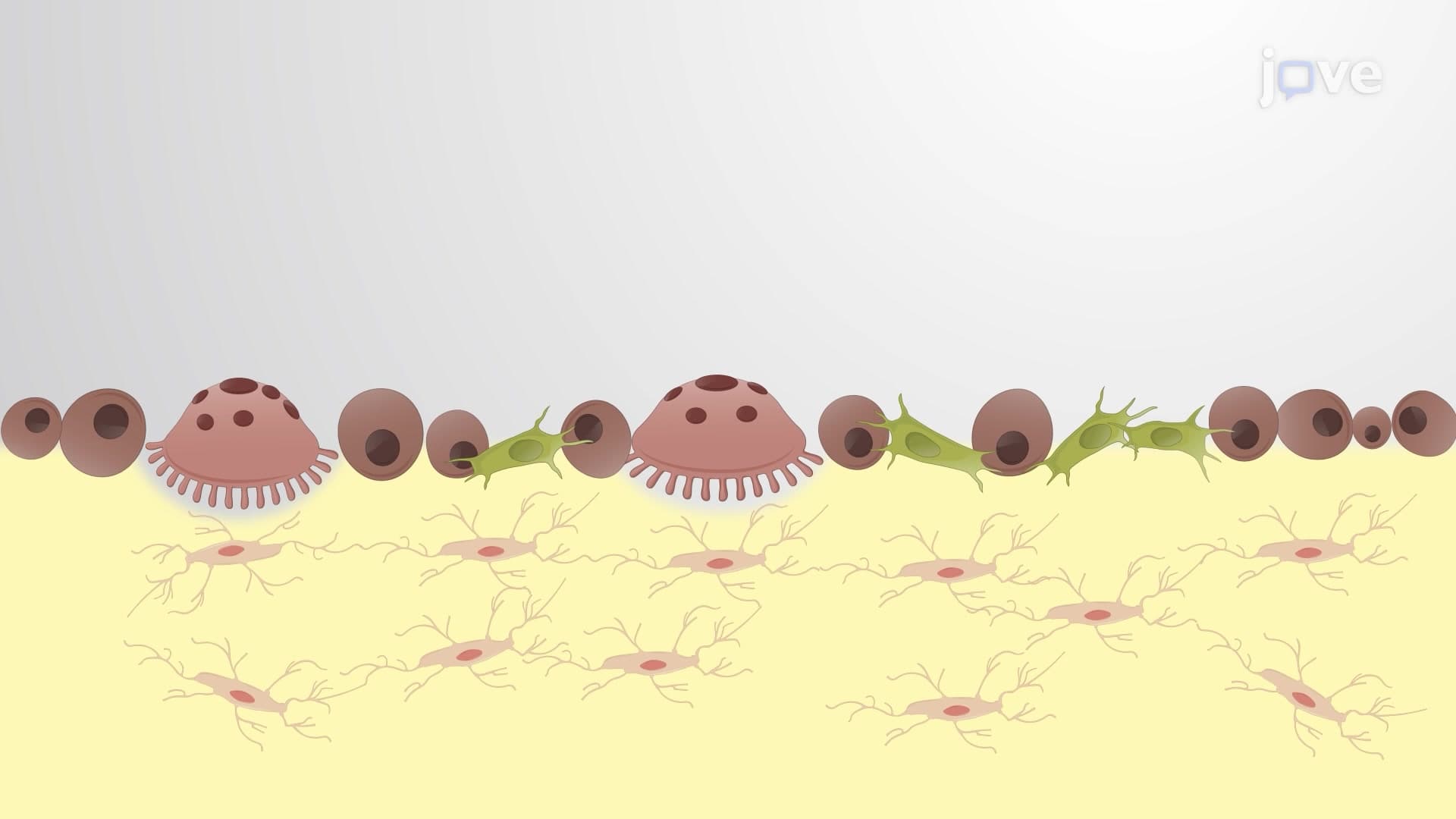
The process begins when clusters of mesenchymal cells receive developmental signals to become osteoblasts, the bone-building specialists. These osteoblasts immediately start secreting osteoid, an unmineralized organic matrix rich in collagen fibers and proteins. Think of osteoid as the scaffolding upon which minerals will crystallize. Within days, calcium phosphate crystals begin depositing on this organic framework, creating the characteristic hardness we associate with bone tissue.
Students preparing for the MCAT or AP Biology exams should note that this mineralization process involves hydroxyapatite formation, giving bone its incredible strength-to-weight ratio. As osteoblasts continue their work, some become trapped within their own matrix secretions, transforming into osteocytes-the mature bone cells responsible for maintaining bone tissue throughout life.
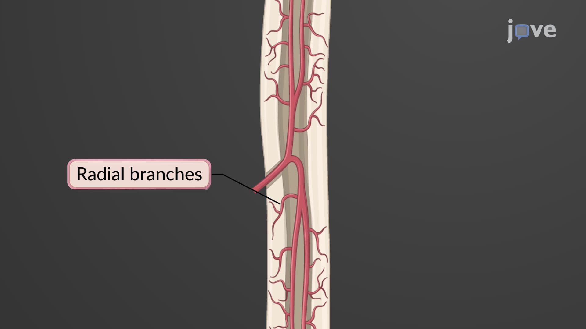
Multiple ossification centers develop simultaneously across the forming bone, each growing outward in a radial pattern. This bone formation membrane explained process critically depends on blood vessel integration. Unlike cartilage, which lacks blood supply, developing bone tissue requires constant nutrient delivery and waste removal. Blood vessels not only nourish the growing tissue but also bring additional mesenchymal cells that can differentiate into more osteoblasts.
For college anatomy students, understanding this vascular relationship explains why bone fractures heal faster than cartilage injuries-bones have superior blood supply supporting repair processes.
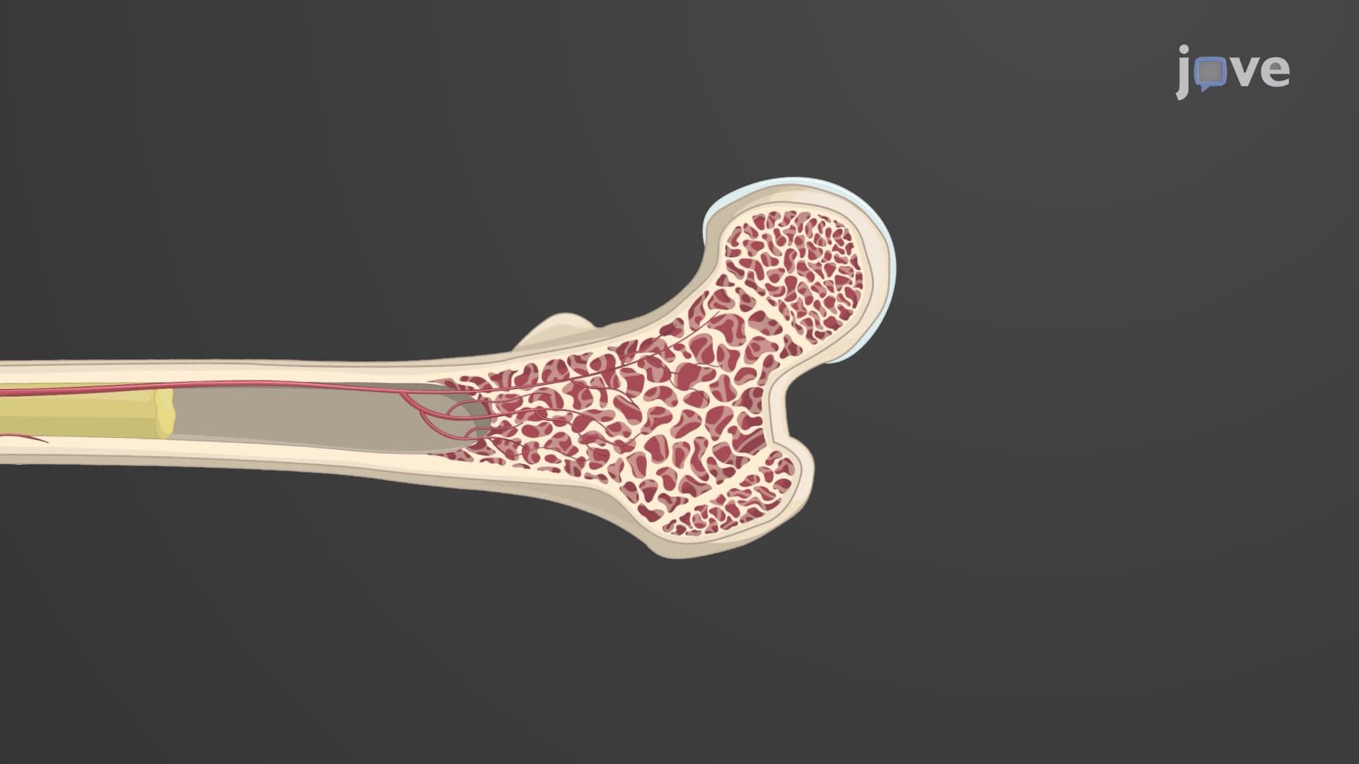
As neighboring ossification centers expand and merge, they create the characteristic spongy bone architecture with its network of bony spicules and interconnected spaces. Near the bone's surface, osteoblasts organize more systematically, laying down concentric layers that form compact bone. This outer compact layer provides structural strength, while the inner spongy region remains metabolically active and houses bone marrow.
The surrounding mesenchyme develops into the periosteum, a vital membrane containing blood vessels, nerves, and stem cells capable of generating new bone tissue throughout life. This periosteal layer becomes crucial for bone repair and remodeling, concepts frequently tested on nursing exams like the NCLEX and HESI A2.
Related Micro-courses