Loading subject page…


132,626 views


Video Summary: What are Classification of Bones
Ever wondered why your thigh bone looks so different from your skull? Bone classification types organize the 206 bones in your skeleton into four distinct categories based on their unique shapes and functions. From the long femur that helps NCAA basketball players jump to the flat skull bones protecting your brain, each bone type serves specific biomechanical purposes. Understanding what are classification of bones reveals how form perfectly follows function in human anatomy. Watch the full video on JoVE Coach to master this concept with expert-led visuals and step-by-step explanations.
Bone classification types represent one of the most fundamental organizing principles in human anatomy, categorizing all 206 adult bones into four distinct morphological groups. This systematic approach helps students, healthcare professionals, and researchers understand how skeletal structure directly correlates with biomechanical function. The classification system emerged from centuries of anatomical study, beginning with Andreas Vesalius's groundbreaking work in the 1500s and refined through modern orthopedic research at institutions like the Mayo Clinic and Johns Hopkins.
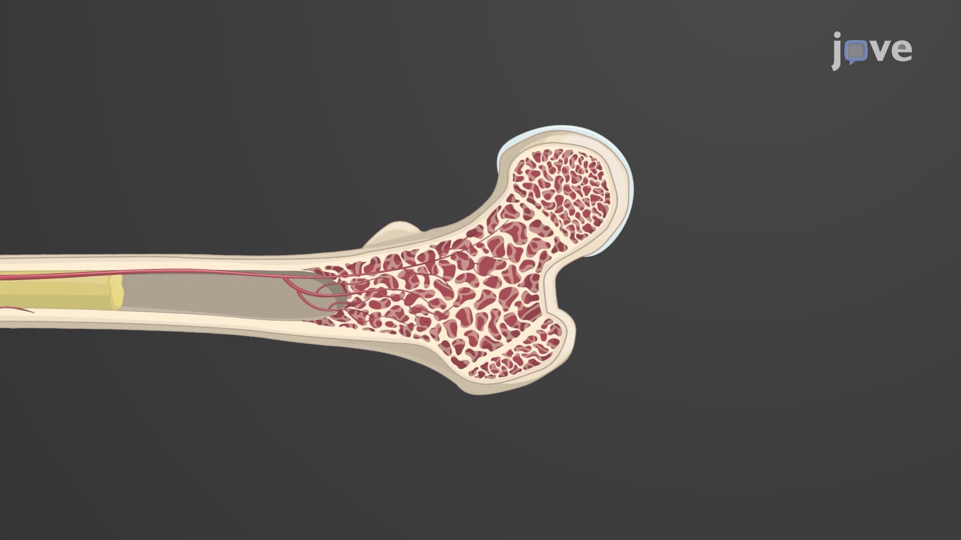
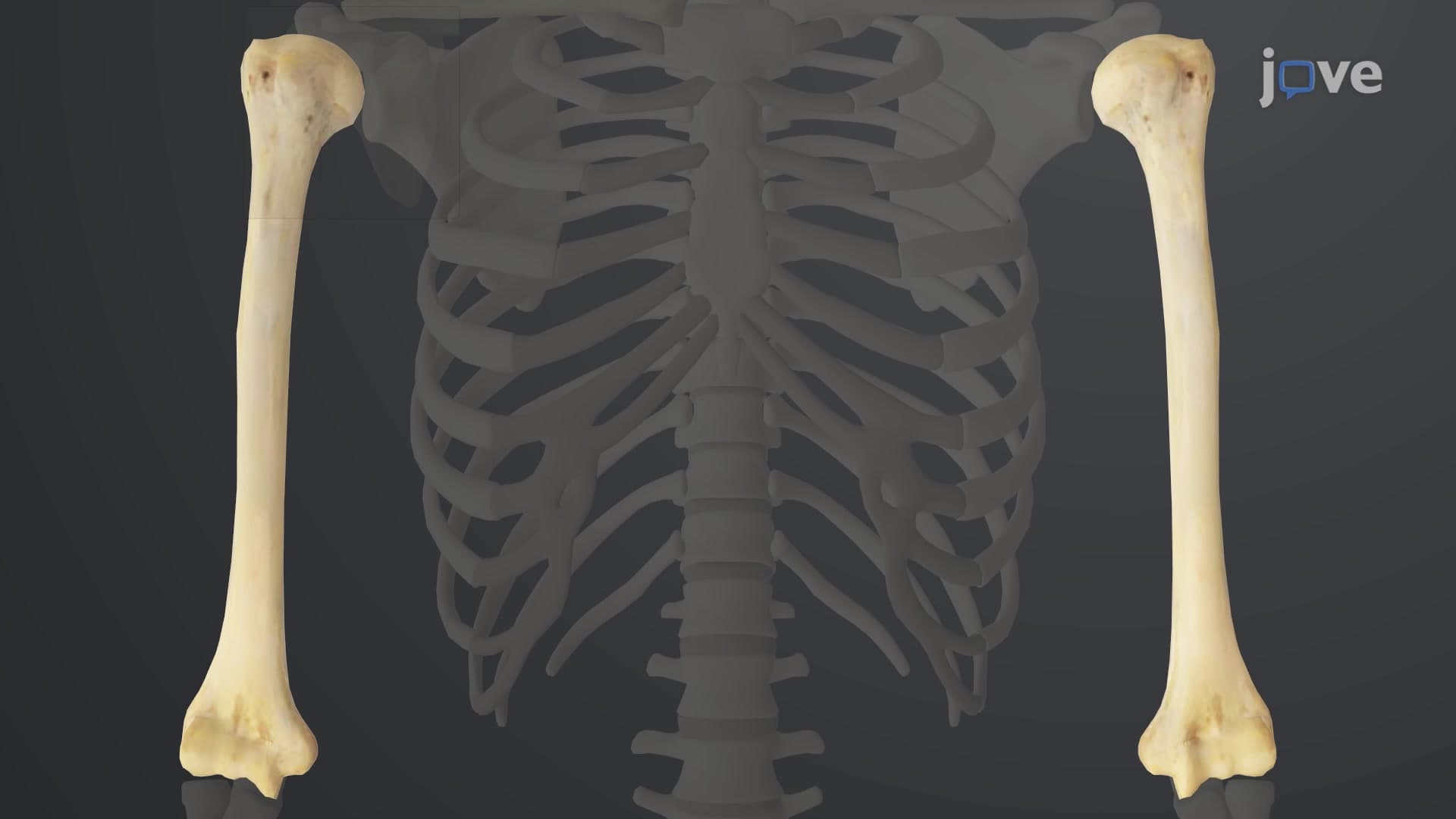
Long bones constitute the primary weight-bearing and locomotion structures in your appendicular skeleton. These cylindrical bones, including the femur (thighbone), humerus (upper arm), and phalanges (finger bones), share common architectural features: a central shaft (diaphysis), expanded ends (epiphyses), and growth plates (metaphyses) during development. The femur, measuring approximately 18 inches in average adults, represents the longest and strongest bone, capable of withstanding forces exceeding 1,700 pounds per square inch during activities like running or jumping. This classification frequently appears on AP Biology exams and MCAT questions, particularly regarding bone growth mechanisms and fracture patterns.
Bones classified in anatomy as short bones exhibit cube-like proportions with roughly equal dimensions, primarily located in weight-bearing regions requiring stability over mobility. The eight carpal bones in your wrist and seven tarsal bones in your ankle exemplify this category, functioning as shock absorbers during impact activities. Sesamoid bones, a specialized subset, develop within tendons and muscles to reduce friction and alter force directions. The patella (kneecap) represents the largest sesamoid bone, while smaller ones exist in hands and feet. Understanding sesamoid bone development proves crucial for USMLE Step 1 preparation, as these structures commonly present diagnostic challenges in radiology.
Flat bones, despite their name, exhibit gentle curves optimized for muscle attachment and organ protection. The skull's parietal and frontal bones shield your brain, while the sternum guards vital cardiovascular structures. These bones contain red bone marrow throughout life, making them primary sites for hematopoiesis (blood cell production). Irregular bones possess complex, composite shapes reflecting multiple functional requirements. Vertebrae combine flat surfaces for weight distribution, curved arches for spinal cord protection, and multiple processes for muscle and ligament attachment. The sphenoid bone in your skull demonstrates ultimate irregular complexity, featuring over 15 distinct anatomical landmarks that medical students must master for neuroanatomy courses and NCLEX-RN examinations.
Related Micro-courses