Loading subject page…


132,610 views


Video Summary: What Is Bone Markings
Ever wonder why a football player's shoulder can withstand crushing tackles? Bone surface markings are specialized features that allow bones to form powerful joints and anchor muscles. From the rounded head of your femur that fits perfectly into your hip socket to the ridges where your bicep attaches to your humerus, these anatomical landmarks determine how your skeletal system functions. What are bone markings becomes clear when you see how they enable everything from a pitcher's fastball to a dancer's leap. Watch the full video on JoVE Coach to master this concept with expert-led visuals and step-by-step explanations.
Bone surface markings represent specialized structural features that transform bones from simple calcium deposits into sophisticated mechanical components. These anatomical landmarks serve as attachment points, joint surfaces, and passageways that enable the complex movements and functions of the human skeleton. In AP Biology and college anatomy courses, students must master these features to understand how bones interact with muscles, ligaments, and other tissues.
The most prominent bone markings are articulating projections that form joints. Condyles are rounded, knuckle-like projections found on bones like the femur, where they create the knee joint with the tibia. These smooth surfaces allow for rolling and gliding movements essential for walking and running. Facets provide flat articulating surfaces, such as those found on vertebrae that allow spinal flexibility while maintaining stability.
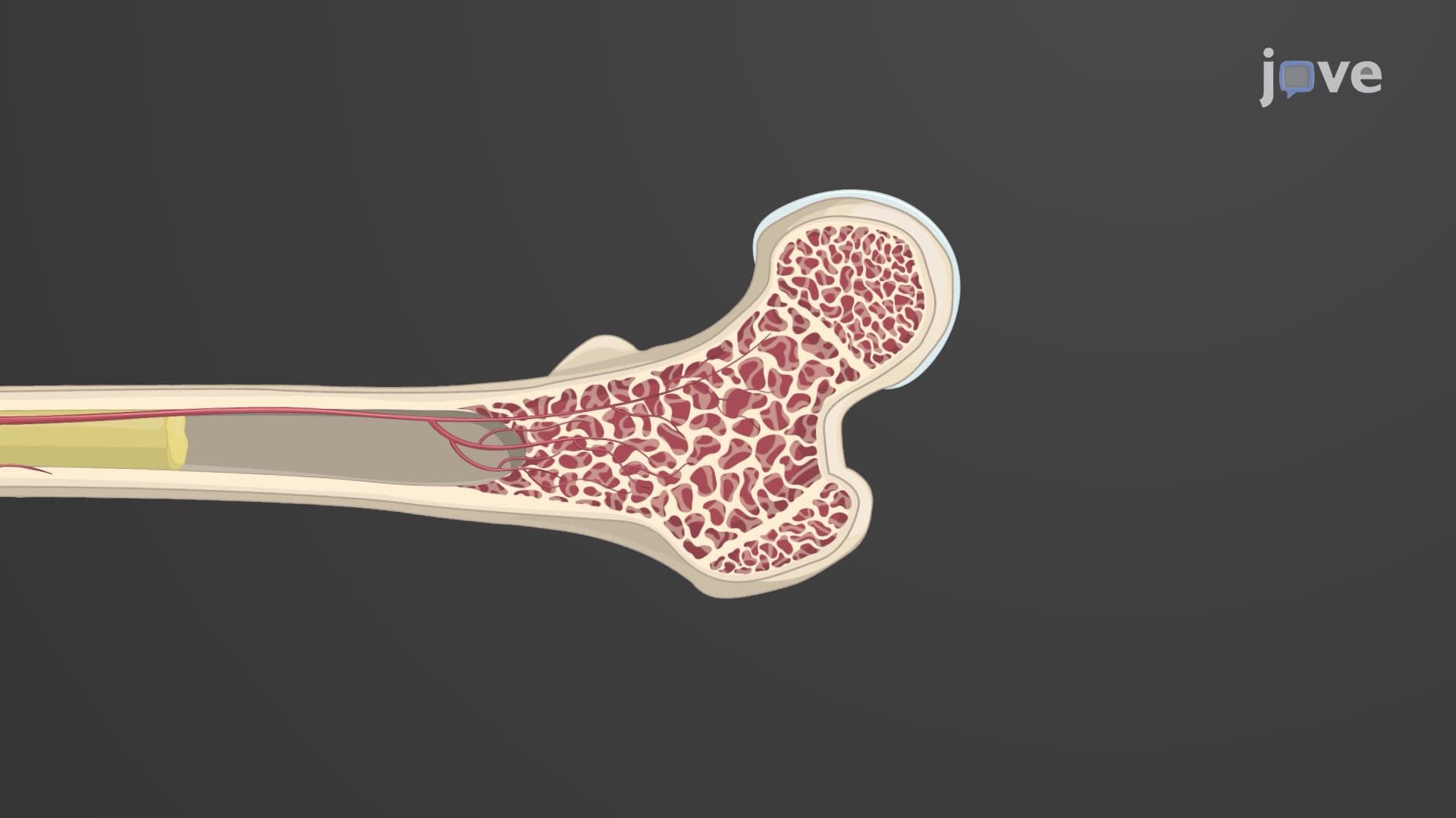
The femur exemplifies how major long bones use a rounded head supported by a narrow neck to create ball-and-socket joints. This design, seen in the hip joint, provides maximum range of motion while distributing weight effectively-crucial knowledge for MCAT students studying biomechanics.
Bones feature various processes that serve as attachment sites for muscles, ligaments, and tendons. Crests are prominent ridges like the iliac crest of the hip bone, where multiple abdominal muscles attach. Spines are sharp, slender projections such as the spine of the scapula, which serves as an attachment point for the trapezius and deltoid muscles.
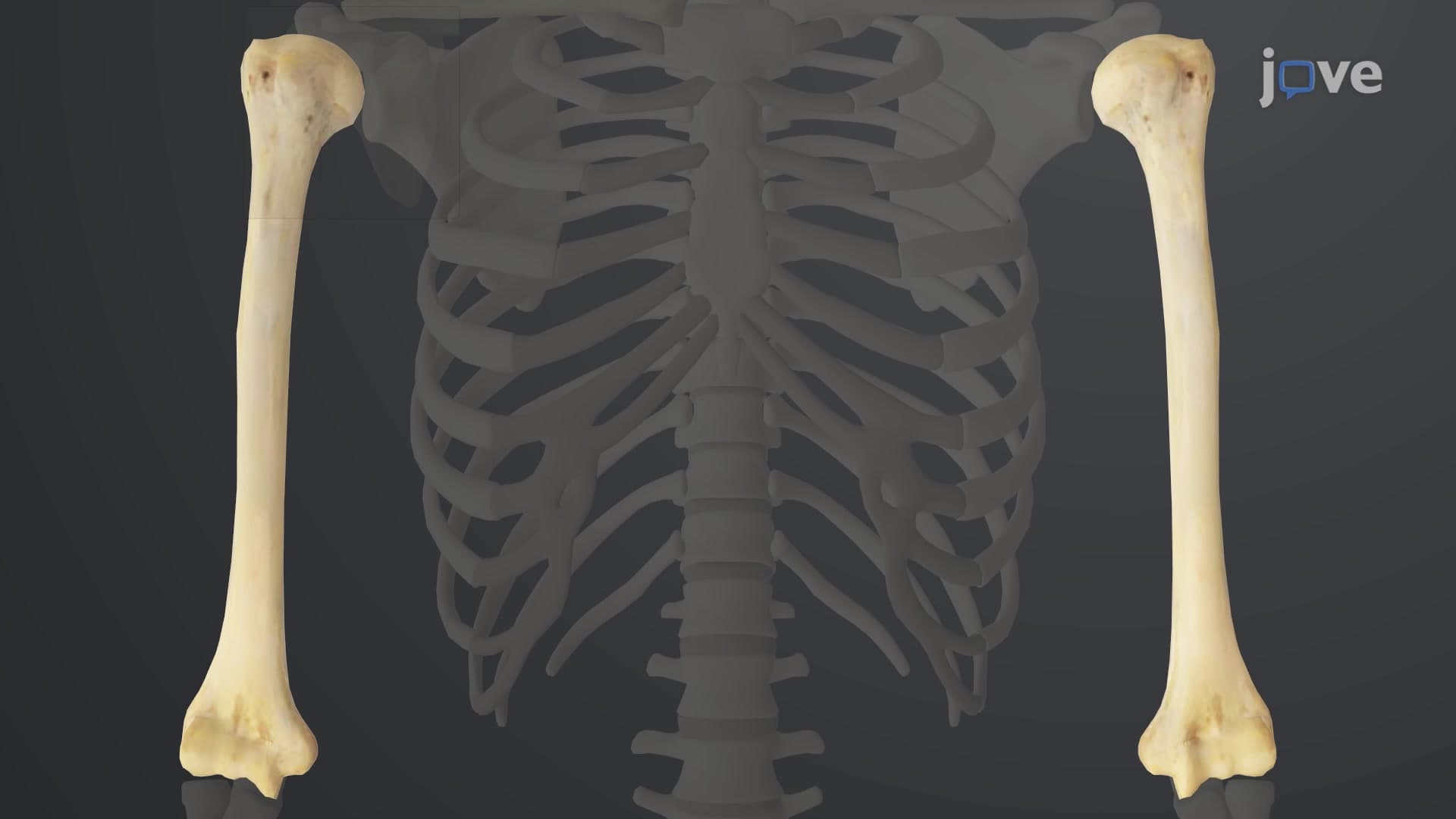
Epicondyles are small, rounded prominences located on condyles, like the medial and lateral epicondyles of the humerus where forearm muscles originate. Tubercles and tuberosities are roughened projections of varying sizes-the greater tubercle of the humerus anchors rotator cuff muscles, while the deltoid tuberosity provides attachment for the powerful deltoid muscle.
Bone depressions accommodate and protect other structures. A fossa is a broad, shallow depression like the glenoid fossa of the scapula, which forms the shoulder socket. Sulci are narrow grooves that house nerves and blood vessels, such as the intertubercular sulcus of the humerus.
Various openings allow passage of vital structures. Foramina are round openings like the foramen magnum in the skull, through which the spinal cord passes. Fissures are narrow, slit-like openings that accommodate nerves and vessels in tight spaces.
Related Micro-courses