Loading video…


 19 Concepts
 132.6K views

Video Summary: What Is Fractures Bone Repair
Did you know that your bones can completely rebuild themselves after a break? The bone fracture repair stages involve a remarkable three-phase healing process that transforms a shattered bone back to its original strength. Consider a basketball player at UCLA who breaks their tibia during practice - their body immediately launches into fracture bone repair, starting with blood clot formation and ending with complete bone reconstruction. Watch the full video on JoVE Coach to master this concept with expert-led visuals and step-by-step explanations.
Bone fracture repair stages represent one of the most sophisticated healing mechanisms in the human body. When University of Michigan researchers studied over 1,000 fracture cases, they found that understanding these stages helps predict healing outcomes and prevent complications. This process transforms what appears to be devastating tissue damage into complete structural restoration.
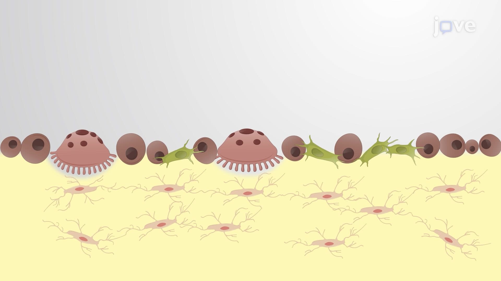
The fracture healing process explained begins immediately after injury with the reactive phase. Within minutes of a bone break, damaged blood vessels create a fracture hematoma - essentially a specialized blood clot that acts as the foundation for repair. This hematoma callus formation disrupts normal circulation, causing nearby bone cells to die and creating an inflammatory response. Emergency departments at hospitals like Johns Hopkins see this phase lasting 1-2 weeks, during which phagocytes and osteoclasts arrive to clean cellular debris. Students preparing for the MCAT should remember that inflammation here is beneficial, not harmful - it's the body's way of clearing the construction site.
The reparative phase demonstrates how do fractures heal during bone repair through cellular migration and tissue formation. Fibroblasts and chondroblasts move from healthy bone regions into the fracture gap, creating collagen fibers and fibrocartilage. This fibrocartilaginous callus stage produces what orthopedic surgeons call "soft callus" - a flexible bridge connecting bone fragments. Cleveland Clinic studies show this phase typically lasts 6-8 weeks. As healing progresses, osteoprogenitor cells differentiate into osteoblasts, beginning bony callus bone repair by forming spongy bone trabeculae. The soft callus gradually transforms into hard callus, creating a bulky but functional repair.
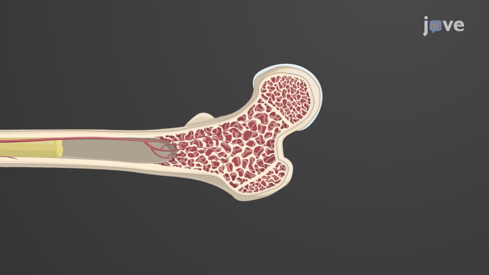
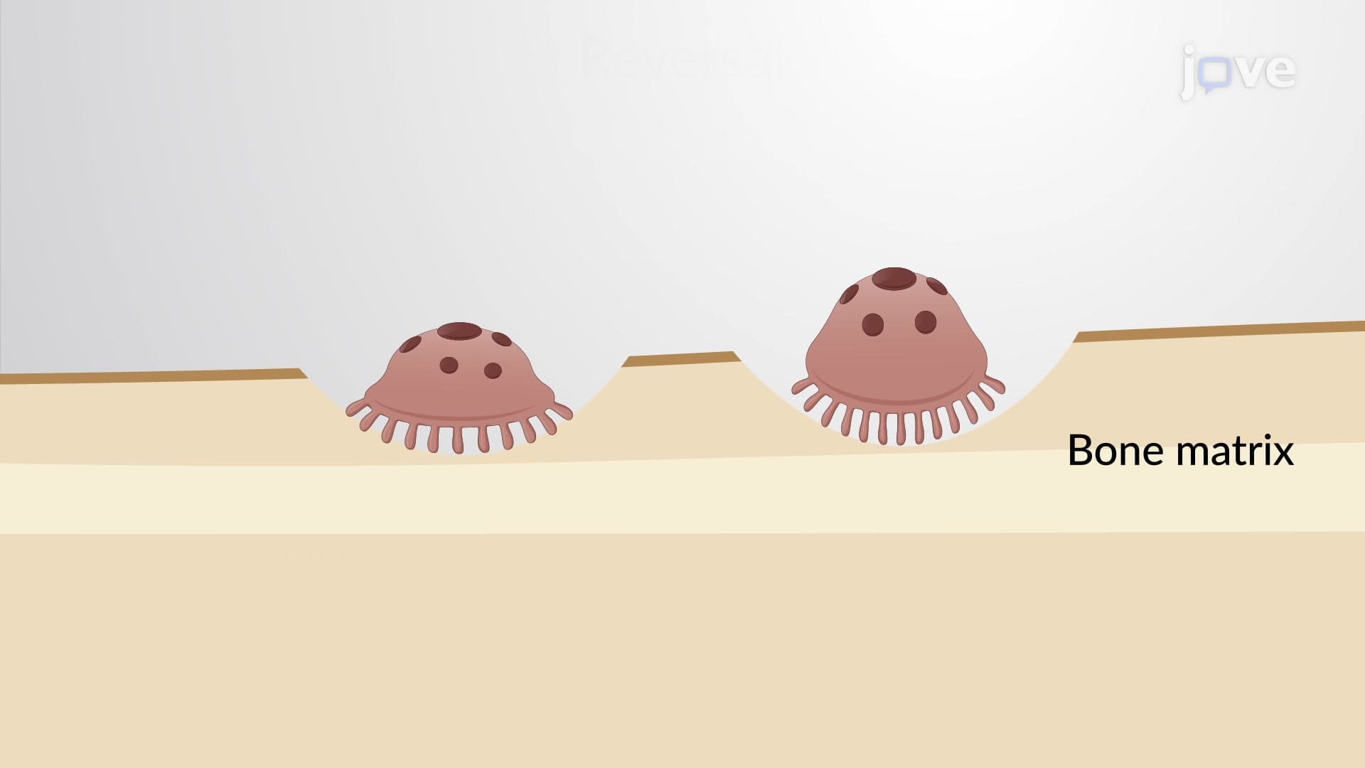
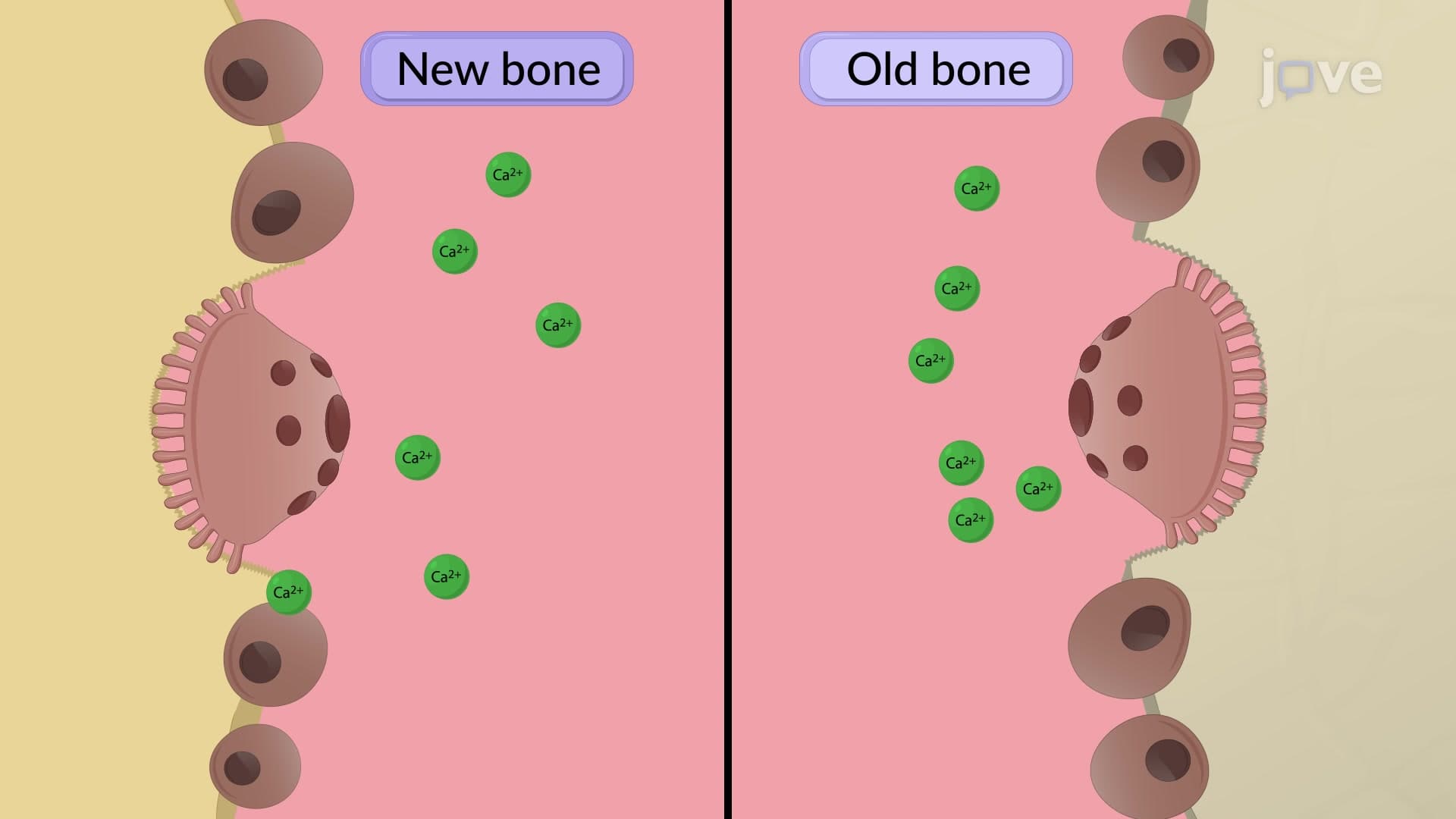
The final fracture remodeling stage can last months to years, depending on the patient's age and fracture severity. Mayo Clinic orthopedic specialists observe how osteoclasts and osteoblasts work together to replace spongy bone with compact bone, restoring the original bone architecture. This phase is crucial for AP Biology students to understand because it demonstrates bone's dynamic nature - constantly breaking down and rebuilding tissue. The remodeling process explains why properly healed bones often become stronger than before the injury, a phenomenon documented in Stanford University biomechanics research.
Related Micro-courses