Loading video…


 19 Concepts
 132.6K views

Video Summary: What Is Gross Anatomy of Bone
Ever wonder why your femur can support your entire body weight during a basketball jump shot? The bone gross anatomy overview reveals a sophisticated structural design where each component serves a specific mechanical function. From the thick-walled diaphysis that acts like a steel beam to the spongy epiphyses that distribute joint forces, understanding what is the gross anatomy of a bone explains how human bones achieve remarkable strength while remaining lightweight. Watch the full video on JoVE Coach to master this concept with expert-led visuals and step-by-step explanations.
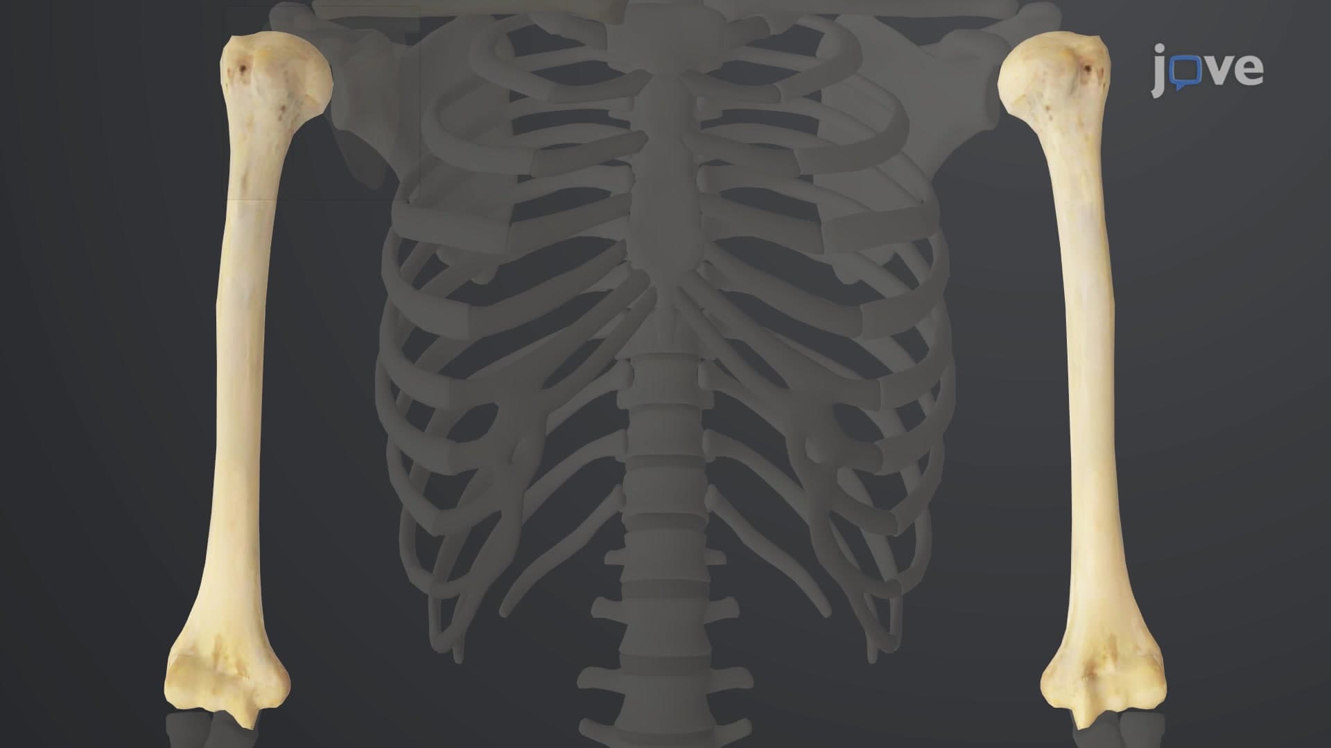
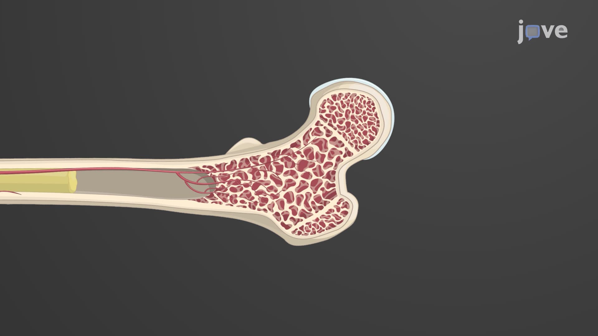
The gross anatomy of bone is best understood through long bones like the femur, humerus, or tibia-bones that serve as the primary load-bearing structures in your appendicular skeleton. These bones demonstrate a sophisticated architectural design that engineers often study when developing lightweight yet strong materials for bridges and aircraft.
The diaphysis forms the central shaft and represents the bone's primary structural column. Its thick walls of compact bone create a hollow cylinder-an engineering marvel that maximizes strength while minimizing weight. This design principle appears in everything from airplane wings to construction beams. The metaphysis serves as the transition zone where the narrow diaphysis flares outward, distributing mechanical stress over a larger area before reaching the epiphyses.
The epiphyses function as the bone's shock absorbers and joint interfaces. Their spongy bone internal structure creates a honeycomb-like framework that can compress under load and spring back, protecting joints during high-impact activities like running or jumping. This is why athletes rarely fracture their epiphyses during normal sports activities, despite the enormous forces generated.
The periosteum serves dual roles that are crucial for understanding bone biology on exams like the AP Biology or MCAT. Its outer fibrous layer provides attachment points for muscles, ligaments, and tendons-essentially serving as the bone's connection hub to the muscular system. The inner osteogenic layer contains stem cells that activate during fracture repair, making it essential for clinical applications in orthopedic medicine.
Understanding these relationships helps explain why periosteum preservation is critical during surgical procedures at major medical centers like Johns Hopkins or Mayo Clinic.
The medullary cavity's contents change dramatically with age, transitioning from red hematopoietic marrow to yellow adipose marrow. This transition explains why bone marrow biopsies in adults typically target the pelvis or sternum rather than long bone shafts. In medical school anatomy courses and USMLE preparation, students must understand how this distribution affects both normal physiology and pathological conditions like leukemia or aplastic anemia.
Related Micro-courses